Introduction
Atrioventricular septal defect (AVSD) represents a spectrum of defects of the endocardial cushion with variable deficiency of the atrial and ventricular septa, common atrioventricular junction, and abnormalities of the atrioventricular valves.Reference Ebels, Anderson, Devine, Debich, Penkoske and Zuberbuhler1–Reference Allen, Driscoll, Shaddy and Feltes3 Within this spectrum, lesions are traditionally categorised as complete or incomplete atrioventricular septal defects, the latter encompassing partial and so-called transitional forms, based on ventricular septal involvement assessed by echocardiography and/or surgical inspection.Reference Anderson, Ho, Falcao, Daliento and Rigby2–Reference Calkoen, Hazekamp and Blom4 Classic morphologic classifications have consistently emphasised that the shared annular hinge point defines the lesion.Reference Becker and Anderson5–Reference Buratto, McCrossan and Galati9 Despite this fundamental principle, terminology applied to the incomplete atrioventricular septal defects spectrum remains inconsistent in contemporary clinical practice.Reference Rigby6
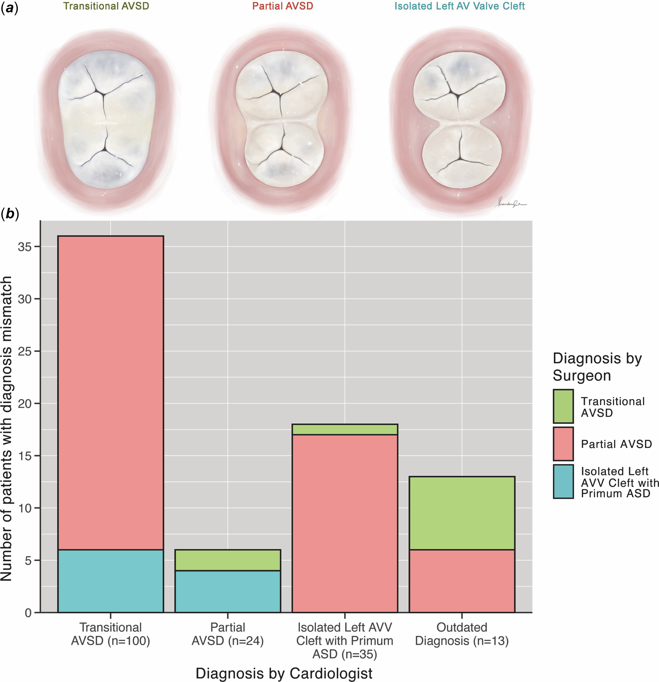
Differences in diagnostic perspectives arise between cardiologists and surgeons working collaboratively.Reference Rigby6 Ecocardiography classifies the AVSD primarily by assessment of both atrial and ventricular septal defects, presence of a common or divided annulus, and annular attachments. In contrast, surgical classification relies on direct intraoperative visualisation of the common annulus, bridging leaflet attachments, and inlet septal morphologyReference Calkoen, Hazekamp and Blom4,Reference Espinola-Zavaleta, Mũoz-Castellanos, Kuri-Nivón and Keirns7,Reference Craig10 (Figure 1a). As a result, lesions that share a common anatomic substrate may receive different diagnostic labels depending on the modality and discipline interrogating the anatomy. This ambiguity is particularly pronounced in lesions classified as transitional atrioventricular septal defects.Reference Rigby6
(a) Morphologic continuum of incomplete AVSD illustrated enface view of the leaflets of atrioventricular junction. The left is transitional AVSD, the middle is partial AVSD, and the right is isolated LAVV cleft with primum ASD. (b) Alternative diagnostic classifications assigned by preoperative echocardiographic and intraoperative anatomic diagnosis in discordant AVSD cases.

Figure 1. Long description
Panel A: The image shows three illustrations of atrioventricular valve leaflets. From left to right, these are labeled as Transitional AVSD, Partial AVSD, and Isolated Left AV Valve Cleft. Each illustration depicts the leaflets of the atrioventricular junction from an enface view. Panel B: The bar graph displays the number of patients with diagnosis mismatches between cardiologists and surgeons. The x-axis is labeled ‘Diagnosis by Cardiologist’ and includes categories: Transitional AVSD (n=100), Partial AVSD (n=24), Isolated Left AVV Cleft with Primum ASD (n=35), and Outdated Diagnosis (n=13). The y-axis is labeled ‘Number of patients with diagnosis mismatch’. The graph uses different colors to represent diagnoses by surgeons: green for Transitional AVSD, red for Partial AVSD, and blue for Isolated Left AVV Cleft with Primum ASD. The bars show the distribution of mismatches across these categories.
The persistent diagnostic variability has important clinical implications that are frequently overlooked. Nomenclature directly influences operative planning, expectations regarding atrioventricular valve repair, perioperative risk stratification, and the way patients are coded within institutions and multicentre databases.Reference Craig10–Reference Agarwal, Lakhdhir, Reveler and O’Sullivan13 Morphologic atrioventricular septal defects, including its incomplete forms, is uniformly associated with intrinsic abnormalities of the left atrioventricular valve and carries a well-recognised risk for postoperative valve dysfunction and the need for reintervention.Reference Ebels, Anderson, Devine, Debich, Penkoske and Zuberbuhler1,Reference Murashita, Kubota, Oba, Aoki, Matano and Yasuda14–Reference Alsoufi, Al-Halees and Khouqeer16 Clear communication, rather than semantic precision alone, is therefore essential for meaningful interdisciplinary care and outcome comparison.
The purpose of this study was to evaluate the frequency and clinical significance of diagnostic discrepancies within the spectrum of incomplete atrioventricular septal defects. Specifically, we aimed to (1) quantify concordance between preoperative echocardiographic diagnosis, intraoperative diagnosis based on surgeon’s visulisation, and standardised morphology-based review, and (2) determine whether diagnostic discordance is associated with differences in either early postoperative recovery, atrioventricular valve performance, reintervention rates, or long-term ventricular function. By analysing diagnostic transitions within a consecutive surgical cohort, we sought to characterise contemporary classification variability and its clinical relevance, rather than to propose a new taxonomy.
Methods
We retrospectively reviewed paediatric patients who underwent surgical repair of incomplete atrioventricular septal defects at a single tertiary referral centre between November 2000 and February 2025. Complete atrioventricular septal defects and single ventricle pathway diagnoses were excluded.
The design of the study was approved, and a waiver of consent was obtained by the Children’s National Hospital Institutional Review Board (Pro00015566). Demographic data, preoperative and postoperative echocardiographic studies, operative reports, and clinical outcomes encompassing early postoperative recovery and late reintervention were reviewed from institutional records to identify subtype-specific factors influencing postoperative course.
Three independent diagnostic classifications were evaluated for each patient:
-
1. Preoperative echocardiographic diagnosis was defined as a diagnosis documented in the preoperative echocardiography report.
-
2. Intraoperative anatomic diagnosis was defined as the lesion classification recorded by the operating surgeon in the operative note. Each operative report has a specified section that highlights operative findings, which highlights the pathology of the lesion.
-
3. The researcher diagnosis was assigned by a congenital cardiac surgeon who performed a secondary, blinded review based exclusively on operative anatomic descriptions. This classification was an intentionally morphology-based description of the operative findings and did not incorporate echocardiographic shunt physiology or functional orifice appearance.
Morphology-based classification, which served as the reference definition throughout the results and discussion, was defined as follows:
Transitional atrioventricular septal defects – Common atrioventricular junction with ± ventricular septal defect, which may be partially closed by accessory valve tissue. Bridging leaflets fusion partially divides the atrioventricular orifice, but the annulus remains morphologically common and at the ventricular septal crest. Presence of primum atrial septal defect.
Partial atrioventricular septal defects – Common atrioventricular junction with ± ventricular septal defect. Bridging leaflet fusion is well formed at the ventricular septal crest, giving the appearance of a divided annulus. Presence of primum atrial septal defect and cleft left atrioventricular valve.
Isolated LAVV cleft with primum atrial septal defect Primum atrial septal defect with cleft left atrioventricular valve. Although the valve may appear phenotypically similar to mitral leaflet morphology with a cleft, the spatial orientation instead is more appropriately that of a shared atrioventricular valve, resulting in the appearance of separate annuli. Complete fusion at the crest. No ventricular septal defect.
Fenestration was used selectively in patients with elevated pulmonary vascular resistance, borderline ventricular compliance, or concern for postoperative left atrial hypertension. Prior surgical leadership positioned the patch at all times to leave the coronary sinus in the left atrium in order to minimise the risk of injury to the conduction axis and nodal triangle. In our current practice the coronary sinus is left draining to the right atrium when with the atrial septal defect patch closure.
Diagnostic concordance and discordance among these three classifications were assessed.
Descriptive and comparative analyses were performed using R version 4.3.3 (R Foundation for Statistical Computing, Vienna, Austria). Patients were grouped into three subtypes of incomplete atrioventricular septal defects according to the surgeon’s operative diagnosis. Categorical variables are presented as counts and percentages, and continuous variables as medians with interquartile ranges or means with standard deviations, as appropriate. A Sankey diagram was constructed to visually depict transitions between preoperative cardiology diagnosis and intraoperative surgical diagnosis. All statistical tests were two-sided, with a predefined significance level of α = 0.05. P-values are reported to three decimal places, and 95% confidence intervals are provided when appropriate. Due to minimal missing data, a complete-case analysis was performed. Outdated diagnoses referred to nonspecific or legacy terms (e.g., “endocardial cushion defect”) that could not be reliably mapped to contemporary atrioventricular septal defect subtypes and were analysed separately.
Results
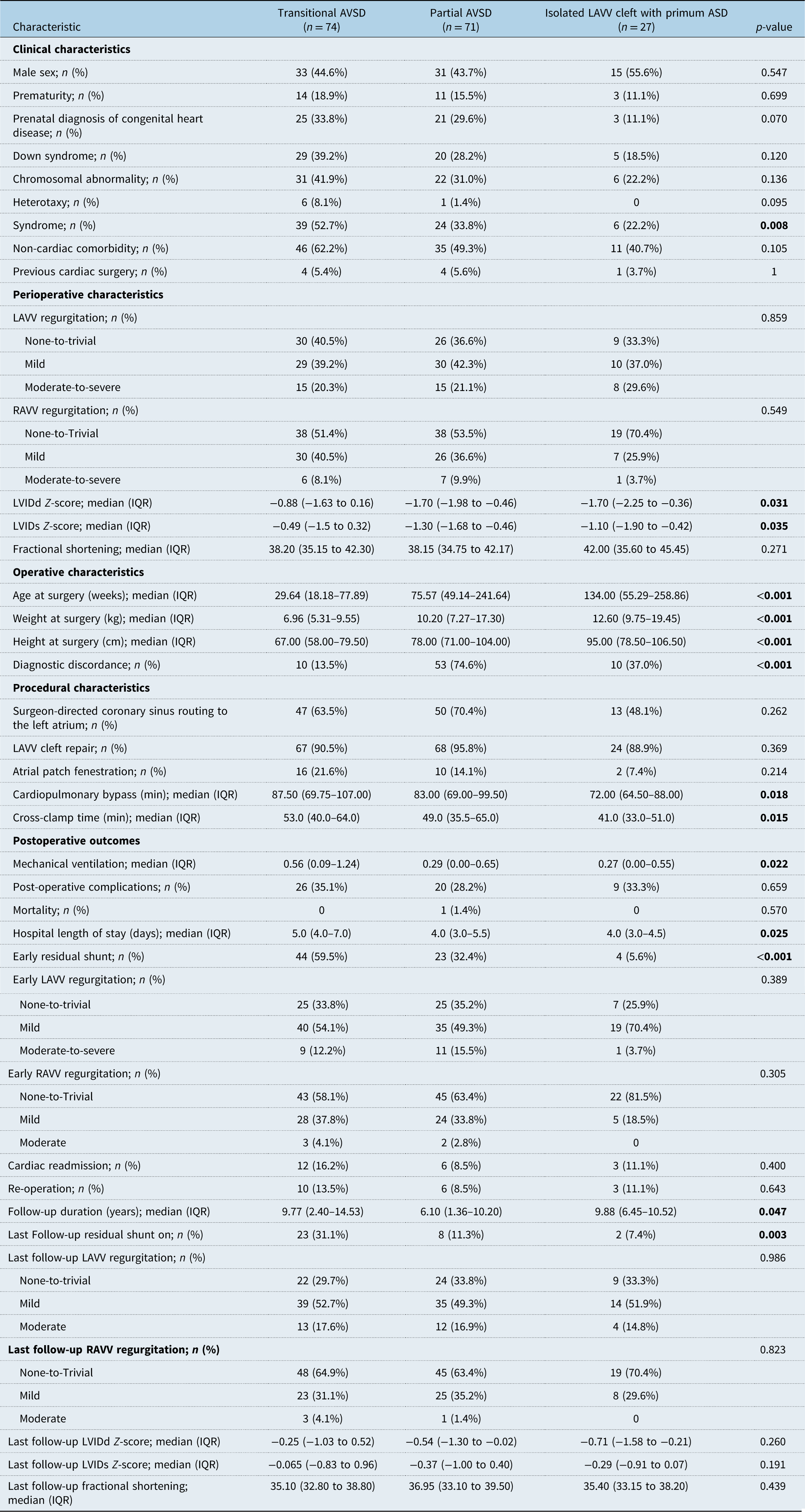
A total of 172 patients who underwent surgical repair of incomplete atrioventricular septal defects were classified by intraoperative surgeon-diagnosis as transitional atrioventricular septal defects (n = 74), partial atrioventricular septal defects (n = 71), and isolated LAVV cleft with primum atrial septal defect (n = 27). Baseline demographics, including sex distribution and prematurity, were similar across diagnostic subtypes (Table 1). Non-cardiac comorbidity, defined as any pre-existing medical condition unrelated to cardiac anatomy or function, was also comparable between the groups. The prevalence of syndromic conditions was significantly different between groups, with Down syndrome followed by heterotaxy being the most associated syndrome. Transitional atrioventricular septal defects were associated with a higher frequency of syndromic diagnosis when compared with partial atrioventricular septal defects and isolated LAVV cleft with primum atrial septal defect (52.7% versus 33.8% versus 22.2%, p = 0.008).
Baseline and perioperative characteristics by intraoperative anatomic diagnosis of AVSD subtype.

Table 1. Long description
The table compares baseline and perioperative characteristics by intraoperative anatomic diagnosis of AVSD subtype. It has 27 rows and 12 columns. The columns are labeled: Characteristic, Transitional AVSD (n = 14), Partial AVSD (n = 71), Isolated LAWV left with primum ASD (n = 27), and p-value. The rows include various characteristics such as sex, prematurity, Down syndrome, chromosomal abnormality, heterotaxy, syndromic, non-cardiac comorbidity, previous cardiac surgery, LAWV regurgitation, RAWV regurgitation, LVIDD Z-score, and LVIDs Z-score. Each row provides specific data points for each AVSD subtype and a p-value indicating statistical significance. Notable trends include the prevalence of syndromic conditions being significantly different between groups, with Down syndrome being the most associated syndrome.
LVIDd = Left Ventricular Internal Diameter (Diastolic); LVIDs = Left Ventricular Internal Diameter (Systolic).
Age, weight, and height at surgery differed markedly across diagnostic subtypes. Patients with transitional atrioventricular septal defects underwent repair at a significantly younger age and lower body size compared with the other groups (p < 0.001), reflecting earlier clinical presentation and surgical indication in this subtype. Operative procedures were similar across diagnostic groups, including rates of cleft repair, atrial septal defect fenestration, and coronary sinus positioning. However, transitional atrioventricular septal defect repairs were associated with greater operative complexity, with longer cardiopulmonary bypass times and aortic cross-clamp durations comparable with partial atrioventricular septal defects, and shortest in isolated LAVV cleft with primum atrial septal defect (both p < 0.005).
Preoperative echocardiographic assessment demonstrated greater ventricular dilation in transitional atrioventricular septal defects, with higher left ventricular end-diastolic dimension Z-scores compared to partial atrioventricular septal defects and isolated left atrioventricular valve cleft with primum atrial septal defect (−0.88 versus −1.70 versus −1.70, p = 0.031). A similar pattern was observed for left ventricular end-systolic dimension Z-scores (p = 0.035), while preoperative ventricular systolic function as assessed by fractional shortening did not differ significantly. Overall, moderate-to-severe left atrioventricular regurgitation was more frequently observed than right atrioventricular valve involvement across all diagnostic subtypes.
Diagnostic discordance
Diagnostic discordance varied significantly by subtype and reflected systematic reclassification patterns rather than random disagreement. Although discordance was most frequently observed among cases ultimately classified as partial atrioventricular septal defects (74.6%) and isolated LAVV cleft with primum atrial septal defect (37.0%) (p < 0.001), this pattern primarily reflected overclassification of transitional atrioventricular septal defects on preoperative echocardiography.
Among patients labelled as transitional atrioventricular septal defects by cardiologists, only 58.7% were confirmed intraoperatively. In comparison, 27.5% were reassigned to partial atrioventricular septal defects and 5.5% to primum atrial septal defect with mitral cleft, accounting for the majority of discordant cases (Figure 1b).
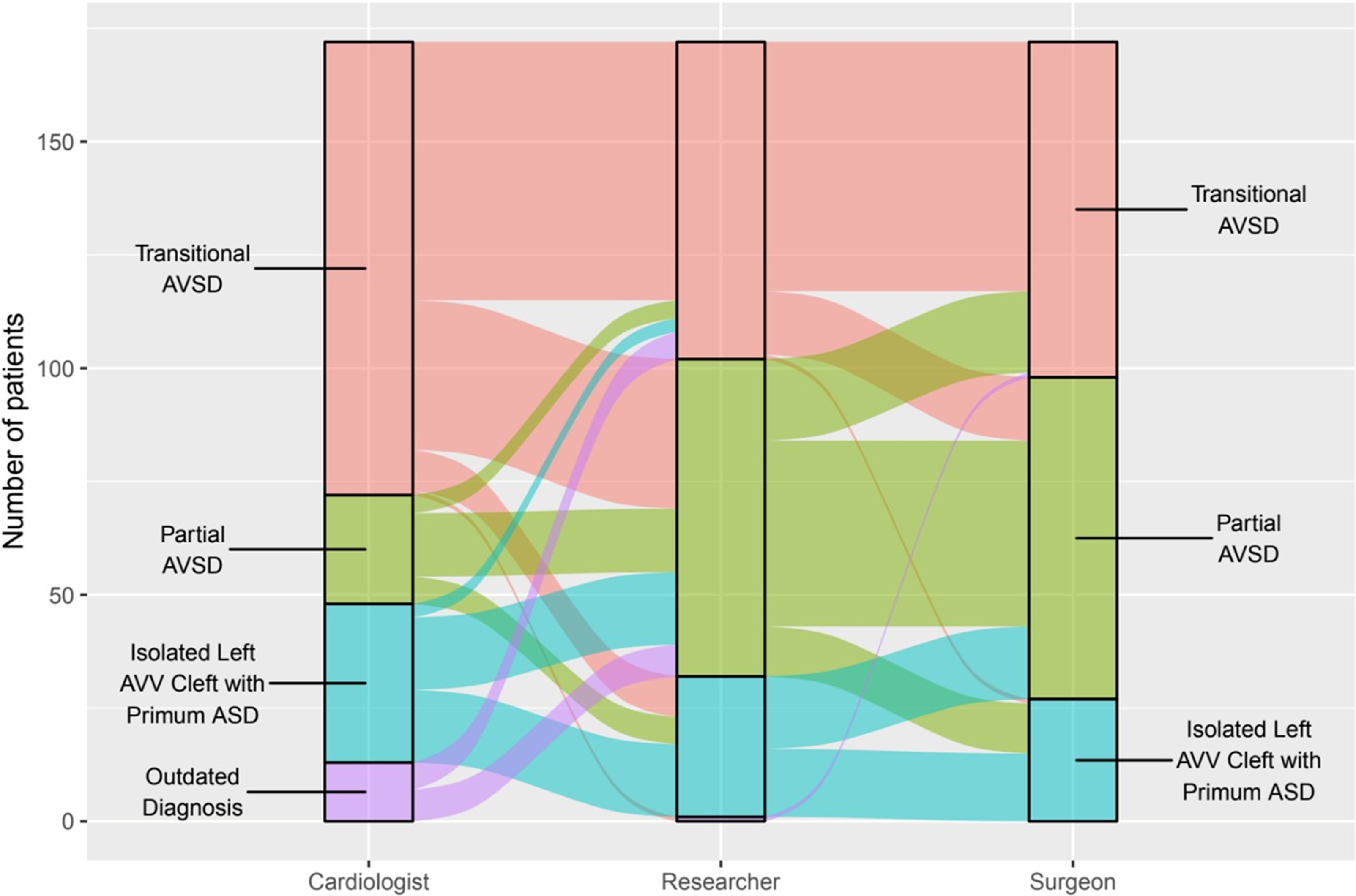
As illustrated in Figure 2, the greatest diagnostic drift occurred between preoperative echocardiographic assessment and intraoperative surgical classification, with relatively stable agreement thereafter. Once operative anatomic features were incorporated at the researcher’s diagnosis, diagnostic categories showed minimal shift, indicating preservation of anatomic concordance between surgical and morphology-based review. Transitional atrioventricular septal defects functioned as a boundary category in preoperative echocardiographic labelling, whereas partial atrioventricular septal defects and isolated LAVV cleft with primum atrial septal defect demonstrated substantial overlap in preoperative diagnostic assignment.
Diagnostic transitions across preoperative cardiology, intraoperative surgery, and researcher-assigned AVSD classification.

Once the operative anatomic details were systemically incorporated at the researcher level, diagnostic categories stabilised with minimal shifts (37.2%) between surgeon and researcher classifications; among these, 57.8% reflected reclassification between transitional and partial atrioventricular septal defects, while the remaining 42.2% involved disagreement between partial atrioventricular septal defects and isolated LAVV cleft with primum atrial septal defect.
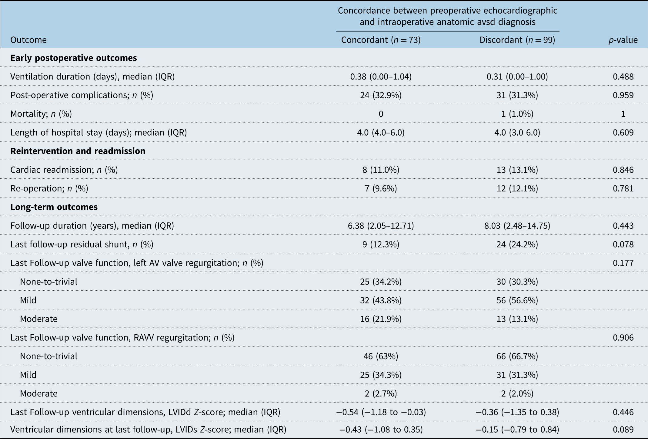
When patients were stratified by diagnostic concordance between preoperative cardiology and intraoperative surgical assessment, no significant differences were observed in early postoperative outcomes, reintervention rates, or long-term ventricular function (Table 2). However, a trend toward higher residual shunt at last follow-up was observed in discordant cases compared with concordant cases (24.2% versus 12.3%, p = 0.078).
Patient outcomes stratified by concordance between preoperative echocardiographic and intraoperative anatomic AVSD diagnosis

Table 2. Long description
The table presents patient outcomes stratified by concordance between preoperative echocardiographic and intraoperative anatomic AVSD diagnosis. It has 18 rows and 4 columns. The columns are labeled ‘Outcome’, ‘Concordant (n = 73)’, ‘Discordant (n = 99)’, and ‘p-value’. The rows are grouped into three main categories: Early postoperative outcomes, Reintervention and readmission, and Long-term outcomes. Each category includes specific metrics and their corresponding values for concordant and discordant groups, along with p-values indicating statistical significance. Notable metrics include ventilation duration, post-operative complications, mortality, length of hospital stay, cardiac readmission, re-operation, follow-up duration, residual shunt, valve function, and ventricular dimensions.
LVIDd = Left Ventricular Internal Diameter (Diastolic); LVIDs = Left Ventricular Internal Diameter (Systolic).
Reintervention and outcomes
Patients with transitional atrioventricular septal defects required longer durations of mechanical ventilation (0.56 [0.09–1.24] versus 0.29 [0.00–0.65] versus 0.27 [0.00–0.55] days, p = 0.022) and experienced longer hospital stays (5.0 [4.0–7.0] versus 4.0 [3.0–5.5] versus 4.0 [3.0–4.5] days, p = 0.025) compared with partial atrioventricular septal defects and isolated LAVV cleft with primum atrial septal defect, respectively. Post-operative complications occurred in 26 (35.1%), 20 (28.2%), and 9 (33.3%) patients across the three diagnostic groups (p = 0.659). Complications were predominantly arrhythmia, most commonly junctional ectopic tachycardia and other postoperative arrhythmias, several of which required temporary pacing and medical management during the early postoperative period. Additional complications included complete heart block requiring permanent pacemaker implantation (n = 4), sepsis (n = 1), pulmonary hypertensive crisis episode (n = 1), acute respiratory distress requiring reintubation (n = 2), cardiac arrest necessitating extracorporeal membrane oxygenation cannulation (n = 1), and cardiac tamponade (n = 1). Overall mortality was low, with one death (0.6%) occurring in the partial atrioventricular septal defects group, attributable to sepsis complicated by multiorgan failure.
Rates of reoperation specific to the index pathology were low and comparable across diagnostic subtypes, occurring in 19 patients (11.0%) overall (p = 0.643). Seven (9.6%) concordant and 12 (12.1%) discordant patients required reoperation (p = 0.781) (Table 2). Among patients undergoing reintervention, persistent moderate-to-severe left atrioventricular valve regurgitation was present in seven cases, indicating continued risk for future valve-related procedures despite reintervention.
Median duration of follow-up differed modestly between diagnostic groups, with the shortest follow-up observed in partial atrioventricular septal defects (6.10 [1.36–10.20] years) compared with transitional atrioventricular septal defects (9.77 [2.40–14.53] years) and isolated LAVV cleft with primum atrial septal defect (9.88 [6.45–10.52] years, p = 0.047). At last follow-up, the prevalence of residual shunt declined substantially in all groups, with approximately halving in transitional atrioventricular septal defects (59.5% to 31.1%) and a threefold reduction in partial atrioventricular septal defects (32.4% to 11.3%); whereas patients with isolated LAVV cleft with primum atrial septal defect demonstrated a persistently low prevalence of residual shunt with minimal change over time (5.6% to 7.4%). Residual shunt remained more prevalent in transitional atrioventricular septal defects (31.1%) compared with partial atrioventricular septal defects and isolated LAVV cleft with primum atrial septal defect (p = 0.003). Most residual shunts were small and haemodynamically insignificant, not requiring reintervention.
Long-term atrioventricular valve regurgitation severity, ventricular dimensions, and systolic function did not differ significantly between diagnostic subtypes, indicating preserved ventricular performance irrespective of initial classification.
Comment
Despite more than four decades of refinement in nomenclature and imaging, the classification of incomplete atrioventricular septal defects remains conceptually unsettled. From Anderson and Becker’s 1982 “What’s in a name?” editorial to contemporary echocardiographic practice, the central challenge has not changed: translating a complex, three-dimensional morphologic substrate, defined by a common atrioventricular junction and variable leaflet attachments, into discrete diagnostic labels.Reference Calkoen, Hazekamp and Blom4–Reference Rigby6,Reference Jacobs, Burke, Quintessenza and Mavroudis17 The current work is likely one of the most in-depth studies on this topic. Our findings demonstrate that contemporary diagnostic discordance reflects limitations of terminology rather than deficiencies in anatomic understanding, as the minimal shifts between surgeon and researcher diagnoses indicate preserved anatomic concordance. Even when operative anatomy is clearly defined, diagnostic labels may vary by interpretation, underscoring that discordance is often semantic rather than cognitive or anatomical. In contrast, the greatest diagnostic drift occurred between preoperative echocardiographic labelling and intraoperative findings, highlighting the modality-dependent nature of classification rather than true disagreement regarding anatomy. One important mechanism for this difference of opinion could be the left-to-right shunt across the cleft. While it is close to impossible to differentiate echocardiographically, the restrictive left-to-right shunt at the crest of the ventricular septum could represent a small ventricular communication at the cleft that crossed the septum. The surgeon usually finds in this situation a gap between the superior and inferior bridging leaflets that is closed by just one suture. Most surgerons wouldn’t call this a ‘restrictive’ ventricular septal defect, although this small ventricular communication is a sine qua non of a transitional atrioventricular septal defect.
Inconsistencies in terminology persist even within operative surgical reports (Figure 2). Although surgeons accurately identify cardiac anatomy intraoperatively, the diagnostic labels applied may vary according to training, institutional convention, or reporting framework. This distinction is particularly relevant when interpreting surgical databases and retrospective studies, where labels often replace detailed morphologic descriptions. While surgeons consistently recognise atrioventricular septal defect morphology through direct inspection, descriptive terms such as transitional atrioventricular septal defects remain imprecise. This instability likely contributes to favourable outcomes despite diagnostic variability, while simultaneously impairing interdisciplinary communication and shared understanding across care teams.Reference Jacobs, Jacobs and Mavroudis11,Reference Adachi, Uemura, McCarthy and Ho18
Long-term atrioventricular valve regurgitation severity, ventricular dimensions, and systolic function did not differ significantly across groups, indicating preserved ventricular performance irrespective of diagnostic subtype. The absence of meaningful differences in early and mid-term outcomes reinforces the concept that incomplete atrioventricular septal defect variants share a common surgical substrate, even when labelled differently. Rather than representing discrete entities, partial atrioventricular septal defects, transitional atrioventricular septal defects, and primum atrial septal defect with left atrioventricular valve cleft appear to reflect complete and incomplete expressions along a continuous morphologic spectrum. In this context, the enduring question of “What’s in a name?” reflects the intrinsic complexity of the atrioventricular septal defects phenotype and the limitations of applying rigid taxonomic frameworks to a continuous anatomic process.Reference Rigby6
Nearly half of lesions labelled as isolated left atrioventricular valve cleft with primum atrial septal defect were reclassified as partial atrioventricular septal defects based on operative morphology, supporting the view that these entities overlap substantially along the same embryologic–anatomic continuum. The distinction between cleft left atrioventricular valve with primum atrial septal defect and partial atrioventricular septal defects may have evolved over time institutionally, contributing to the apparent splitting of the cleft group demonstrated in Figure 2. Importantly, reoperation rates were comparable among primum atrial septal defect with cleft, partial atrioventricular septal defects, and transitional atrioventricular septal defects, emphasising primum atrial septal defect should not be regarded as a simple lesion but as part of the broader atrioventricular septal defect spectrum with similar long-term risk.
A key finding of this study is that diagnostic discordance observed in 42% of cases between preoperative echocardiographic and intraoperative surgical diagnosis did not translate into worse clinical outcomes. Length of stay, complications, mortality, reintervention, and functional status were similar between concordant and discordant groups. This suggests that experienced congenital teams adapt operative strategy to intraoperative anatomy regardless of preoperative labels. In practice, operative decision-making appears to be driven more by morphology than nomenclature, with multidisciplinary teams converging on a shared anatomic understanding even when diagnostic labels differ.
Consistent with our findings, prior surgical series have demonstrated excellent early and long-term outcomes following atrioventricular septal defect repair, independent of subtype designation.Reference Buratto, McCrossan and Galati9,Reference Minich, Atz and Colan19 Collectively, these data indicate that incomplete atrioventricular septal defect variants share a highly successful surgical substrate, with long-term morbidity driven primarily by valve-related sequelae.Reference Strickland, Riehle-Colarusso and Jacobs20 While reassuring, this should not be interpreted as evidence that nomenclature is inconsequential, particularly for communication, research classification, and longitudinal follow-up.
The highest rate of diagnostic discordance occurred in lesions classified as transitional atrioventricular septal defects by echocardiography, supporting the concept that “transitional” often functions as a boundary term when anatomy falls between traditional categories (Figure 2). Imaging of this region is inherently challenging, as views such as the apical plane are prone to signal dropout when the septum is oriented parallel to the ultrasound beam. Identification of a ventricular septal defect typically relies on colour Doppler demonstration of interventricular shunting. However, accurate delineation may be confounded by the presence of a concurrent left-to-right shunt across an adjacent primum atrial septal defect, as well as by regurgitant flow across one or both atrioventricular valves, which together can obscure or mimic ventricular-level shunting. This instability highlights the inherent limitations of echocardiography in defining atrioventricular junctional morphology. Although advanced modalities such as three-dimensional echocardiography improve anatomic characterisation, they do not fully eliminate these challenges in preoperative assessment.Reference Rigby6,Reference Kutty and Smallhorn21
Our findings support openly acknowledging these limitations and framing diagnostic discordance as an expected consequence of applying rigid terminology to a continuous embryologic and anatomic spectrum. Improved awareness may enhance interdisciplinary communication and research interpretation without altering clinical outcomes. Finally, the high discordance observed in lesions ultimately classified as partial atrioventricular septal defects reflects differing diagnostic frameworks rather than uncertainty in anatomy. From a surgical perspective, interpretation emphasises embryologic and junctional continuity, leading to intraoperative reclassification when a shared substrate is recognised despite physiologic emphasis on preoperative imaging.
In conclusion, these findings suggest that diagnostic discordance in incomplete atrioventricular septal defects is an expected consequence of applying categorical terminology to a continuous embryologic and anatomic spectrum. Importantly, this discordance was not associated with adverse clinical outcomes, underscoring that patient management is driven by morphology rather than nomenclature. Nonetheless, persistent variability in diagnostic labelling remains relevant for interdisciplinary communication, interpretation of multicentre databases, and research comparability. Recognising and explicitly acknowledging these differences may improve shared understanding across disciplines without altering clinical care.
Acknowledgements
We thank Sofia Hanabergh for the visual designs.
Financial support
None.
Competing interests
Yves d’Udekem is a consultant for Bayer Pharmaceutcals.
Ethical standard
The authors assert that all procedures contributing to this work comply with the ethical standards of the on human experimentation in the United States and the Helsinki Declaration of 1975, as revised in 2008, and has been approved by the Children’s National Hospital Institutional Review Board.