Introduction
Nematodes are ancient, biologically diverse and the most abundant metazoans on earth, widely spread in almost all habitat types, including within and on host animals and plants (Laznik et al., Reference Laznik, Ross, Toth, Lakatos, Vidrih and Trdan2009; Blaxter & Denver, Reference Blaxter and Denver2012). It is presumed that the association between nematodes and terrestrial gastropods is both ancient and widespread (Morand et al., Reference Morand, Wilson, Glen and Barker2004). Recent studies indicated that nematodes use terrestrial gastropods as either intermediate or definitive hosts (Diano et al., Reference Diano, Dalan, Singh and Sumaya2022; Gimond et al., Reference Gimond, Poullet, Braendle, Haspel and Hart2022; Schurkman et al., Reference Schurkman, Dodge, Mc Donnell, Tandingan De Ley and Dillman2022).
Caenorhabditis species of the Elegans group are among the well-studied nematodes that are morphologically similar but also show high divergence at genetic level (Félix & Braendle, Reference Félix and Braendle2010) through acquiring changes (mutations) during the course of evolution. One gonochoristic member species is Caenorhabditis brenneri Sudhaus & Kiontke, 2007 (Nematoda: Rhabditida), which is unique among Caenorhabditis as it has the highest within-species molecular diversity (hyperdiverse) among eukaryotes (Dey et al., Reference Dey, Chan, Thomas and Cutter2013). It has been recorded to thrive in soil detritus (Fitch et al., Reference Fitch, Emmons and Baird1994) and was associated with the giant African land snail, Achatina fulica Bowdich, 1822 (Gastropoda: Achatinidae) from the Philippines (Diano et al., Reference Diano, Dalan, Singh and Sumaya2022).
This report of association between C. brenneri and terrestrial gastropod Philippinella moellendorffi Collinge, 1899 (Stylommatophora: Ariophantidae) is the first record of this nematode species and a slug. Furthermore, this study represents the first record of P. moellendorffi in Mindanao, Philippines.
Materials and methods
Slug collection and nematode isolation
Philippinella moellendorffi were collected from Imelda, Zamboanga Sibugay, Mindanao, Philippines (7°40′1.9986″N, 122°59′4.9992″E) from the undergrowth of Arachis pintoi (Fabales: Fabaceae) with an average air temperature of 26°C. Slug samples were thoroughly washed with distilled water to remove adhering dirt or debris and individually placed in sterile containers, fed with surface-sterilized carrots and misted with water daily until death. The first dead slug specimen was examined for nematode occurrence following the method described by De Ley & Mundo-Ocampo (Reference De Ley, Mundo-Ocampo, Chen, Chen and Dickson2004). For this, the slug cadaver was washed with water and placed on nutrient agar media M001 HiMedia®(Maharashtra, India)(0.5% peptone and sodium chloride, 0.15% beef and yeast extracts and 1.5% agar, 7.4 ± 0.2 pH at 25°C) in a 90 mm × 15 mm Petri plate, which served as the seed culture. After 32 h of incubation, the plate was examined and the presence of nematodes was recorded. Up to ~60 individuals of nematodes were first observed in the seed culture plate. Agar blocks with live nematodes were transferred onto freshly plated agar. To obtain a pure culture of nematode, a series of sub-culturing was performed by picking and transferring one gravid female onto another freshly plated agar, previously inoculated with Alcaligenes faecalis, a bacterial strain isolated from P. moellendorffi cadaver.
Morphological and molecular analysis
For morphological and morphometric characterization, the nematode samples were processed based on the protocol of Seinhorst (Reference Seinhorst1959) as modified by De Grisse (Reference De Grisse1969). Fixed slide mounts of nematodes were photographed and measured using an Olympus (Olympys Corporation,Tokyo, Japan) light microscope BX53 equipped with Olympus DP27 microscope digital camera and cellSens imaging software (Olympus Corporation,Tokyo, Japan).
One gravid female from the pure culture was picked into 1.5 ml Eppendorf collection tubes with distilled water, frozen and crushed with blunted pipette tips. The extraction of the total genomic DNA (gDNA) was based on the manufacturer's protocol (Dongsheng Biotech, Inc.,Guangdong, China). Extracted gDNA were stored in 1.5 ml Eppendorf tubes, labelled and sent to Macrogen, South Korea, for polymerase chain reaction (PCR) amplification and sequencing. For 18S ribosomal DNA (rDNA), SSU18A 5′-AAAGATTAAGCCATGCATG-3′ and SSU26R 5′-CATTCTTGGCAAATGCTTTCG-3′ was used with the following reaction conditions: initial preheating at 94°C for 5 min, 35 cycles of (94°C for 1 min; 52°C for 2 min; 68°C for 2 min) and final extension step at 68°C for 10 min (Mwangi et al., Reference Mwangi, Gichuki, Wanjohi, Runo and Maina2016). The D2–D3 segment of the 28S rDNA was amplified by the primer pair, D2A 5′-ACAAGTACCGTGAGGGAAAGTTG-3′ and D3B 5′-TCCTCGGAAGGAACCAGCTACTA-3′ (Nunn, Reference Nunn1992) with the following PCR conditions: denaturation at 95°C for 5 min, followed by 40 cycles of denaturation at 94°C for 30 s, annealing at 55°C for 45 s and extension at 72°C for 1 min; the final extension was performed at 72°C for 10 min (Ye & Giblin-Davis, Reference Ye and Giblin-Davis2013).
Generated sequences were BLASTn-searched to compare with all sequences deposited in the National Centre for Biotechnology (NCBI) (Altschul et al., Reference Altschul, Gish, Miller, Myers and Lipman1990) for closely related species. All obtained sequences were trimmed, annotated and then deposited at the NCBI GenBank. Phylogenetic analysis was done with other identified closely related species based on the BLASTn output. The nematode Steinernema carpocapsae voucher SMF7 (KY914571) and S. carpocapsae strain NCR (KJ950292) were used as outgroups for the phylogenetic relationship construction using the 18S rDNA and D2–D3 segment of 28S rDNA sequences. Multiple sequence alignment was made using MUSCLE in MEGA version 11 (Tamura et al., Reference Tamura, Stecher and Kumar2021) followed by manual trimming of the poorly aligned ends. Phylogenetic analysis using the 18S rDNA and D2–D3 segment of 28S rDNA sequences was constructed using Bayesian phylogenetic analysis (MrBayes 3.2.7, https://nbisweden.github.io/MrBayes/links.html) and carried out using GTR + I + G model; analyses were run under 1 × 10⁶ generations (four runs), Monte Carlo Markov chains were sampled every 100 generations and 20% of the converged runs were regarded as burn-ins (Huelsenbeck & Ronquist, Reference Huelsenbeck and Ronquist2001).
Results and discussion
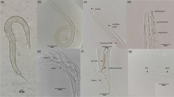
Morphological characteristics of the nematode comprise that of a typical rhabditid, such as a well-developed stoma (cheilostom, gymnostom and stegostom), median and basal bulb, vulva at mid-body filiform tails for females, peloderan-type bursa for males, with spicule longer than gubernaculum (fig. 1). Male: Testis single. Tail enveloped with a closed bursa, sustained by nine pairs of bursal rays. Precloacal hook long and slender; posterior cloacal lip with tongue-like appendage with two cloacal sensilla. Spicules long and slender with evenly, somewhat ventrally curved blade and with a pointed tip. Gubernaculum also slender. Bursa half heart-shaped in lateral view, covering the rod-like papillae. Female: Gonadal system amphidelphic, anterior and posterior gonads are symmetric with each other with visible developing eggs. Long filiform tail occupies 14.8% of body length. Anus and rectum clearly visible. Resolution into species level using morphology based on light microscopy does not guarantee a robust identification; thus, molecular analysis limited to the use of small subunit (18S) and large subunit (D2–D3 expanded segments of 28S) rDNA was also performed.
Caenorhabditis brenneri strain IZSP female (a) and male (b); anterior region showing stoma, median bulb and terminal bulb (c); cheilostom, gymnostom and stegostom (d); female vulva region with embryo (e); male caudal region showing spicule, gubernaculum and bursa (f); female caudal region showing anus and filiform tail (g).

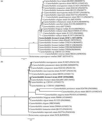
An 18S rDNA gene sequence was generated (MT130970) and was found to be 99.8% closely related to C. brenneri strain Kbcn01 (MT039632) from the Philippines (Diano et al., Reference Diano, Dalan, Singh and Sumaya2022) and was designated as C. brenneri strain IZSP; 99.88% similar with C. brenneri isolates CFB234 (OK086052), CFB135 (OK086050), CFB104 (OK086049) and CFB50 (OK086048) from Vietnam, and 99.40% similar with C. brenneri CB5161 (U13930) (formerly Caenorhabditis sp. CB5161) from Trinidad (Fitch et al., Reference Fitch, Emmons and Baird1994). This was also supported by the D2–D3 expansion segments gene sequence of 28S rDNA (MT043808) with 99.5% similarity to C. brenneri strain Kbcn001 (MT150592) from the Philippines (Diano et al., Reference Diano, Dalan, Singh and Sumaya2022); 99.5% similarity with C. brenneri (AY602175) (formerly Caenorhabditis sp. CB5161) and 98.58% similarity with Caenorhabditis sp. 1 CB5161 (DQ059062) (Hong et al., Reference Hong, Villwock and Sommer2005).
The phylogenetic relationship of the C. brenneri strain IZSP inferred from the 18S rDNA sequences showed that it is closely related and forms a monophyletic clade with C. brenneri strain Kbcn01 (MT039632) from the Philippines, as well as with C. brenneri isolates CFB234 (OK086052), CFB135 (OK086050), CFB104 (OK086049), CFB50 (OK086048) from Vietnam and C. brenneri CB5161 (U13930) from Trinidad (fig. 2a). This is also supported using the D2–D3 expansion segments of 28S rDNA sequence, C. brenneri strain IZSP (MT043808) forms a monophyletic clade with C. brenneri strain Kbcn001 (MT150592) from the Philippines and together forms a sister clade with Caenorhabditis sp. CB5161 (AY602175) and Caenorhabditis sp. 1 CB5161 (DQ059062) (fig. 2b).
Phylogenetic relationships inferred by the 18S (a) and D2–D3 segment of 28S rDNA (b) provide strong evidence that Caenorhabditis brenneri strain IZSP (bold) is closely related to C. brenneri strain Kbcn001, having posterior probability (PP) values of 0.95 PP and 0.97 PP by Bayesian analysis using GTR + I + G model. Scale bar represents the number of nucleotide substitution per siterespectively.

Caenorhabditis brenneri species are distributed pantropically, localized along the equator and regions between the Tropic of Cancer and Capricorn with tropical climates, and are commonly isolated in various habitats, ranging from compost, soil, sometimes in animal dung and oftentimes forming associations with invertebrates (Sudhaus & Kiontke, Reference Sudhaus and Kiontke2007). Though commonly found in environments with abundant concentrations of bacterial food sources (e.g. compost, detritus), Caenorhabditis species were also recorded in association with gastropods such as Caenorhabditis briggsae in snails (Kiontke & Sudhaus, Reference Kiontke and Sudhaus2006; Guerrero et al., Reference Guerrero, Rincon-Orozco and Delgado2018), Caenorhabditis remanei in snails (Baird, Reference Baird1999; Kiontke & Sudhaus, Reference Kiontke and Sudhaus2006) and slugs (Sudhaus, Reference Sudhaus1974; Tandingan De Ley et al., Reference Tandingan De Ley, McDonnell, Lopez, Paine and De Ley2014; Petersen et al., Reference Petersen, Hermann, Barg, Schalkowski, Dirksen, Barbosa and Schulenburg2015; Schurkman et al., Reference Schurkman, Dodge, Mc Donnell, Tandingan De Ley and Dillman2022), Caenorhabditis elegans in slugs (Petersen et al., Reference Petersen, Hermann, Barg, Schalkowski, Dirksen, Barbosa and Schulenburg2015), Caenorhabditis fruticicolae in the snail Fruticola seiboldiana (Shinohara, Reference Shinohara1960), Caenorhabditis formosana and Caenorhabditis oncolmelanie with the snail Oncomelania hupensis (Yokoo & Okabe, Reference Yokoo and Okabe1968) and recently a strain of C. brenneri in A. fulica (Diano et al., Reference Diano, Dalan, Singh and Sumaya2022).
In this study, C. brenneri was isolated from the cadaver of P. moellendorffi, suggesting that the nematode survives the action of radula and the gut system of the gastropod. This observation was also recorded in other free-living Caenorhabditis species such as C. elegans and C. briggsae that were repeatedly isolated in various developmental stages from intestines of slugs (Félix & Duveau, Reference Félix and Duveau2012; Petersen et al., Reference Petersen, Hermann, Barg, Schalkowski, Dirksen, Barbosa and Schulenburg2015). This could be an adaptation for dispersal to a new environment via endophoresy (Sudhaus, Reference Sudhaus2018). It is also possible that C. brenneri hitches a ride on slugs, waits until the vector's death and then feeds on the cadaver. In Caenorhabditis species, obligate parasitic lifestyle is unheard of, even if some species survive in the guts of gastropods. Facultative parasitism can be regarded as a potential transition phase from free-living to parasitic lifestyle. This switching depends on environmental conditions such as food source availability. This could serve as a pre-adaptation towards opportunistic or facultative parasitism (Luong & Mathot, Reference Luong and Mathot2019). Despite these assumptions on the association of C. brenneri strain IZSP with P. moellendorffi, and since only one slug was investigated, further tests are needed to confirm the nature of association by fulfilling Koch's Postulates (Berman, Reference Berman and Berman2019) to prove pathogenicity of C. brenneri strain IZSP on P. moellendorffi or other gastropods. This recovery of C. brenneri from P. moellendorffi is the first report of any Caenorhabditis species isolated from the cadaver of a terrestrial slug.
In conclusion, this study showed for the first time that C. brenneri associates with terrestrial gastropods, further confirming that gastropods are reservoirs for several nematode species. However, the nature of association with P. moellendorffi is unclear, and may be a form of adaptation from free-living to a parasitic lifestyle. Furthermore, this is the first report of C. brenneri isolated from a terrestrial slug.
Acknowledgment
We would like to thank Dr. C. Demayo for his assistance during our microscopy works and to Dr. J.G. Martinez and Dr. M. Villacorte-Tabelin for their technical support. We wish to also thank the anonymous reviewers for their valuable comments and suggestions.
Financial support
This research was supported by the Accelerated Science and Technology Human Resource Development Program-National Science Consortium (ASTHRDP-NSC) of the Department of Science and Technology (DOST), with Master's degree scholarship to L.B.Dalan and M.A.Diano.
Conflicts of interest
None.
Ethical standards
The authors assert that all procedures contributing to this work comply with the ethical standards of the relevant national and institutional guides on the care and use of laboratory animals.