Introduction
Parabronema skrjabini (Habronematidae) is a nematode that parasitizes the abomasum of ruminants and is widely distributed in some Mediterranean countries, Asia and Africa. Many of these countries, including Mongolia (Sharkhuu, Reference Sharkhuu2001), Saudi Arabia (El-Azazy, Reference El-Azazy1990; Magzoub et al., Reference Magzoub, Omer, Haroun and Mahmoud2000), Namibia (Krecek et al., Reference Krecek, Boomker, Penzhorn and Scheepers1990), Turkey (Umur and Yukari, Reference Umur and Yukari2005), India (Yadav et al., Reference Yadav, Sengar and Chauhan1987), Iran (Hasheminasab, Reference Hasheminasab2015; Hasheminasab et al., Reference Hasheminasab, Jalousian and Meshgi2016) and China (Huang and Li, Reference Huang and Li2002; Yang et al., Reference Yang, Yang, Liu, Zhang and Zhang2004), have reported that P. skrjabini is mainly parasitic in ruminants such as goats, sheep, camels, giraffes and elephants, among which camels are the preferred hosts. The prevalence rate in camels in Iran has reportedly reached 28% (Mirzayans and Halim, Reference Mirzayans and Halim1980). In China, a study including 30 camels found a 100% P. skrjabini infection rate; furthermore, the highest number in an infected camel reached as many as 7611 nematodes (Yang et al., Reference Yang, Yang, Liu, Zhang and Zhang2004). Infected animals suffer from diarrhoea and chronic malnutrition, and eventually die. Meanwhile, the occurrence of parabronemosis is difficult to detect unequivocally due to factors such as the uncertainty regarding the vectors of the nematode, because of which parabronemosis has caused great losses in the economy of the camel breeding industry. Hence, it is of paramount importance to identify the insect vectors of the nematode in order to define preventive and control measures for parabronemosis.
At present, available information on P. skrjabini, especially with regards to its vectors and the life history of parabronemosis in China, is very scarce, although seemingly, in Mongolia, the vectors may be members of Haematobia (H. irritans and H. titillans) and Paregle alatavensis (Ivashkin and Khromova, Reference Ivashkin and Khromova1983), while in Uzbekistan, at least one report suggested they likely are Stomoxys, H. irritans and H. titillans (Saparov et al., Reference Saparov, Dadaev and Azimov2005). However, the infection status and parasitic status of P. skrjabini in vectors are currently lacking detailed information. Moreover, which stage of vectors infects the nematode and how do it develop in vectors? And in what way do vectors transmit the nematode to the host to complete its full life cycle? These existing related issues also need to be further explored in the future. Previously, we conducted a preliminary screening of the likely vectors of parabronemosis that allowed us to find suspected nematode larvae in the bodies of two species of horn flies, which we tentatively identified by visual observation only.
While researching putative vectors, adult nematodes can usually be identified by morphological characteristics (Yang et al., Reference Yang, Wang, Zhang, Ma and Qin1990), but it is difficult to accurately identify larvae. ITS-rDNA sequences provide genetic markers for accurate identification of distantly and closely related nematode species (Jacobs et al., Reference Jacobs, Zhu, Gasser and Chilton1997; Zhu et al., Reference Zhu, Jacobs, Chilton, Sani, Cheng and Gasser1998; Traversa et al., Reference Traversa, Giangaspero, Galli, Paoletti, Otranto and Gasser2004). In addition, accurate identification of fly species is an important experimental basis for the unequivocal determination of the vectors of parabronemosis. Mitochondrial-DNA cytochrome oxidase subunit I (mtDNA COI) gene sequencing is a common method to identify different fly species (Zhang and Hewitt, Reference Zhang and Hewitt1997; Vincent et al., Reference Vincent, Vian and Carlotti2000; Harvey et al., Reference Harvey, Dadour and Gaudieri2003; Cai et al., Reference Cai, Liu, Ying, Deng, Dong, Zhang, Tao, Pan, Yan and Liao2005). Furthermore, both ITS-rDNA-based and COI-based sequencing complement the limitations of individual morphological observation.
The aim of this study was to identify the insect vectors of P. skrjabini. Thus, we first determined the species of flies through morphological observation; then, we applied ITS-rDNA sequencing to identify the putative nematode larvae found in the flies autopsied, followed by COI sequencing to identify the fly species, and finally, we confirmed the identity of the insect vectors of parabronemosis. Our findings lay the foundation for further exploration of the life history of P. skrjabini and the control of parabronemosis in camels.
Materials and methods
Collection areas
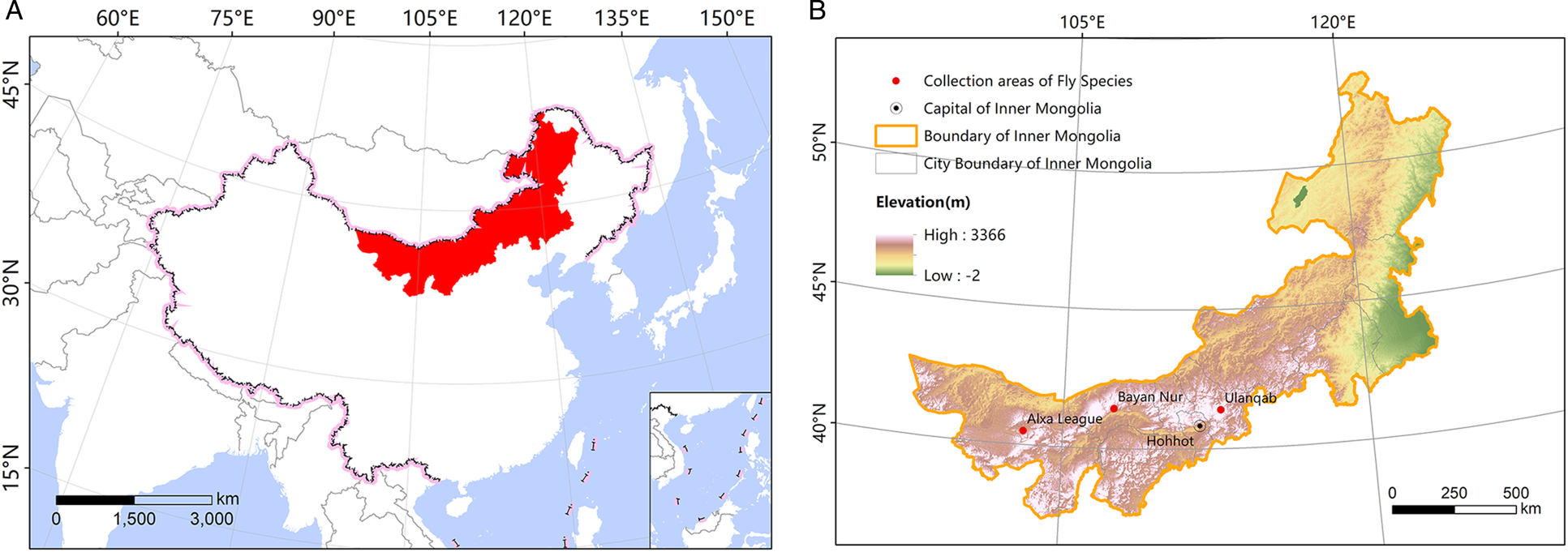
Inner Mongolia (97°12′–126°04′E, 37°24′–53°23′N) is located in the northern part of China and belongs to Eurasia continent (Fig. 1a). The vast area, long and narrow terrain results in climate diversity. Its continental climate transitioning from arid to humid from west to east affects the distribution of fly species. The three collection areas of fly species in this study are arranged in order from west to east in Inner Mongolia: Alxa League, Bayan Nur and Ulanqab (Fig. 1b). Among them, Alxa League belongs to the inland high plain area, the terrain is high in the south and low in the north, the desert Gobi is surrounded by mountains, and the temperature difference between day and night is large. It is a typical continental climate. Bayan Nur has relatively low terrain relief and is a typical mid-temperate continental monsoon climate. Ulanqab is located inland, which is a typical mid-temperate semi-arid continental monsoon climate. The annual average temperature of the three areas is 7.7–9.8, 3.7–7.6 and 0–6°C, and the annual average precipitation is 32.8–208.1, 100–300 and 150–450 mm, respectively.
(a) Location of Inner Mongolia and (b) the main collection areas of fly species.

Sample collection
We captured flies from the body surface of camels randomly in three of the main camel-producing areas (Alxa League, Ulanqab and Bayan Nur) in Inner Mongolia, China. The flies were washed with normal saline and soaked for 1 h, and then sterilized with 70% alcohol.
We obtained camel stomachs at the local official slaughterhouse, dissected the abomasum, looked for adult P. skrjabini, picked out by teasing needle, counted and calculated the infection rate, washed with saline and stored at −20°C in 75% alcohol.
Morphological identification
Under a stereoscopic microscope (Stemi DV4, Carl Zeiss, Germany), horn fly appearance and the parts with important identification significance such as head, wing vein and arista were carefully observed (Xue and Zhao, Reference Xue and Zhao1996; Fan, Reference Fan1997; Cai, Reference Cai2001). The purpose of this stage was to conduct preliminary morphological identification of the species of horn flies. Then dissected the horn flies under the microscope and looked for nematodes or eggs in the head, chest, abdomen and contents of the horn flies. Finally, each of the identified species of flies were stored in 75% alcohol. Subsequently, larvae from different species of flies were collected and stored at −80°C and 75% alcohol for further use.
Scanning electron microscopy procedure
The larvae found in the flies stored in 75% alcohol were washed with an ultrasonic cleaner (45 Hz, 15–20 min) for three times, and the cleaned larvae were fixed with a 2.5% glutaraldehyde solution for 48 h. The specimens were then dehydrated in a graded series of ethanol and then immersed into isoamyl acetate for 15–20 min. With liquid carbon dioxide as the drying medium, the sample was put into the critical point dryer for drying and then mounted on stubs. The sample was sprayed with platinum on the ion sputtering apparatus. Finally, the micrographs were prepared using a scanning electron microscope (S-530, Hitachi, Japan).
DNA extraction, PCR and sequencing
Genomic DNA of suspected nematode larvae, fly tissue and adult P. skrjabini nematodes was extracted using a commercial DNeasy Blood and Tissue kit (Qiagen, Hilden, Germany) according to the manufacturer's instructions. Extracted DNA was stored at −20°C until use. ITS-rDNA from suspected nematode larvae was amplified using the primer pair designed by primer 5.0, based on the ITS genome sequence of P. skrjabini rDNA. The forward primer was STM: 5′-TTTTACAAGAGGGATACGCC-3′ and the reverse primer was STN: 5′-GGTATCACAAACTTATCGGG-3′. The mtDNA COI of flies was amplified using universal primers. The forward primer was UEA-3: 5′-TATAGCATTCCCACGAATAAATAA-3′ and reverse primer was UEA-10: 5′-TCCAATGCACTAATCTGCCATATTA-3′ (Zhang and Hewitt, Reference Zhang and Hewitt1997). Primers were synthesized by Shanghai Sangon Biological Engineering Technology and Services Company. Polymerase chain reaction (PCR) amplifications of the above regions were performed using 25 μL of reaction mixture containing 12.5 μL of Premix Taq (TaKaRa), 0.5 μL of each primer (20 μM) and 2 μL of template DNA (20 ng μL−1); DNase/RNase-Free deionized water was added to make up the volume to 25 μL. A positive control (adult P. skrjabini DNA) and a negative control (without DNA) were included in each PCR reaction. Amplification conditions for PCR reactions were as follows: initial denaturation at 94°C for 5 min, followed by 35 cycles consisting of denaturation at 94°C for 30 s, annealing at 50°C for 30 s and extension at 72°C for 1 min. A final elongation step at 72°C for 10 min was carried out at the end of the PCR amplification procedure. The PCR products were analysed on 1% agarose gels and visualized using SYBR Green I. PCR products were purified using a QIAquick PCR purification Kit (Qiagen, Hilden, Germany) according to the manufacturer's recommendations and subsequently sent to TaKaRa Company for sequencing.
Sequence analysis
The sequences obtained were compared to those registered in GenBank (www.ncbi.nlm.nih.gov) using the Basic Local Alignment Search Tool (BLAST). DNA sequencing results were analysed using MegAlign 7.1.0 software.
Results
Morphology
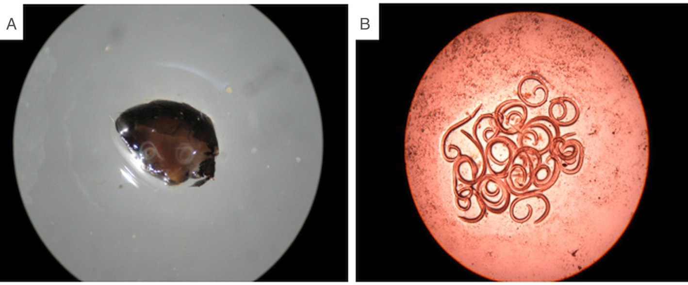
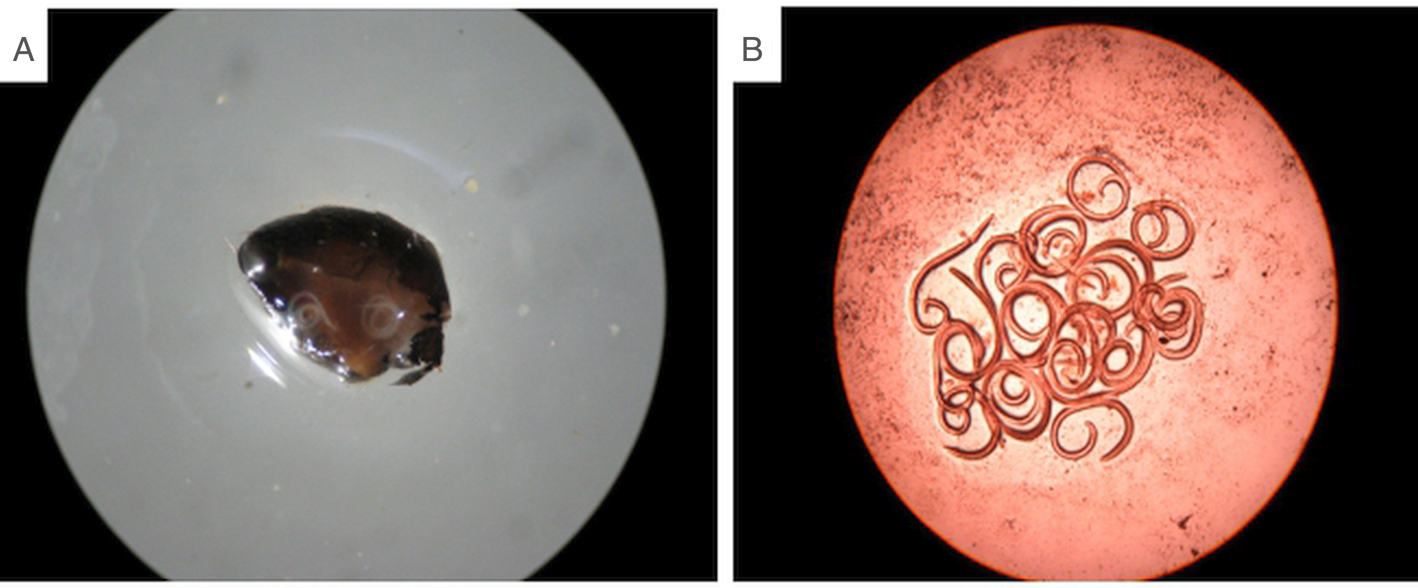
We collected a total of 6776 flies, of which Alxa League, Bayan Nur and Ulanqab were 1564, 2360 and 2852 respectively. All the blood-sucking diptera specimens collected were dissected and investigated under a stereo microscope (Stemi DV4, Carl Zeiss, Germany). Subsequently, we found two species of flies carrying nematode larvae inside their abdomen, curled into spirals of different lengths (Fig. 2). The average infection rate of the P. skrjabini larvae in horn flies in the three regions was 16.8%, among which Bayan Nur was 26.5%, Ulanqab was 7.1% and Alxa League was 0%. In addition, we dissected camel rumps from three regions, and the infection rates of adult worms were: 90.7% (Alxa League), 74.1% (Bayan Nur) and 51.9% (Ulanqab).
(a) Larvae found in the abdomen of a fly (40×) and (b) larvae inside a fly (40×).
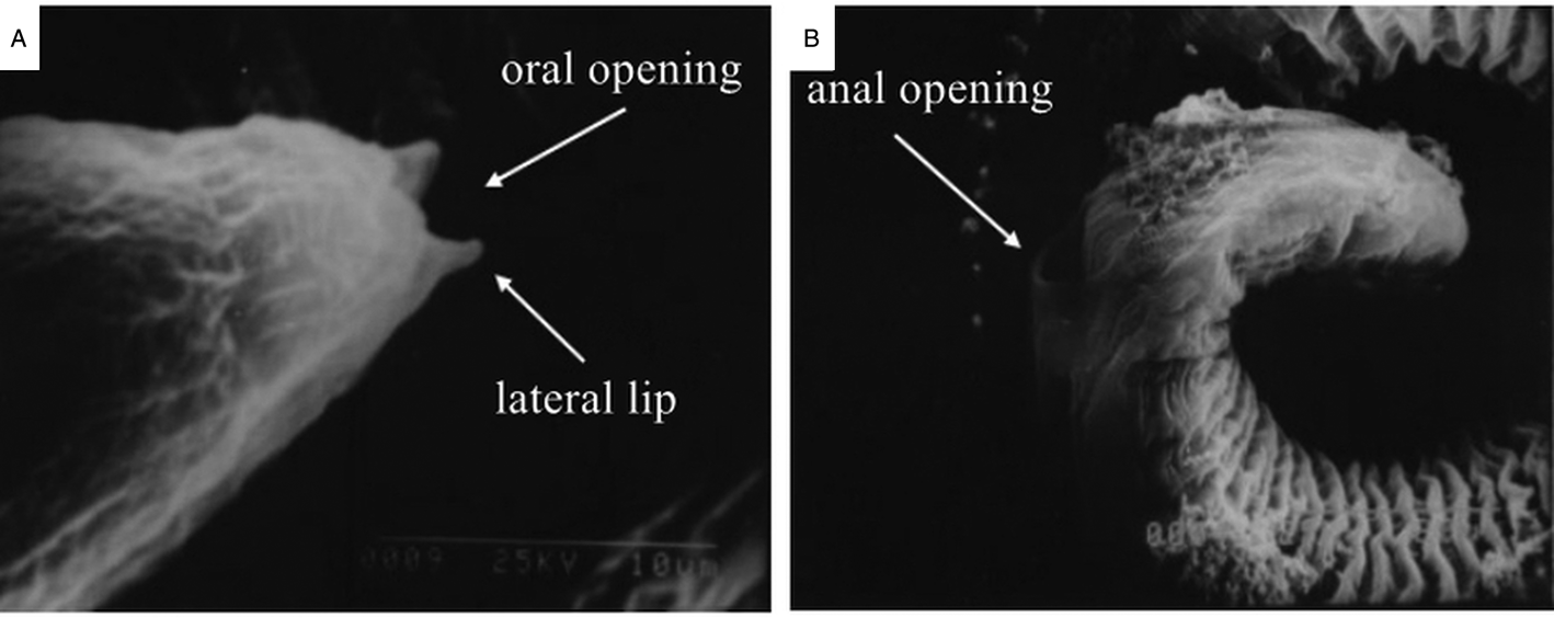
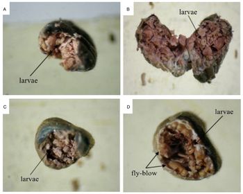
Using scanning electron microscopy (S-530, Hitachi, Japan), we observed that these larvae had many horizontal stripes on their body surface, which were formed by the cable-like ridges of the cuticle. The oral opening and anus were clearly visible, with the mouth surrounded by two lateral lips and the anus transversely split, the front was curved, and the back was flat, extending along the length of the body. Owing to incomplete development, the auxiliary mating organs and the sexual mastoid process could not be observed (Fig. 3). From an anatomical perspective, the larvae should be mainly parasitic in the digestive tract of flies (Fig. 4). Furthermore, we measured the length of piercing mouthparts and found that the average length of the oral organ was 964.63 μm, the average width at the widest point was 226.18 μm and the average width at the narrowest point was 85.10 μm.
(a) Head of a nematode larva (300×) and (b) tail of a nematode larva (300×).

(a) Cross-section of the abdomen of a H. irritans fly (40×); and (b) longitudinal section of the abdomen of a H. irritans fly (40×) and (c) and (d) cross-sections of the abdomen of a H. titillans fly (40×).

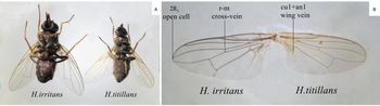
Two species of flies, H. titillans and H. irritans, were identified by morphological observation. As shown in Fig. 4, the end of cu1 + an1 wing vein in H. titillans is succise and the veins do not gradually taper towards the end, whereas in H. irritans it is the exact opposite. The distance between the cu1 + an1 wing vein and the wing margin of H. titillans and H. irritans is approximately 2 and 0.5 times its own length, respectively. The 2R5 opening cell of H. titillans and H. irritans are 2 and 1.5 times greater than the r-m cross-vein, respectively (Fig. 5a, b).
(a) H. irritans and H. titillans and (b) veins of H. irritans and H. titillans.

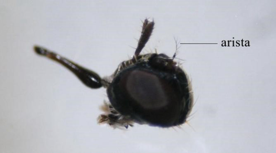
In general, H. irritans is larger than H. titillans; however, owing to the differences in species and gender, the size of the individual cannot be used as the primary criterion for identification. When the wings are damaged, or the wing veins are blurred, they can be identified by observing the number of aristae. Haematobium titillans has 5–7 aristae, while H. irritans has 9–11 (Fig. 6). We initially identified the two species of flies using this morphological observation.
Arista of H. titillans.

Molecular analysis
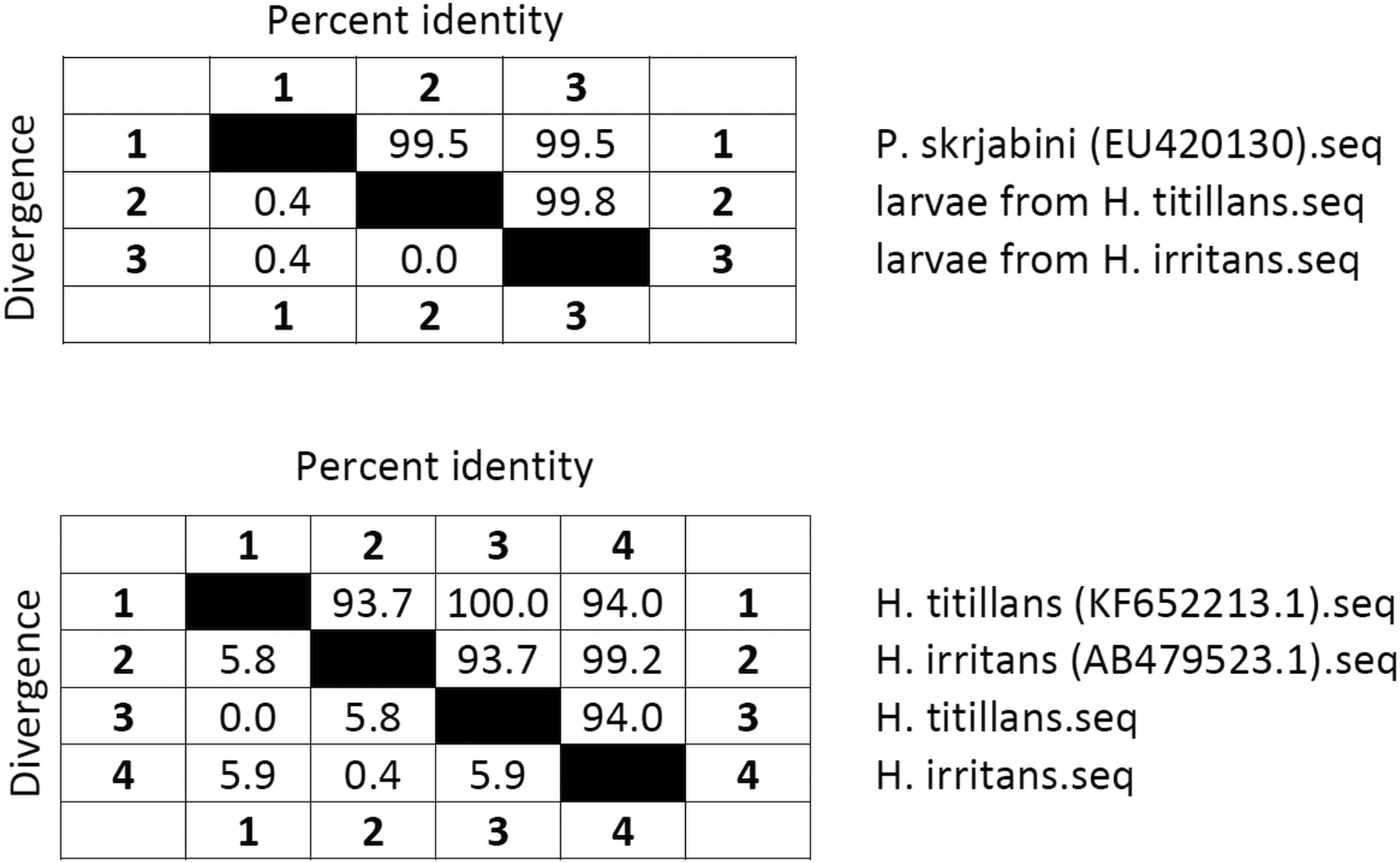
We amplified and sequenced rDNA ITS genes of the nematode larvae detected in H. titillans and H. irritans, all of which showed a sequence length of 551 bp. The homology of rDNA ITS sequences from nematode larvae found in H. titillans and H. irritans was 99.8%. Subsequently, on comparing with the rDNA ITS sequences of P. skrjabini in GenBank (EU420130), the homology was 99.5%, and in combination with morphological identification, we unequivocally confirmed that the nematode larvae found in the digestive tract of the fly species H. titillans and H. irritans were P. skrjabini larvae (Fig. 7a).
(a) ITS sequence homology between EU420130 and the larvae from Haematobia spp. and (b) mtDNA COI sequence homology between KF652213.1, AB479523.1, and Haematobia spp.

The mtDNA COI sequences of the fly species under study were also amplified and sequenced. PCR amplicons 1295 bp in length were obtained for both H. titillans and H. irritans. We compared the sequences with the mtDNA COI sequence of H. titillans (KF652213.1) and H. irritans (AB479523.1) deposited in GenBank and found that the homology was 100 and 99.2%, respectively (Fig. 7b). Thus, morphological identification along with molecular identification allowed species determination.
Discussion
Camels are greatly susceptible to infection by P. skrjabini (Yang et al., Reference Yang, Yang, Liu, Zhang and Zhang2004). In a previous study, we autopsied 76 camels (2–15 years old) in Alxa League, Inner Mongolia and found that the average infection rate of P. skrjabini was 86.9%, with the highest infection intensity being more than 900 worms per hump (Zhao et al., Reference Zhao, Wang, Guo, Yang, Wang, Zhao, Ao, Liu and Yang2012); therefore, it is crucial to detect and identify insect vectors of P. skrjabini.
The conventional strategy to identify an insect vector is via biological autopsy. Accordingly, many suspected vectors were captured in the environment and examined one by one for the presence of the suspected parasite larvae, which allowed us to successfully identify two insect vectors of P. skrjabini. However, unequivocal identification of the parasite larvae by this method is difficult and often results in false positive detection, because the morphological characteristics of larvae are not the same as those of adults. In the early stage of our research on the vectors of parabronemosis, nematode larvae were found in flies, but due to poor primer specificity, larval species identification was occasionally not reproducible. In addition, the identification of flies by morphological observation was not sufficiently precise. In this study, we adopted a strategy that combined molecular and morphological identification to classify the vectors. In addition to overcoming the limitations of morphological identification described above, this strategy effectively prevents false positive detection caused by human error, making the results accurate and reliable.
Recent investigations indicated that the vectors of P. skrjabini might be blood-sucking flies (Ivashkin and Khromova, Reference Ivashkin and Khromova1983; Saparov et al., Reference Saparov, Dadaev and Azimov2005). In our study, larvae of P. skrjabini were found in two species of fly whose morphology was very similar. We identified the species of these two flies by combining morphology and molecular identification. However, previous studies showed that P. alatavensis was not distributed in Inner Mongolia to a considerable extent. Another species of blood-sucking fly was captured in the environment where camels live; it was identified as Stomoxys calcitrans and did not carry any nematodes. Furthermore, an unknown nematode was found in Chaetopleurophora asiatica; this nematode was morphologically different from the larvae of P. skrjabini and was clearly another species of nematode. Therefore, we cannot discard the possibility that these flies might also be vectors of parabronemosis.
According to the length and width measurements of the oral organ, we found that the width is enough for nematode larvae to pass. Therefore, we inferred that P. skrjabini, like other parasites, spreads through blood-sucking insects, entering the blood of the final host through the oral organ of the insect vector and then reaching the abomasum of camels through a specific migration path, a hypothesis that remains to be experimentally tested.
In this study, flies were captured in three major camel-producing areas in Inner Mongolia: Alxa League, Ulanqab and Bayan Nur. In these three areas, the average infection rate of the P. skrjabini larvae in horn flies was 16.8%, among which Bayan Nur was 26.5%, Ulanqab was 7.1%. It is worth noting that the analysis of the infection rate and infection intensity of the adult P. skrjabini we examined through the camels’ abomasum showed that Alxa League had the highest rate among the three regions. However, no H. titillans or H. irritans flies were caught in this case, likely because the climate of Alxa League is different from that in the other two main producing areas, and it has a higher average temperature and less precipitation, which leading to the flies appearing and disappearing and the peak periods of activities being different, thereby affecting the amount of fly capture. Additionally, other potential vector species may exist in these areas. Moreover, during the process of necropsy, the presence of sexually mature larvae was also detected in camels from November through January of the following year, indicating that the seasonal dynamics of P. skrjabini growth and development in camels in Inner Mongolia may differ from region to region, as these results are different from those of Ivashkin V M (April to November) (Ivashkin, Reference Ivashkin1955). The specific influencing factors warrant further study. The growth and development of flies are restricted by various environmental conditions, competitive species and natural enemies. Indeed, biological control might prove to be a useful approach in managing the populations of vector flies to compensate for the limitations of physical control and drug resistance.
In conclusion, our data support a successful procedure for finding nematode larvae in flies and identifying flies as vectors of parabronemosis. Our findings lay the foundation for further exploration of the life history of P. skrjabini and the control of parabronemosis in camels. Future research will focus on the study of flies and other potential vectors, including the development, living habits and home-range size in different climatic regions. Furthermore, we will strive to study the infection mechanism of P. skrjabini and the physiological and biochemical characteristics of the host infection site in detail.
Acknowledgements
We would like to thank Editage (www.editage.cn) for English language editing.
Financial support
This work was supported by the National Natural Science Foundation of China (grant number 30660142).
Conflict of interest
None.
Ethical standards
All experiments were performed according to the regulations of the Administration of Affairs Concerning Experimental Animals in China. However, the relevant document number is not available at Inner Mongolia Agricultural University. Permission was obtained from the local official slaughterhouse before collection of the specimens.