Implications
Expression of appetite regulators was detected in the fetal lambs at 75 days of gestation, but expression did not differ due to maternal fatness at this time of development. Increased body fatness of ewes during early to mid-gestation does not appear to be accompanied by disregulated expression of the hypothalamic appetite regulators. Changes in expression of the estrogen receptor α gene may affect energy balance and/or reproduction.
Introduction
The prenatal uterine environment influences the future well-being and health of an individual. Under- or over-nutrition during critical periods of fetal development increases the risk for adult-onset obesity and disease (Muhlhausler et al., Reference Muhlhausler, Adam, Marrocco, Findlay, Roberts, McFarlane, Kauter and McMillen2005 and Reference Muhlhausler, Adam, Findlay, Duffield and McMillen2006; Fischer-Posovszky et al., Reference Fischer-Posovszky, Wabitsch and Hochberg2007).
Both maternal diet and adiposity potentially influence offspring health and well-being. Maternal obesity in sheep at conception and increased nutrient intake during gestation cause increased adiposity and plasma concentrations of leptin, disregulated blood glucose and increased feed intake in the adult offspring (Long et al., Reference Long, George, Uthlaut, Smith, Nijland, Nathanielsz and Ford2010). Maternal obesity or over-nutrition during the first half of gestation affects offspring development and predisposes offspring to obesity through potentially different mechanisms, including changes in muscle fiber density (Zhu et al., Reference Zhu, Han, Tong, Ma, Kimzey, Underwood, Xiao, Hess, Ford, Nathanielsz and Du2008), glucose production and insulin signaling (Ford et al., Reference Ford, Zhang, Zhu, Miller, Smith, Hess, Moss, Nathanielsz and Nijland2009), fat cell hypertrophy (Lewis et al., Reference Lewis, Bertrand, McMahan, McGill, Carey and Masoro1986) and/or changes in appetite regulation (Muhlhausler et al., Reference Muhlhausler, Adam, Findlay, Duffield and McMillen2006).
Developments of the processes that regulate feed intake are species specific (Grove et al., Reference Grove, Grayson, Glavas, Xiao and Smith2005; McMillen et al., Reference McMillen, Adam and Muhlhausler2005). Perturbations of these processes in rats permanently affect food intake, potentially leading to hyperphagia and obesity in adult offspring (Kirk et al., Reference Kirk, Samuelsson, Argenton, Dhonye, Kalamatianos, Poston, Taylor and Coen2009). In utero exposure to a high-fat maternal diet stimulated neural proliferation and differentiation in the fetal rat third ventricle with a greater proportion of the newly formed neurons expressing orexogenic peptides (Guo-Qing et al., Reference Guo-Qing, Gaysinskaya, Karatayev and Leibowitz2008). Plane of maternal nutrition during late pregnancy in sheep also influenced appetite regulators. Ewes fed a diet with excess metabolizable energy during late pregnancy gave birth to offspring with increased hypothalamic expression of pro-opiomelanocortin (POMC). These lambs had greater weight gain and adipose deposition by day 30 postnatal (Muhlhausler et al., Reference Muhlhausler, Adam, Findlay, Duffield and McMillen2006). Effects that may occur earlier in gestation have not been investigated.
The objective of this study was to examine the effect of dam obesity on the development of hypothalamic appetite regulators during the first half of gestation in the sheep. A second objective was to determine whether differences in dam obesity were associated with inherent differences in the expression of hypothalamic appetite regulators among individual ewes. Individual differences in the neurophysiology of either appetite regulators or the brain-reward mechanisms could result in overeating or altered metabolic signaling among individuals.
Material and methods
Animal care and use procedures were approved by the University of Wyoming Animal Care and Use Committee.
Animal protocol
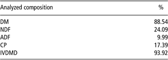
Multiparous, white-faced crossbred ewes in moderate body condition 60 days before mating were weighed and fed differing quantities of the same highly palatable diet to meet (100%; control, n = 7) or exceed (150%; obese, n = 7) recommended nutrient requirements as specified by the National Research Council for total digestible nutrients based on metabolic BW (BW0.75) of individual ewes (National Research Council (NRC), 1990; Table 1). Ewes were weighed at weekly intervals and rations were adjusted for weight gain. Body condition scores (BCSs) were obtained at monthly intervals to determine change in maternal body fatness. A BCS from one (emaciated) to nine (obese) was assigned by the same two trained observers after palpation of the transverse and vertical processes of the lumbar vertebrae (L2 to L5) and the region around the tail head (Sanson et al., Reference Sanson, West, Tatman, Riley, Judkins and Moss1993).
Composition of diet fed to ewes 60 days before breeding through day 75 of gestation. Control ewes received 100% of NRC recommendations while obese ewes received 150% NRC
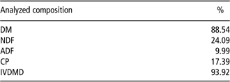
NRC = National Research Council; DM = dry matter; IVDMD = in vitro DM disappearance.
On day 75 of pregnancy, ewes were weighed and evaluated for BCS. Ewes were then fasted overnight (24 h), sedated with ketamine (22.2 mg/kg BW, i.v.) and anesthetized by isofluorane inhalation (2.5%). Anesthetized ewes were euthanized by heart puncture and fetuses were collected. Fetal weights, sex and organ weights were recorded. From the original ewes placed on treatment, only twin-bearing ewes with one male and one female fetus were utilized for fetal tissue analysis in this study (n = 3 ewes and six fetuses per group).
Potential differences in hypothalamic expression of appetite regulators were also contrasted in tissues from the most obese ewes (n = 4; BCS = 8.2 ± 0.4) to those of ewes in moderate body condition (n = 4; BCS 5.7 ± 0.4), regardless of fetal number or sex.
Tissue collection
Brains were removed from the fetal cranium, sectioned mid-sagitally, and the hypothalamus was dissected using surface landmarks. In fetuses, a diencephalic block extending laterally approximately 3 mm and dorsally from the anterior margin of the optic chiasm to the anterior margin of the mammillary body was dissected. In ewes, the medial basal hypothalamus including the arcuate nucleus was dissected from the preoptic area at the caudal margin of the optic chiasm (Moss et al., Reference Moss, Crowder and Nett1981). Tissues were snap frozen and stored at −80°C until RNA extraction.
RNA isolation
Total cellular RNA was isolated using the TRI reagent (Sigma Aldrich Chemical, St. Louis, MO). Briefly, 100 mg of brain tissue (fetal hypothalamus and dam medial basal hypothalamus) was homogenized in 1 ml of TRI reagent and incubated at room temperature for 10 min before adding 0.2 ml of chloroform, followed by incubation at room temperature for 10 min. The homogenate was centrifuged for 15 min at 4°C at 12 000 × g. The aqueous layer was transferred and RNA was precipitated using isopropanol and centrifugation. The pellet was washed with 70% ethanol and suspended in RNase-free water. Concentration of RNA was determined using a NanoDrop spectrophotometer (ND-1000 Spectrophotometer, Nanodrop Technologies, Wilmington, DE, USA) and 10 μg was purified further using RNEASY (Qiagen Inc., Santa Clara, CA, USA) with on-column DNase digestion.
Real-time PCR
RNA (2 μg) was mixed with 4 μl of reverse transcription buffer (5X) and 1 μl of IScript reverse transcriptase (Bio-Rad Laboratories, Richmond, CA, USA). The mixture was placed in a thermocycler for 5 min at 25°C, 30 min at 42°C, 5 min at 85°C and held at 4°C. The cDNA was diluted with 100 μl of nuclease-free water and stored at −20°C until semi-quantitative real-time PCR was performed.
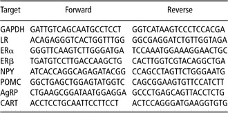
Primers were designed using Primer 3 software (Rozen and Skaletsky, Reference Rozen and Skaletsky2000) with 100 bp amplicons. Real-time PCR was performed by mixing 10 μl of diluted cDNA with 12.5 μl of SYBR green Supermix (Bio-Rad Laboratories, Inc., Hercules, CA, USA), 1 μl/500 pmol each forward and reverse primer and 0.5 μl of nuclease-free water in each well of a 96-well plate. Amplification was performed using the IQ5 and 40 cycles of 95°C for 30 s and 60°C for 30 s. Melting curve analysis was performed post amplification to ensure the quality of PCR products as noted by the presence of a single peak. The PCR plate was heated to 95°C for 3 min, cooled to 55°C and then the temperature increased by 0.5°C/s up to 95°C. Real-time PCR reactions from each primer set yielded a single band on an agarose gel, which was extracted, purified and submitted for DNA sequencing (Davis Sequencing, Davis, CA, USA). Each sequence was queried using NCBI/BLAST and was confirmed to be homologous to the gene of interest. Ovine glyceraldehyde 3-phosphate dehydrogenase (GAPDH) was used as the reference gene and all gene expression levels were quantified and reported relative to GAPDH expression using the 2−▵▵CT method (Livak and Schmittgen, Reference Livak and Schmittgen2001). Real-time PCR was performed in duplicates for each sample using primers for ovine leptin receptor (LR), neuropeptide Y (NPY), agouti-related protein (AgRP), POMC, bovine cocaine- and amphetamine-regulated transcript (CART), ovine estrogen receptor alpha (ERα), and estrogen receptor beta (ERβ). A list of primers and sequences is shown in Table 2.
Forward and reverse PCR primers used for quantitative PCR

PCR = polymerase chain reaction; GAPDH = glyceraldehyde 3-phosphate dehydrogenase; LR = leptin receptor; ERα = estrogen receptor alpha; ERβ = estrogen receptor beta; NPY = neuropeptide Y; POMC = pro-opiomelanocortin; AgRP = agouti-related protein; CART = cocaine- and amphetamine-regulated transcript.
Statistical analysis
Data were analyzed as a complete randomized design utilizing GLM procedures of SAS (version 9.1, Cary, NC, USA). Preplanned mean comparisons were analyzed utilizing PDIFF option of GLM; least squares means ± s.e.m. are reported. Beginning and ending ewe weights and BCS were analyzed for change with the Paired-T analysis (SAS version 9.1, Cary, NC, USA). Correlations between fetal and maternal relative expression of appetite regulators were determined using the CORR procedures of SAS (version 9.1).
Results
BWs and BCS of ewes did not differ (P > 0.05) at the initiation of treatment but were greater (P < 0.01) in obese ewes at the time of tissue collection as reported by Zhu et al. (Reference Zhu, Han, Tong, Ma, Kimzey, Underwood, Xiao, Hess, Ford, Nathanielsz and Du2008). The current report is focused on appetite regulators in a subpopulation of these ewes and their offspring. In this subpopulation, obese fed ewes with the most extreme body condition had greater (P = 0.004) BCS than in control ewes (8.2 ± 0.4 v. 5.7 ± 0.4, respectively). BW at 75 days of gestation was greater (P = 0.01) in obese than in control ewes (104.9 ± 5.4 kg v. 77.0 ± 5.4 kg).
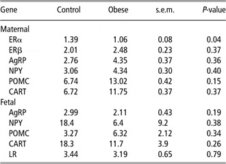
Expression of the housekeeping gene, GAPDH, did not differ (P = 0.44) by ewe diet. Maternal hypothalamic mRNA expression of ERβ, NPY, POMC, CART or AgRP did not differ (P ⩾ 0.2) in the most obese compared with control ewes (Table 3). Relative gene expression of hypothalamic ERα, however, was greater (P = 0.04) in obese than in control ewes (Table 3).
Relative mRNA gene expression of hypothalamic appetite regulatory genes in control and obese ewes and their fetuses at 75 days of gestation

ERα = estrogen receptor alpha; ERβ = estrogen receptor beta; AgRP = agouti-related protein; NPY = neuropeptide Y; POMC = pro-opiomelanocortin; CART = cocaine- and amphetamine-regulated transcript; LR = leptin receptor.
Relative gene expression of AgRP, NPY, CART or POMC did not differ (P ⩾ 0.16) among fetuses from control and obese ewes (Table 3). Fetal expression of CART was negatively correlated (r = −0.86, P = 0.03) with the expression of CART in their dams. Expression of LR did not differ (P = 0.8) among fetuses from control or obese dams (Table 3) and was not correlated with LR expression in control (P = 0.06) or obese (P = 0.09) ewes. Fetal expression of POMC and CART or orexogenic NPY and AgRP were not influenced by sex (P ⩾ 0.44) or by any treatment by sex interaction (P ⩾ 0.42).
Discussion
Circulating proteins are accessible to the arcuate nucleus through breaks in the blood–brain barrier at this circumventricular organ. The arcuate nucleus is situated at the base of the hypothalamus and is highly interconnected to the dorsal medial and lateral hypothalamus (Clarke, Reference Clarke2008; Morris and Rui, Reference Morris and Rui2009). Appetite-stimulating (orexogenic) and appetite-suppressing (anorexogenic) neurons co-exist within the arcuate nucleus.
Obese ewes in the current experiment had greater feed intake leading to increased body fat (Ford et al., Reference Ford, Zhang, Zhu, Miller, Smith, Hess, Moss, Nathanielsz and Nijland2009) and increased plasma concentrations of leptin (George et al., Reference George, Uthlaut, Long, Zhang, Ma, Smith, Nathanielsz and Ford2010). Predisposition to hyperphagia may be due to the expression of appetite regulators in these ewes as central injection of AgRP and NPY increased feed intake in ad libitum-fed sheep (Wagner et al., Reference Wagner, McMahon, Marks, Daniel, Steele and Sartin2004). However, results of the current study do not support the hypothesis that differences in orexogenic (AgRP or NPY) or anorexogenic (POMC or CART) neuropeptide expression contribute to variations in obesity in ewes that voluntarily increase caloric intake when provided a palatable diet. Differences in obesity may simply reflect individual differences in inherent efficiency to accumulate or maintain energy stores. Discreet differences in appetite regulators, however, may not have been detected in the current study as mRNA was extracted from a subsection of a larger hypothalamic mass. Although ewes were fasted 24 h before tissue collection to facilitate anesthesia, this abbreviated fast would not be expected to significantly affect gut fill (Tatman et al., Reference Tatman, Judkins, Krysl and Moss1991) or affect the expression of regulators of appetite in the ruminant animal.
Differences in expression of the LR would influence how the orexogenic neurons respond to increased obesity associated with increased plasma concentrations of leptin (Morris and Rui, Reference Morris and Rui2009). Leptin may influence feed intake in the sheep as intra-cerbroventricular injections of leptin decreased voluntary food intake (Adam and Findlay, Reference Adam and Findlay2010). Although the obese ewes had increased plasma concentrations of leptin (George et al., Reference George, Uthlaut, Long, Zhang, Ma, Smith, Nathanielsz and Ford2010), changes in expression of the LR were not noted. It is possible that obese ewes have compromised transport of circulating leptin across the blood–brain barrier (Adam and Findlay, Reference Adam and Findlay2010), which limits availability of leptin to its receptor.
Reproductive function of estradiol is generally attributed to activation of ERα (Hewitt et al., Reference Hewitt, Deroo, Hansen, Collins, Grissom, Afshari and Korach2003) and obese females often exhibit impaired reproduction (Norman and Clark, Reference Norman and Clark1998). In this study, ERα expression was up-regulated in the hypothalamus of obese dams compared with dams in moderate body condition. Estradiol 17β rapidly increased phosphorylation of the IGF-1 receptor in vitro in the presence of ERα (but not ERβ; Kahlert et al., Reference Kahlert, Nuedling, Van Eickels, Vetter, Meyer and Grohe2000). Although overt differences in insulin sensitivity are not noted in obese ewes (George et al., Reference George, Uthlaut, Long, Zhang, Ma, Smith, Nathanielsz and Ford2010), concentrations of glucose, insulin and cortisol are increased in obese ewes at day 75 of gestation (Ford et al., Reference Ford, Zhang, Zhu, Miller, Smith, Hess, Moss, Nathanielsz and Nijland2009). Even though only ewes with established pregnancies were included in the present experiment, reproductive function does not appear to be impaired in the obese ewes as approximately 95% conceived during the breeding season. Although long-term effects are not clear, increased expression of hypothalamic ERα has the potential to disrupt reproduction and/or energy balance in obese ewes.
Perirenal fat was increased in fetuses from obese ewes at day 75 of gestation (George et al., Reference George, Uthlaut, Long, Zhang, Ma, Smith, Nathanielsz and Ford2010), but differences in fetal expression of the appetite regulators due to maternal obesity were not observed in the current study. In rat, intrauterine growth restriction affected hypothalamic differentiation and organization (Coupe et al., Reference Coupe, Amarger, Grit, Benani and Parnet2010), indicating that the maternal environment can clearly influence neural development. Species differences as well as the timing and duration of maternal nutrient disruption may be important in programming the fetus for adult-onset obesity. Organization of the appetite regulatory system occurs postnatally in rodents and during the prenatal period in longer gestational species, such as humans and sheep (McMillen et al., Reference McMillen, Adam and Muhlhausler2005). Expression of genes important for the regulation of appetite has been reported to occur by 110 days of gestation in the fetal lamb (Muhlhausler et al., Reference Muhlhausler, McMillen, Rouzaud, Findlay, Marrocco, Rhind and Adam2004), but earlier expression has not been investigated.
Adult offspring from ewes obese at conception and maintained on obesogenic diets throughout pregnancy had increased feed intake when provided an ad libitum diet (Long et al., Reference Long, George, Uthlaut, Smith, Nijland, Nathanielsz and Ford2010), but changes in the appetite regulators were not reported. Because of the nondiscreet timing of the maternal over-nutrition reported by Long et al. (Reference Long, George, Uthlaut, Smith, Nijland, Nathanielsz and Ford2010), it is not known whether early or late gestational nutrition excess contributed to the increased appetite. Muhlhausler et al. (Reference Muhlhausler, Adam, Findlay, Duffield and McMillen2006) reported that in sheep, over-nutrition during late gestation resulted in increased milk intake and subcutaneous fat accumulation accompanied by an unpredicted increased expression of POMC, but not CART mRNA at 30 days postnatally. Differences in expression of orexogenic NPY and AgRP were not detected. In lambs from control-fed, but not obese ewes, CART mRNA expression was positively correlated with total fat mass, plasma glucose, non-esterified fatty acids and leptin (Muhlhausler et al., Reference Muhlhausler, Adam, Findlay, Duffield and McMillen2006). In the current study, hypothalamic expression of CART was negatively correlated among dams and their fetuses. However, CART expression did not change systematically with increased obesity in the dams. Therefore, maternal obesity at conception or over-nutrition during the first half of gestation may have limited impact on the development of the hypothalamic appetite regulatory system in the sheep. Alternatively, influence of the early maternal environment may not be evident until later life through epigenetic change (Stevens et al., Reference Stevens, Begum, Cook, Connor, Rumball, Oliver, Challis, Bloomfield and White2010). The positive expression of these appetite regulators clearly indicates that these systems are maturing at this early fetal age. How vulnerable the appetite system is to the maternal uterine environment at this developmental age is not clear.
Differences in relative expression of LR mRNA were not detected among fetuses from obese and control dams. Although a strong correlation was not detected in the current study likely due to the limited sample size, a positive correlation between plasma leptin concentrations in the ewe and fetus in well-fed and under-nourished conditions during late pregnancy has been reported (Fowden and Forhead, Reference Fowden and Forhead2009). It is not known whether this relationship is due to placental transfer of leptin or effects on the secretory capacity of leptin in the fetus.
Expression of appetite regulators was detected in the fetal lambs at 75 days of gestation, but expression did not differ due to maternal fatness at this developmental time point. Increased body fatness of ewes during early to mid-gestation does not appear to be accompanied by disregulated expression of the hypothalamic appetite regulators.
Acknowledgement
This work was supported by INBRE P20 RR016474-04.