INTRODUCTION
The family Taeniidae (Phylum Platyhelminthes, Class: Cestoda) contains two genera, Taenia and Echinococcus, which have been studied extensively owing to their medical importance and the economic losses that they cause in some species of livestock. The significant taeniids of veterinary and medical importance include Taenia solium, T. saginata, T. ovis, Echinococcus granulosus and E. multilocularis. Considerable attention has been directed towards the study of the ontogeny, life-history and ecology of the various species of Taenia and Echinococcus. Many contemporary studies of these parasites have been driven by the substantial challenges that these parasites pose for public health and food safety globally and the increasing interest in promoting socio-economic development in poor nations (Craig and Pawlowski, Reference Craig and Pawlowski2002; Hoberg, Reference Hoberg2002). Adult taeniids inhabit the small intestine of their hosts and are usually of little pathological significance, whereas larval stages develop in the tissues of intermediate hosts, causing morbidity and mortality in humans and economic losses to farmers and the livestock industry.
Control of infection with taeniid cestodes may, logically, be achieved through an alteration in human behaviour patterns and improved public hygiene. The transmission of cystic hydatidosis, caused by E. granulosus, can be curtailed through improvements in public education related to sanitation, the regular deworming of dogs, together with other measures which restrict dogs having access to hydatid-infected offal (Craig and Larrieu, Reference Craig and Larrieu2006). The control of porcine cysticercosis, caused by T. solium, can be achieved through improvements in the economic situation of endemic countries, which is reflected in improved farming practices and the sanitary disposal of human sewage. However, changing people's customs and food habits is difficult, and few deliberate control programmes for cysticercosis or hydatidosis have been successful (Craig and Larrieu, Reference Craig and Larrieu2006).
Much attention has been focused on the development of new tools to improve the effectiveness of hydatid and cysticercosis control programmes. A number of practical vaccines against these diseases have been developed and been shown to be highly effective in preventing infection in the intermediate hosts (Lightowlers et al. Reference Lightowlers, Flisser, Gauci, Heath, Jensen and Rolfe2000; Lightowlers, Reference Lightowlers2006). The most effective of these vaccines have been those that have utilized antigens obtained from the oncospheral stage (Lightowlers et al. Reference Lightowlers, Gauci, Chow, Drew, Gauci, Heath, Jackson, Dadley-Moore and Read2003, Reference Lightowlers2006).
DEVELOPMENT OF PRACTICAL RECOMBINANT VACCINES BASED ON ONCOSPHERE ANTIGENS
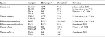
Studies undertaken during the 1950–70s indicated that oncospheres were a potent source of host-protective antigens (reviewed by Rickard and Williams, Reference Rickard and Williams1982). These studies provided strong evidence indicating the susceptibility of the early stages in the development of taeniid cestodes to immune attack and provided evidence that practical vaccines might be developed. The advent of recombinant DNA technology saw this vision fulfilled, with the production of effective, recombinant vaccines based on the use of oncospheral antigens produced against T. ovis (Johnson et al. Reference Johnson, Harrison, Lightowlers, O'hoy, Cougle, Dempster, Lawrence, Vinton, Heath and Rickard1989; Harrison et al. Reference Harrison, Heath, Dempster, Gauci, Newton, Cameron, Robinson, Lawrence, Lightowlers and Rickard1996), E. granulosus (Lightowlers et al. Reference Lightowlers, Lawrence, Gauci, Young, Ralston, Maas and Health1996a), T. saginata (Lightowlers et al. Reference Lightowlers, Rolfe and Gauci1996b), E. multilocularis (Gauci et al. Reference Gauci, Merli, Muller, Chow, Yagi, Mackenstedt and Lightowlers2002), T. solium (Flisser et al. Reference Flisser, Gauci, Zoli, Martinez-Ocana, Garza-Rodriguez, Dominguez-Alpizar, Maravilla, Rodriguez-Canul, Avila, Aguilar-Vega, Kyngdon, Geerts and Lightowlers2004) and T. multiceps (Gauci et al. Reference Gauci, Vural, Oncel, Varcasia, Damian, Kyngdon, Craig, Anderson and Lightowlers2008) (Table 1).
Host-protective recombinant oncosphere antigens of taeniid cestodes

a Homology group was assigned based on a high level of amino acid identity between antigens with reference to the first antigen of the group to be characterized.
b Protection reflects the optimum level achieved in vaccination and challenge infection trials.
c Results of vaccination trials using the TSA-9 and TSA-18 antigens together.
d Based on mortality following challenge infection.
Modified from Lightowlers (2006).
Host-protective oncosphere antigens are expressed exclusively in the oncospheral stage and are secreted glycoproteins
Investigations have been undertaken using gene-specific primers in reverse transcription polymerase chain reaction to identify the life-cycle stages in which the genes encoding host-protective antigens To45W, To16 and To18 of T. ovis (Waterkeyn et al. Reference Waterkeyn, Gauci, Cowman and Lightowlers1997; Gauci et al. Reference Gauci, Verastegui, Gilman and Lightowlers2006), TSOL18 and TSOL16 of T. solium (Gauci et al. Reference Gauci, Verastegui, Gilman and Lightowlers2006) and EG95 of E. granulosus (Chow et al. Reference Chow, Gauci, Cowman and Lightowlers2004) are transcribed. In each case, the associated mRNAs appeared to be present exclusively in the oncosphere. Typically, the mRNAs were found in gravid, but not non-gravid, segments of the adult tapeworm. The mRNAs occur particularly in the eggs (containing the inactive oncosphere) and display increased transcription in oncospheres following activation in vitro. Such activation mimics the physiological conditions of the gastric and intestinal environments which see the parasite hatch from its protective egg shell and become active and motile, a process assumed to be associated with the parasite penetrating the intestinal mucosa.
There is considerable evidence available to indicate that the host-protective antigens of taeniid oncospheres are secreted glycoproteins (reviewed by Lightowlers et al. Reference Lightowlers, Gauci, Chow, Drew, Gauci, Heath, Jackson, Dadley-Moore and Read2003). Much of this evidence derives from studies undertaken on the so-called excreted/secreted products of oncospheres maintained in culture. However, none of these investigations has excluded the possibility that the components present in the culture could not have been derived from somatic components of a proportion of oncospheres that had become non-viable during culture in vitro.
WHAT IS THE ULTRASTRUCTURAL LOCATION OF THE HOST-PROTECTIVE ONCOSPHERE ANTIGENS?
There have been no convincing reports of the localization of host-protective vaccine antigens of taeniids or how these antigens are secreted. Silverman and Maneely (Reference Silverman and Maneely1955) and Rajasekariah et al. (Reference Rajasekariah, Mitchell and Rickard1980a, Reference Rajasekariah, Rickard and Mitchellb) hypothesized that the secretions contained in ‘blebs’ produced from the penetration glands of activated oncospheres may be a source of host-protective antigens. However, Furukawa et al. (Reference Furukawa, Niwa and Miyazato1981) showed that the antibody-mediated complement-dependent attack on oncospheres of Hymenolepis nana first occurs on the surface membrane that corresponds to the oncospheral tegument, and was apparent as early as 8 h after infection. These results suggest that secretions are not the primary source of host-protective antigens, at least with respect to H. nana.
The oncospheral tegument of freshly hatched oncospheres of T. taeniaeformis (Engelkirk and Williams, Reference Engelkirk and Williams1982; Nieland, Reference Nieland1968), T. ovis (Harris et al. Reference Harris, Heath, Lawrence and Shaw1987) and E. granulosus (Holcman et al. Reference Holcman, Heath and Shaw1994) has been described as being ‘thrown’ into a series of folds, but within a short period after activation, the surface structure has undergone a marked transformation. In the case of T. taeniaeformis, the oncospheral surface is transformed within 24 h into one covered in long, branching microvilli (Engelkirk and Williams, Reference Engelkirk and Williams1982). Harris et al. (Reference Harris, Heath, Lawrence and Shaw1987) described a similar transformation in T. ovis 48 h after activation in vitro. In addition to long microvillar protrusions, a series of electron-lucid vesicles were seen stacked along their distal region. It has been proposed that these microvilli may be involved in stimulating protective immunity especially as they could actively be ingested by the host's phagocytic cells (Engelkirk and Williams, Reference Engelkirk and Williams1982).
Each of the host-protective oncosphere proteins has been shown to be present in oncospheres while they are in their inactive state and, hence, prior to the formation of the surface microvilli or microvillary vesicles, although this does not necessarily preclude the microvilli as source of host-protective antigens during an infection. Host-protective antigens are known to be present in different life-cycle stages of taeniid cestodes other than the oncosphere (reviewed by Rickard and Williams, Reference Rickard and Williams1982). Heath (Reference Heath1973, Reference Heath1976) presented evidence that antigens from T. pisiformis larvae up to two weeks old could be used to immunize rabbits against infection. These data, together with the evidence which indicates that those recombinant host-protective oncosphere antigens studied to date are expressed exclusively in the oncosphere, suggest there may be additional host-protective antigens produced by post-oncospheral larvae. This view is consistent with the observations made by Bogh et al. (Reference Bogh, Rickard and Lightowlers1988), who showed that extracts of both oncospheres and metacestodes of T. taeniaeformis contain antigens capable of protecting mice against an infection with this parasite but that the two sources of antigens appeared to have their host-protective effects by killing parasites in a challenge infection at different stages in their development.
The first attempt to localize particular oncospheral proteins was made by Harrison and Parkhouse (Reference Harrison and Parkhouse1986), who produced a number of monoclonal antigens against components of T. saginata oncospheres. One of these monoclonal antibodies, HP6, was later found to be directed against the TSA18 host protective antigen (Lightowlers et al. Reference Lightowlers, Rolfe and Gauci1996b). Harrison and Parkhouse (Reference Harrison and Parkhouse1986) used HP6 and immuno-fluorescence on whole activated oncospheres of T. saginata and concluded that the associated antigen was present in the penetration glands. This study was restricted to the use of whole oncospheres fixed on slides and examined using a fluorescence microscope; hence, the ultrastructural detail available to the authors was limited. Subsequently, the same research group, using an immuno-gold labelling technique, reported that TSA18 was present in the penetration glands and on the hooks (Benitez et al. Reference Benitez, Garate, Harrison, Kirkham, Brookes and Parkhouse1996), although the published images did not allow specificity to be confirmed. The only other study which has provided some data on the localization of an oncosphere protein has been that by Mayta et al. (Reference Mayta, Hancock, Levine, Gilman, Farfan, Verastegui, Lane, Garcia, Gonzalez and Tsang2007), who showed that the protein Tso31 was expressed on the limiting membrane (i.e. oncospheral tegument) of hatched and activated oncospheres of T. solium. This antigen does not appear to have the host-protective capacity of TSOL45 or TSOL18 and is not discussed further here.
While there has been much progress over the past twenty years with the development of highly effective vaccines based on oncosphere antigens, it is clear that very little is known about the ultrastructural localization of these antigens within the parasites. Many highly sensitive immunological methods have been developed that should allow the precise ultrastructural localization of the host-protective antigens in and on taeniid oncospheres.
METHODS SUITABLE FOR THE LOCALIZATION OF HOST-PROTECTIVE ANTIGENS OF TAENIIDS
Immuno-histochemistry is a powerful exploratory tool in biology which can provide detailed information about antigen localization that complements knowledge about parasite ultrastructure. A large number of methods and technologies are available for immuno-histochemical studies which can provide spatial and temporal information. A high degree of topological resolution has been made possible with the assistance of confocal and electron microscopy. These techniques provide unique information that cannot be readily obtained employing biochemical, immuno-chemical or morphological methods alone. In addition, the localization of mRNA associated with the expression of antigens can also be achieved, and such data can complement those obtained using immuno-histochemical techniques. In situ hybridization is a ‘localizing technique’ for either mRNA within the cytoplasm or DNA within the chromosomes of the nucleus. Data on tissue localization of gene transcription provides complementary information to immuno-chemical localization (Brahic and Haase, Reference Brahic and Haase1978).
CURRENT KNOWLEDGE OF STRUCTURE/ULTRASTRUCTURE OF TAENIID ONCOSPHERES
In order to achieve an understanding of the ultrastructural localization of oncosphere antigens, a detailed knowledge of structure/ultrastructure of the oncosphere is required. A number of reports have been published on the ultrastructure of non-taeniids, particularly species of Hymenolepis. However, knowledge of the ultrastructure of early stages of taeniids is very limited. Research on the structure/ultrastructure of taeniid cestodes is a topic which has been rather neglected, while many studies have been undertaken on the immuno-biology and molecular biology of this group of parasites.
Embryonic development of taeniids
The eggs of taeniid cestodes, when examined in utero in mature proglottids, contain an embryo which can be in different stages of its embryonic development, whereas the gravid proglottids contain mainly mature, infective oncospheres. Each oncosphere in utero is surrrounded by four or five oncospheral or egg envelopes (Conn and Swiderski, Reference Conn and Swiderski2008). When taeniid eggs are released into the external terrestrial environment, the outer egg envelope rapidly disappears and the infective oncospheres are surrounded and protected mainly by their embryophores. The structure of taeniid oncospheres is not well described other than in a single study by Swiderski (Reference Swiderski1983), who used serial semi-thin and ultra-thin sections to reconstruct oncospheres of E. granulosus. This study concentrated particularly on the hook muscle system of the oncosphere. Much of our current understanding has been derived from interpretations made by various authors concerning the nature of the various cell types. An attempt has been made to synthesize current knowledge and this is summarized in Fig. 1.
Schematic diagram of a taeniid oncosphere illustrating the structure and bilateral symmetry in the pattern of hooks and cellular organization of the hexacanth. (A) Embryophore of a typical taeniid egg having polygonal embryophoric blocks and granular layer; both belong to the inner envelope. (B) Cellular organisation of the hexacanth. Abbreviations: BSC, binucleated subtegumental cell; EM, embryophore; HRM, hook-region membrane; GC, germinative cells; GL, granular layer; IE, inner envelope; NC, nerve cells; OM, oncospheral membrane; OT, oncospheral tegument; PG1, penetration gland, type one; PG2, penetration gland, type two; SC, somatic cells. Oncospheres are approximately 25×30 μm.

The embryophore (Morseth, Reference Morseth1965; Nieland, Reference Nieland1968) is thick and composed of numerous keratin blocks, giving it a striated appearance in optical sections. The oncosphere has been defined as “bilaterally symmetrical, spherical or oval embryo, armed with three pairs of hooks, containing a pair of glands and surrounded by embryonic envelopes” (Rybicka, Reference Rybicka1966). This definition has been confirmed and extended significantly by more recent transmission electron microscopic (TEM) studies (Swiderski, Reference Swiderski1972; Swiderski and Tkach, Reference Swiderski and Tkach2002; Swiderski et al. Reference Swiderski, Ndiaye, Miquel, Tkach, Marchand, Chomicz, Grytner-Ziecina and Sereda2002; Mlocicki et al. Reference Mlocicki, Swiderski, Miquel, Eira and Conn2006; Conn and Swiderski, Reference Conn and Swiderski2008).
Three stages of embryonic development can be distinguished during oncospheral differentiation: cleavage, the pre-oncospheral phase and the mature oncosphere. The period of early segmentation of the ovum is referred to as the cleavage phase. Cleavage results in three types of blastomeres classified by size and their ulrastructural features; macromeres, mesomeres and micromeres (Rybicka, Reference Rybicka1966; Swiderski, Reference Swiderski, Clark and Adams1981; Swiderski and Tkach, Reference Swiderski and Tkach2002; Mlocicki, Reference Mlocicki2006).
The pre-oncospheral phase is one of intense cell multiplication and differentiation, indicated by high metabolic activity in the embryo and resembling organogenesis in higher animals. The pre-oncospheral phase involves morphological processes which include: (1) the formation of embryonic envelopes; (2) apoptosis or degeneration of some micromeres leading to evident reduction in the number of the oncospheral cells; (3) differentiation of somatic and germinative cells from the remaining micromeres; (4) development of glandular regions and (5) development of hooks, hook muscles and somatic musculature (Swiderski, Reference Swiderski, Clark and Adams1981; Swiderski and Tkach, Reference Swiderski and Tkach2002; Mlocicki et al. Reference Mlocicki, Swiderski, Conn, Eira, Miquel and Baranowska-Korczyc2005; Mlocicki, Reference Mlocicki2006). The morphology of the oncosphere is relatively constant until hatching. When eggs are shed from taeniid tapeworms the oncosphere is fully mature and potentially capable of further development. No further development occurs until the egg is ingested by a suitable intermediate host (Rybicka, Reference Rybicka1966).
In a mature taeniid egg, the embryophore is surrounded by a simple unit membrane and hexagonal blocks consisting of an electron-dense, amorphous substance (Fig. 1A). The embryophoric blocks contain numerous round lacunae, which are mostly empty, but sometimes contain remnants of membranes (Schramlova and Blazek, Reference Schramlova and Blazek1982). According to some authors, these lacunae relate to circular bodies (Morseth, Reference Morseth1965) or remnants of mitochondria (Nieland, Reference Nieland1968). Between the individual embryophoric blocks, there is a cement substance which appears in TEM as an electron-lucent homogeneous material (Morseth, Reference Morseth1965; Schramlova and Blazek, Reference Schramlova and Blazek1982). Examination of the chemical composition of the embryophores of T. hydatigena, T. pisiformis and T. ovis revealed that the infrared absorption pattern was almost identical to that of keratin (Morseth, Reference Morseth1966). Beneath the embryophore is a highly granular-cytoplasmic layer of variable thickness, which is rich in free ribosomes (Fig. 1A). It is limited by a membrane separating it from the oncospheral membrane situated below it. The oncospheral membrane surrounding the embryo is a thin, amorphous layer composed of two closely apposed double-unit membranes. The hook region membrane is formed from the pre-oncospheral cytoplasmic cap which covers the somatophoric pole of the oncosphere. This structure is directly attached to the oncospheral tegument (inner most layer). The surface of the oncospheral tegument (Fig. 1B) has filamentous processes/microvilli of various lengths (Schramlova and Blazek, Reference Schramlova and Blazek1982; Swiderski, Reference Swiderski1983).
Cellular structure
At the end of early cleavage, the number of embryonic cells initially increases in the early embryos; later, it decreases significantly due to apoptosis or the degeneration of some micromeres, leading to a reduction in the number of the oncospheral cells (Rybicka, Reference Rybicka1966; Swiderski, Reference Swiderski, Clark and Adams1981; Mlocicki, Reference Mlocicki2006). Sagittal sections through the middle parts of the oncospheres have revealed a bilateral symmetry of the hexacanth (Swiderski and Tkach, Reference Swiderski and Tkach2002). Swiderski (Reference Swiderski1983) reported 54 nuclei belonging to 4 major cell types. The glandular regions (8 nuclei) are subdivided into three categories: two types of penetration glands and neuro-secretory cells. The muscle cells (34 nuclei) are subdivided into hook and somatic musculature. In addition to the above mentioned types there are two groups of five germinative cells and a binucleated subtegumental cell. The features of the infective taeniid oncosphere, based on published data for E. granulosus, are illustrated schematically in Fig. 1B (after Swiderski, Reference Swiderski1983). Swiderski (Reference Swiderski1983) described two regions in the developing larva of E. granulosus based on the presence of cells following the description by Ogren (Reference Ogren1971) : (1) the ‘somatophore’ containing mainly somatic cells and oncospheral hooks, differentiating rapidly and forming the anterior end of the onchosphere (Fig. 1B); and (2) the germatophore (mesophore), the posterior half, containing some somatic cells and larger embryonic cells referred to as germinative cells together with the penetration glands (Fig. 1B). More recently, Swiderski (Reference Swiderski1972, Reference Swiderski, Clark and Adams1981), Swiderski and Tkach (Reference Swiderski and Tkach1997, Reference Swiderski, Ndiaye, Miquel, Tkach, Marchand, Chomicz, Grytner-Ziecina and Sereda2002) and Mlocicki et al. (Reference Mlocicki, Swiderski, Miquel, Eira and Conn2006) described the morphological differentiation between somatic and germinative cells. The somatic cells represent the myocytons of somatic and hook musculature and are characterized by their connection with long muscle fibres, whereas germinative cells do not have such connections. The former cells are always smaller and their nucleo-cytoplasmic ratio is lower than in the germinative cells. Alternately, the germinative cells have a high nucleo-cytoplasmic ratio and large, lobate granular nuclei containing prominent electron-dense, spherical nucleoli surrounded by numerous irregular heterochromatin islands adjacent to the nuclear membrane (Swiderski, Reference Swiderski1972; Swiderski and Tkach, Reference Swiderski and Tkach1997, Reference Swiderski, Ndiaye, Miquel, Tkach, Marchand, Chomicz, Grytner-Ziecina and Sereda2002; Mlocicki et al. Reference Mlocicki, Swiderski, Miquel, Eira and Conn2006). The germinative cells have a very thin granular layer of cytoplasm which is rich in free ribosomes, whereas this layer is inconspicuous in somatic cells. Swiderski (Reference Swiderski1983) observed five pairs of germinative cells arranged in two lateral groups in E. granulosus (Fig. 1B). He also proposed that the nuclear and cytoplasmic features of germinative cells were indicative of their potential for the further growth and multiplication in post-embryonic stages of the life cycle. The oncospheres of E. granulosus also contain two nuclei of binucleate sub-tegumental cells and six nuclei in two types of penetration glands. In addition, two nerve cells containing neuro-secretory granules in nerve processes (Fig. 1B) are usually present in the somatophore region of the oncosphere (Swiderski and Tkach, Reference Swiderski and Tkach1997, Reference Swiderski, Ndiaye, Miquel, Tkach, Marchand, Chomicz, Grytner-Ziecina and Sereda2002; Mlocicki et al. Reference Mlocicki, Swiderski, Miquel, Eira and Conn2006).
Hooks and musculature
A typical taeniid oncosphere, as those of all Eucestoda, is characterized by three pairs of keratinized oncospheral hooks (Fig. 1B). Hook rudiments appear in the pre-oncospheral phase. Each hook arises intracellularly in a specialized cell, the oncoblast or the hook forming cell. The hook primordium, adjacent to the invaginated part of the nuclear envelope, is surrounded by an abundance of free ribosomes, mitochondria and Golgi-regions. Simultaneously, with its elongation and transformation into a blade, the hook primordium material becomes differentiated to form an electron-dense cortex and a less dense core. Subsequently, the blade of the hook protrudes outside the oncoblast and is surrounded by two electron-dense rings connected by a circular septate desmosome (Nieland, Reference Nieland1968; Swiderski, Reference Swiderski1976; Mlocicki et al. Reference Mlocicki, Swiderski, Conn, Eira, Miquel and Baranowska-Korczyc2005).
Typically, the oncospheral hooks consist of one pair of medial hooks and two pairs of lateral hooks, each lateral pair being composed of one dorso-lateral and one ventro-lateral hook (Fig. 1B). In E. granulosus, each oncopsheral hook is ~10–12 μm in length and consists of the following three parts: (1) a curved blade, (2) a short guard or collar and (3) long shank or handle, which terminates in a knob-like enlargement (Swiderski, Reference Swiderski1983). The hooks of the lateral pairs differ in size and shape; the dorsal are more slender than the larger ventral ones. The blades of medial hooks are more sharply curved than those of lateral hooks. Ultrastructurally, oncospheral hooks are composed of two to four concentric layers, depending on the level of the cross sections (Swiderski, Reference Swiderski1983). The muscle system of E. granulosus has been described as consisting of 34 cells, of which 16 are associated with a somatic muscle system, and 18 represented hook muscle cells (Swiderski, Reference Swiderski1983). The somatic or body muscle system forms wide bands of transverse-encircling muscles localized close to the oncospheral tegument. These muscles are underlain by a dorsal and ventral pair of longitudinal muscles. The somatic musculature functions to maintain oncospheral shape and, by differential contraction, may be important in the burrowing activity of the oncosphere into the host tissues. Hook muscles insert at the collar and base of the hooks and at the basal lamina of the oncospheral tegument (Swiderski, Reference Swiderski1972, Reference Swiderski1976, Reference Swiderski1983; Swiderski and Tkach, Reference Swiderski and Tkach2002).
Penetration glands
Reid (Reference Reid1948) introduced the term “penetration glands”, hypothesizing that the secretion of these oncospheral glands may help oncospheres to penetrate the tissues of the intermediate host. These glands have been described as comprising two cells located symmetrically behind the hooks which are joined together by an isthmus to give an U-shaped syncytial structure. The nuclei lie at the base of the U and the cytoplasm contains numerous secretory granules (Reid, Reference Reid1948). Generally, it is believed that the arms of the U-shaped glands open between the medial and lateral pairs of hooks. Fairweather and Threadgold (Reference Fairweather and Threadgold1981), describing the structure of penetration glands of H. nana, suggested that a single large, round nucleus lies at the base of these glands which is surrounded by the narrow cisternae of endoplasmic reticulum. The cytoplasm of the gland is filled with two types of membrane-limited, secretory granules: (1) one with the tightly packed granular contents within the granules and a uniform pattern of high electron density; (2) a second type of secretory granule appears more swollen and less structured, its coarse granular contents being less homogeneous and of lower electron density. The gland ducts are described as being supported by microtubules and these may prevent any compression of the ducts caused by the vigorous contractions of the body musculature during hatching and penetration. Swiderski (Reference Swiderski1983) observed three different types of glandular regions in oncospheres of E. granulosus. He described two additional pairs of glandular regions between the arms of the typical U-shaped penetration gland (glandular region PG1) of which one pair contained electron-lucent secretory granules while the other possessed small membrane-bound granules of high electron density. Based on the studies published by Fairweather and Threadgold (Reference Fairweather and Threadgold1981) concerning nerve cells in H. nana, the third type of secretory granules of the so-called “glandular regions” (Swiderski, Reference Swiderski1983) are also considered to be neurosecretory granules of nerve cells in E. granulosus and E. multilocularis (Z. Swiderski, unpublished data). Hence, there seems to be a consensus of opinion that there are two different types of penetration glands in E. granulosus, based on differences in the shape and electron density of their secretory granules (Fig. 1B: PG1 and PG2).
Secretions of the penetration gland
Secretions from penetration glands are believed to be involved in the transfer of granules from the penetration gland cells to the external cytoplasm of the oncospheral tegument. It has been suggested that these granules within cytoplasmic blebs are released while the oncosphere penetrates the gut of the intermediate host (Nieland, Reference Nieland1968). It has been reported that H. nana and Mosgovoyia ctenoides release the granules via blebs which become detached from the oncospheral tegument, and it has been suggested that this occurs during penetration of the intestinal epithelial tissue of the intermediate hosts (Furukawa et al. Reference Furukawa, Miyazato, Okamoto and Nakai1977; Miyazato et al. Reference Miyazato, Furukawa, Inoque, Niwa, Inoque and Shimoda1977a, Reference Miyazato, Furukawa, Inoque, Niwa, Inoque and Shimodab; Mlocicki et al. Reference Mlocicki, Swiderski, Miquel, Eira and Conn2006). Other authors speculate that penetration gland secretions are discharged directly to the outside of the oncosphere via penetration gland ducts (Fairweather and Threadgold, Reference Fairweather and Threadgold1981). The secretions have been classified as being merocrine (Lethbridge and Gijsbers, Reference Lethbridge and Gijsbers1974), apocrine (Furukawa et al. Reference Furukawa, Miyazato, Okamoto and Nakai1977; Mlocicki et al. Reference Mlocicki, Swiderski, Miquel, Eira and Conn2006) and holocrine (Fairweather and Threadgold, Reference Fairweather and Threadgold1981). However, the mechanism of secretion from the penetration glands remains unclear and indeed there is neither direct evidence for the contents of the cells being secreted in vivo nor any direct evidence that they play a role in penetration.
Penetration gland material appears heterogeneous ultrastructurally (Lethbridge and Gijsbers, Reference Lethbridge and Gijsbers1974) is periodate-reactive (Lethbridge, Reference Lethbridge1971) and absorbs the cationic dye neutral red (Lethbridge and Gijsbers, Reference Lethbridge and Gijsbers1974). This indicates the presence of acidic carbohydrate-rich mucosubstance and, potentially proteolytic enzymes. A number of descriptions of the secretory products characterize them as including: (1) proteinaceous substance with enzymatic properties (Pence, Reference Pence1970) e.g. serine proteinase (Moczon, Reference Moczon1996); (2) an acid mucoprotein (Fairweather and Threadgold, Reference Fairweather and Threadgold1981); (3) an acidic mucopolysaccharide (Heath, Reference Heath1971); and (4) a polysaccharide (Silverman and Maneely, Reference Silverman and Maneely1955).
Immunological role of penetration glands
Silverman (Reference Silverman1955) speculated as to the potential antigenic nature of penetration gland secretions, but there has been no direct evidence to support this hypothesis. The proposition is supported indirectly by evidence that, during the first few hours of host invasion, the glands constitute a large volume as compared with the total size of the oncosphere, and they secrete macroscopic material (Silverman and Maneely, Reference Silverman and Maneely1955). Rickard and Bell (Reference Rickard and Bell1971) achieved a high level of immunity in sheep against T. ovis in sheep in which they had previously implanted activated oncospheres within diffusion chambers (having a pore size too small to allow intact oncospheres to escape, but large enough to allow the release of excretory/secretory products). Confirmation of the existence of host-protective ‘functional’ oncospheral antigens among secretory products released into in vitro culture medium has been provided by studies of T. pisiformis (Heath, Reference Heath1976; Rickard and Outteridge, Reference Rickard and Outteridge1974), T. saginata (Rickard and Adolph, Reference Rickard and Adolph1976), T. ovis (Rickard and Adolph, Reference Rickard and Adolph1977), T. solium (Patak and Gaur, Reference Patak and Gaur1990) and E. granulosus (Heath et al. Reference Heath, Parmeter, Osborn and Lawrence1981).
Structure of hatched and activated oncospheres of taeniid cestodes
In the process of hatching, the embryophore disintegrates into individual blocks through the apparent dissolution of the interblock cement substance (Laws, Reference Laws1968; Stevenson, Reference Stevenson1983). Hatching can be achieved either through the application of sodium hypochlorite or a combination of artificial gastric and intestinal fluids (e.g. Silverman, Reference Silverman1954; Laws, Reference Laws1968). The latter method not only leads to the hatching but also activation of the oncosphere, such that the organism shows muscular activity and, through the thrusting motion of the parasite's hooks, the oncospheral membrane is torn open and the parasite liberated (Silverman, Reference Silverman1954). Under the light microscope, activated oncospheres can be seen devoid of both the embryophore and the oncospheral membrane (Fig. 1B). Comprehensive investigations by the authors have begun into the ultrastructure of T. ovis oncospheres. Ultrastructurally, non-activated oncospheres of T. ovis are characterized by two main types of oncospheral cells (somatic and germinative), their hooks, musculature, and abundant granules of the predominant two types of penetration glands. The perinuclear cytoplasm of penetration gland cells is dense due to the presence of numerous ribosomes, mitochondria, rough endoplasmic reticulum and Golgi apparatus.
Activated oncospheres are characterized by having rhythmic and vigorous movements which may persist for up to 24 h in vitro. During the first 24 h in culture, droplets can sometime be seen associated with the surface of the oncosphere. These droplets, known as ‘blebs’, appear in the hook region (Holcman et al. Reference Holcman, Heath and Shaw1994). Following the escape of the oncosphere from the oncospheral membrane, there is a release of microvilli from their state of compression, as seen in non-activated oncospheres. An expansion is seen in the tegumental surface around the hook region which expands from a multifolded oncospheral tegument. Other changes observed following the activation of oncospheres in vitro relate to the state of the penetration glands. In activated oncospheres of E. granulosus, the penetration glands have been described as being almost devoid of granules and a number of vesicles, vacuoles and glycogen granules are present in the perikaryon (Holcman et al. Reference Holcman, Heath and Shaw1994). Other authors, working with oncospheres of T. saginata, have described an increase in the number of ribosomes and granular endoplasmic reticulum forming extended cisternae in the cytoplasm of penetration gland cells (Schramlova and Blazek, Reference Schramlova and Blazek1982). Electron-dense granules accumulate at the periphery of these cells and are pressed to the periphery of the embryo where they accumulate in bladders bordered by a simple membrane. The bladders are pressed between filamentous processes under the oncospheral membrane. The granules are composed of a homogeneous substance of moderate electron density with a vacuole in the centre (Schramlova and Blazek, Reference Schramlova and Blazek1982).
Post-oncospheral development
There are few published reports concerning the post-oncospheral development of taeniid oncospheres. The post-oncospheral development in vivo and the ultrastructural characteristics of the polycephalic larva of Taenia parva were described recently by Swiderski et al. (Reference Swiderski, Miquel, Młocicki, Georgiev, Eira, Grytner-Ziecina and Feliu2007). The following descriptions are based on the in vitro post-oncospheral development of T. saginata (Schramlova et al. Reference Schramlova, Blazek, Marhoul and Singhvi1984) and E. granulosus (Harris et al. Reference Harris, Heath, Lawrence and Shaw1989; Holcman et al. Reference Holcman, Heath and Shaw1994) and an in vivo study of T. taeniaeformis (Engelkirk and Williams, Reference Engelkirk and Williams1982; Engelkirk and Williams, Reference Engelkirk and Williams1983).
After a period of 24 h following activation, when observed under the light microscope, the oncosphere is characterized by the presence of a clear zone or ‘halo’. This zone is filled with fine microvilli extending from the surface, which can be seen clearly in electron micrographs (Engelkirk and Williams, Reference Engelkirk and Williams1982; Schramlova et al. Reference Schramlova, Blazek, Marhoul and Singhvi1984). At this early stage of development, the oncosphere consists of a compact mass of cells, rich in ribosomes, rough endoplasmic reticulum, mitochondria and Golgi apparatus. The developing parasite increases in size by day two and contains more nuclei and some differentiation of cells is evident (Holcman et al. Reference Holcman, Heath and Shaw1994). Many of the microvilli extending from the parasite surface are branched and, while their length has not changed from day one, their number increases (Engelkirk and Williams, Reference Engelkirk and Williams1982). In E. granulosus, in the perikaryon of the germinal zone, there are three main types of vesicles: disc-like vesicles with dense granular contents (designated Vd); vesicles with spherical electron-dense granules (Vg1); and vesicles similar in shape and size but with an electron-dense rod-shaped core (Vg2) (Holcman et al. Reference Holcman, Heath and Shaw1994).
In T. taeniaeformis, by day three, the total surface area of the developing parasite increases 7-fold compared with that present at day one, and cellular differentiation results in an increase in the number of nuclei within the developing larva (Engelkirk and Williams, Reference Engelkirk and Williams1982). The cytoplasm of all the cells is rich in ribosomes, mitochondria, rough endoplasmic reticulum and Golgi bodies. There are also lipid droplets and glycogen rosettes present both intracellularly and extracellularly in the sub-tegeumental zone. The tegument in E. granulosus appears to undergo changes more rapidly than described for T. taeniaeformis. In E. granulosus, the tegumental microvilli have been replaced by short microtriches by three days post activation and the first laminated layer surrounding the metacestode appears, being an electron-dense matrix composed of fine microfibrillated material and remnants of sloughed microvilli (Heath and Holcman, Reference Heath and Holcman1997). Microtriches are not seen on the surface of the developing larva of T. taeniaeformis until after eight to nine days of development (Engelkirk and Williams, Reference Engelkirk and Williams1982). In T. taeniaeformis, the hooks have disappeared by day four and the cell number within the parasites has increased substantially. Sub-tegumental cells become separated from each other by clear channels and there were many lipid droplets and glycogen rosettes (Engelkirk and Williams, Reference Engelkirk and Williams1982).
Substitution of the microvilli by microtriches is complete in E. granulosus by day five. At the outer border of the laminated layer, there is a dramatic increase in number of the two types of Vg vesicles forming aggregations. A second laminated layer appears by days six to eight which is more electron dense than the first lamination and is delineated on its outer and inner surfaces by particulate material (Heath and Holcman, Reference Heath and Holcman1997). The developmental sequence that has been described for T. saginata oncospheres in some respects is more similar to that of T. taeniaeformis. On day five, T. saginata the surface of the tegument bears long and branched microvilli. Hook muscles degenerate and the oncosphere is filled with a compact mass of cells. The cells possess a large nucleus with nucleolus and dispersed ribosomes, rough endoplasmic reticulum and ribbon-shaped mitochondria in the cytoplasm (Schramlova et al. Reference Schramlova, Blazek, Marhoul and Singhvi1984).
In T. taeniaeformis, by day seven, the development of the bladder becomes evident with electron-dense matter in the centre, and the tegument contains numerous membrane-bound vesicles and shorter microvilli (Engelkirk and Williams, Reference Engelkirk and Williams1982). There is a replacement of microvilli with microtriches by day eight to nine and the tegument becomes thicker and contains vacuoles, electron-dense microbodies, dumb-bell-shaped vesicles and glycogen rosettes. There is an increase in the size of microtriches by days 10–12, the syncytial cytoplasm of the tegument becomes filled with lipid droplets and the somatic cells degenerate (Engelkirk and Williams, Reference Engelkirk and Williams1983; Schramlova et al. Reference Schramlova, Blazek, Marhoul and Singhvi1984). A striking feature of the microtriches is the appearance of ‘streamers’ which are seen on day 18 but which disappear along with inclusion droplets on day 22 of post-oncospheral development.
RECENT INSIGHTS INTO THE LOCALIZATION ON HOST-PROTECTIVE ANTIGENS OF TAENIIDS
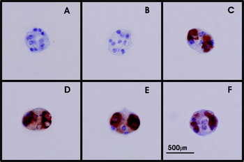
Peroxidase- and fluorescence-based immuno-histochemical methods are being used to determine the localization of host-protective antigens in the oncosphere of T. ovis and preliminary data are becoming available (A. Jabbar, unpublished observations). Fixed T. ovis oncospheres were processed using routine histology procedures and paraffin sections incubated with specific anti-To16 primary antibodies followed by incubation with biotinylated secondary antibodies. A distinctive pattern of bilateral staining involving 2–4 cells was found in non-activated oncospheres stained specifically for the To16 protein (Fig. 2). Only the cytoplasm of the cells containing the antigen contained the brown staining while nuclei were counterstained blue using Gill's haematoxylin. In the same oncospheres, the cytoplasm of all other cells showed no staining (Fig. 2C–F). The cells containing antigen varied in shape from oval or triangular to irregular depending on the plane of section. Occasionally, the cells appeared to be triangular with a long projection. In relation to the location of the oncospheral hooks, these cells were found either postero-lateral to the lateral pairs of hooks or on the poles (Fig. 2C–E) of an oval oncosphere where the hooks were seen in the centre of the oncosphere (Fig. 2D).
Immuno-histochemistry of Taenia ovis non-activated oncospheres showing localization of the To16 host-protective antigen. The eggs were hatched with sodium hypochlorite (1·25%) to get non-activated oncospheres. Following purification of oncospheres with percoll, they were fixed in absolute methanol at −20°C overnight and then embedded in low melting point agarose and processed as per routine histological methods. Sections were reacted with control sera comprising pre-immune serum (A) or polyclonal sera against glutathione S-transferase (GST) (B), or with antisera against To16-GST (C–F, representing different individual oncospheres), followed by biotinylated F(ab)2 fragments of rabbit anti-sheep antibodies, peroxidase-labelled streptavidin and counterstained with Gills hematoxylin. The cytoplasm of cells showing positive reaction stain brown while the nuclei of all cells present in oncosphere stain blue. The To16 antigen shows a bipolar staining pattern, largely restricted to the cytoplasm of the associated cells. Control antisera against GST (B), or non-immune control sera (A), when tested under similar conditions to those used in panel B, show no detectable staining of the oncosphere (A).

The host-protective antigens of T. ovis are also being investigated using immuno-gold labelling in specimens processed for transmission electron microscopy. Cryofixed, non-activated oncospheres were embedded in low temperature resin and ultra-thin sections incubated with primary antibodies followed by colloidal gold particle-conjugated secondary antibodies. The localization of immuno-gold staining in these specimens confirmed the findings of seen with immuno-histochemical staining under light microscopy (Figs. 3–5). Immuno-gold staining was specific for the target antigens as evidenced by the lack of labelling seen with control antisera (Fig. 5). Specific gold labelling was seen in the cytoplasm and the secretory granules of the penetration gland cells (Figs. 4 and 5). Labelling was limited to the cytoplasm of these cells and the nucleus, ribosomes, endoplasmic reticulum and Golgi bodies were devoid of any labelling. No labelling was evident in other types of the cells (somatic, germinative, binucleated subtegumental and nerve cells), hook-region membrane, oncospheral tegument or membrane.
Ultrastructure of a non-activated oncosphere of Taenia ovis. The non-activated oncospheres were pelletted, sandwiched between brass freezer hats (Proscitech) and then frozen using Leica EM High Pressure Freezer (Wien, Austria). Frozen oncospheres were freeze-substituted and embedded in low temperature resin (Polysciences, Warrington, PA, USA, HM20). (A) Whole oncosphere. (B) Higher magnification of a penetration gland cell and the surrounding structures. (C) Secretory granules and the cytoplasm of the penetration gland cell. Abbreviations: GC, germinal cell; H, hook; HRM, hook-region membrane, M, mitochondria; Mv, microvilli; N, nucleus; NSG, neurosecretory granules; OM, oncospheral membrane; OT, oncospheral tegument; PG1&2, penetration glands type one and two; SC, somatic cell; SG1&2, secretory granules types one and two.

Ultrastructural immuno-localization of the host-protective antigen To16 in a non-activated Taenia ovis oncosphere. Ultra-thin sections of cryofixed oncospheres reacted with polyclonal antisera against To16-GST followed by incubation with 12 nm colloidal gold conjugated to rabbit anti-sheep antibodies. Sections were counterstained with uranyl acetate and lead salts. (A) Whole oncosphere. (B) Penetration gland cells. (C) Immuno-gold labelling detected in the secretory granules and the cytoplasm of the penetration gland cell. Abbreviations: L, lipid-like droplets; N, nucleus; PG1&2, penetration glands type one and two; SC, somatic cell; SG1&2, secretory granules types one and two.

Transmission electron micrographs at high magnification showing penetration gland secretory granules and specificity of immuno-gold labelling of To16. Ultra-thin sections of cryofixed oncospheres reacted with sheep antisera against glutathione S-transferase (GST) recombinant antigen (A) or polyclonal sheep antisera against To16-GST (B) followed by incubation with 12 nm colloidal gold-conjugated rabbit anti-sheep antibodies and counterstained with uranyl acetate and lead salts. Clear immuno-gold labelling (arrows) is seen only in the sample reacted with antisera against To16-GST distributed in the secretory granules and also spread throughout the cytoplasm of the penetration gland cell (arrows).

CONCLUSION
Much remains to be revealed concerning the ultrastructure of taeniid oncospheres. A sound understanding of the structure of the organism will be required to understand the implications of antigen-localization studies. Further research is also required to understand the precise localization of the host-protective antigens and the role that these proteins play in the biology of these organisms. Perhaps a more thorough understanding of the role(s) that these proteins play in the biology of taeniid parasites may elucidate the features that make these proteins effective targets of host-protective immune attack. In this respect, the host-protective antigens of taeniid cestodes are unusual in that they provide a weak link in the ability of a taeniid to resist the immune responses in the host. Understanding the functional role(s) that these proteins have in taeniids could provide clues as to the types of proteins that might be able to be exploited successfully in developing effective vaccines against other helminths.
The discovery that the host-protective antigens in T. ovis oncospheres are not located on the parasite tegument comes as a surprise. The principal immunological mechanism by which anti-oncospheral responses have their host-protective effect is through specific antibody and complement-mediated killing of the invading oncospheres (Rickard and Williams, Reference Rickard and Williams1982; Lightowlers et al. Reference Lightowlers, Gemmell, Harrison, Heath, Rickard, Roberts and Yong1992). This capacity can be demonstrated in vitro (Kyngdon et al. Reference Kyngdon, Gauci, Rolfe, Velasquez Guzman, Farfan Salazar, Verastegui Pimentel, Gonzalez, Garcia, Gilmanl, Strugnell and Lightowlers2006) as well as in vivo (Mitchell et al. Reference Mitchell, Goding and Rickard1977). Logically, this mechanism of attack might be expected to have its effects through the tegument. However, in non-activated oncospheres, although there was abundant protein localized within the cytoplasm of so-called penetration gland cells, no evidence was found for the presence of the proteins on the tegument which might provide a target for specific antibody and complement attack. Following activation of oncospheres in the small intestine, the tegument is remodeled rapidly. This remodeling might lead to the presence of the host-protective proteins on the surface of the parasite, but the mechanism by which the proteins would be translocated from penetration gland contents onto the surface is unclear.
Many authors have speculated about the role(s) of the oncospheral penetration glands as a source of host-protective antigens, both in taeniid and non-taeniid cestodes (Rickard and Williams, Reference Rickard and Williams1982; Silverman and Maneely, Reference Silverman and Maneely1955). However to date, there is no unequivocal evidence available to support this speculation. Some researchers have also hypothesized that the secretory granules contained in ‘blebs’, produced from the penetration glands of activated oncospheres, could be a source of host-protective antigens (Rajasekariah et al. Reference Rajasekariah, Mitchell and Rickard1980a, Reference Rajasekariah, Rickard and Mitchellb). These structures might be taken up by host phagocytic cells, which could lead to immune responses against their contents. The bleb-associated antigens could lead to a host-protective attack against the parasite if the antigens contained within the blebs were to be shared with the tegument of the oncosphere.
Clearly, current data and information on the ultrastructure of taeniid oncospheres and the localization of host-protective antigens are incomplete and warrant detailed investigation. In addition to studies of the basic ultrastructural anatomy of the oncosphere, future research should focus on taeniid eggs following their activation and early post-oncospheral development stages. These studies could shed light on the mechanism by which host protective immune attack is effective against taeniids as well as providing clues about how effective vaccines might be developed against other parasitic helminths.
ACKNOWLEDGEMENTS
The authors wish to acknowledge the expert technical assistance of the late Mrs Josie Wilson as well as Mr Paul Benham and Dr Simon Crawford. Funding from National Health and Medical Research Council (grant 350279) is acknowledged.