Introduction
The genus Aporcelinus Andrássy, 2009 is a cosmopolitan dorylaimid taxon, known to occur in all the continents except Antarctica, where it inhabits both virgin and cultivate soils, and less frequently freshwater habitats too. Its general morphology was studied by Peña-Santiago et al. (Reference Peña-Santiago, Abolafia, Nguyen, Álvarez-Ortega and Varela-Benavides2019), who also updated its taxonomy, including 28 valid species and two species inquirendae. Later, three other species were added to its catalogue (Álvarez-Ortega et al., Reference Álvarez-Ortega, Subbotin and Peña-Santiago2020; Mejía-Madrid & Peña-Santiago, Reference Mejía-Madrid and Peña-Santiago2020).
The occurrence of Aporcelinus species in Africa is hitherto restricted to five species – namely, Aporcelinus africanus Álvarez-Ortega & Peña-Santiago, 2013 (Congo, recorded by Andrássy, Reference Andrássy1960), Aporcelinus diadematus (Cobb in Thorne & Swanger, Reference Thorne and Swanger1936) Andrássy, 2009 (Botswana, by De Bruin & Heyns, Reference De Bruin and Heyns1992; South Africa, by Heyns & Lagerway, Reference Heyns and Lagerway1965; Heyns, Reference Heyns1971; Botha & Heyns, Reference Botha and Heyns1990), Aporcelinus granuliferus (Cobb, 1893) Andrássy, 2009 (Congo, by Schuurmans-Stekhoven & Teunissen, Reference Schuurmans-Stekhoven and Teunissen1938; Schuurmans-Stekhoven, Reference Schuurmans-Stekhoven1951; Baqri & Coomans, Reference Baqri and Coomans1973), Aporcelinus kikereensis (Baqri & Coomans, 1973) Álvarez-Ortega & Peña-Santiago, 2013 (Congo, by Baqri & Coomans, Reference Baqri and Coomans1973; Jacobs, Reference Jacobs1984) and Aporcelinus mamillatus (Williams, 1959) Álvarez-Ortega & Peña-Santiago, 2011 (Egypt, by Abu-Gharbieh & Al-Azzeh, Reference Abu-Gharbieh and Al-Azzeh2004). In addition, Williams (Reference Williams1959) also recorded A. granuliferus and A. mamillatus in Mauritius, and Andrássy (Reference Andrássy2004, Reference Andrássy2009, Reference Andrássy and Gerlach2011) recorded Aporcelinus insularis (Andrássy, 2004) Álvarez-Ortega & Peña-Santiago, 2013 and Aporcelinus seychellensis (Andrássy, 2009) Andrássy, 2009 in the Seychelles.
A nematological survey conducted in Nigeria to study the nematode fauna of watermelon fields in south-west Nigeria during 2016/2017 yielded a population of the genus Aporcelinus. Its morphological and molecular characterization revealed that it belonged to an unknown species that is described herein.
Materials and methods
Extraction and processing of nematodes
Rhizosphere soil samples were collected from watermelon fields during a survey conducted in south-west Nigeria during 2016. The extraction of nematodes from soil followed a modified pie pan method (Coyne et al., Reference Coyne, Nicol and Claudius-Cole2007). Nematodes were fixed in a hot 4% formaldehyde solution for morphological observations (Nico et al., Reference Nico, Rapoport, Jiménez-Díaz and Castillo2002) and subsequently mounted in anhydrous glycerine as permanent slides (De Grisse, Reference De Grisse1963). Specimens for molecular analysis were stored in dimethyl sulphoxide, disodium ethylenediamine tetra-acetic acid and saturated sodium chloride solution. Morphological features of nematode specimens were observed, measured and photographed using an Eclipse 80i microscope (Nikon, Tokyo, Japan) equipped with differential interference contrast optics, a drawing tube (camera lucida) and a DS digital camera (Nikon, Tokyo, Japan).
Molecular identification
A single fresh specimen was selected for DNA extraction after examination under a Nikon SMZ 1 500 dissection microscope. The female individual was placed into an Eppendorf tube containing 20 μl of nuclease-free water. DNA was extracted using the modified chelex method (Rashidifard et al., Reference Rashidifard, Marais, Daneel, Mienie and Fourie2019) by adding 25 chelex (5% w/v) and 5 μl of proteinase K, the tube was incubated at 56°C for 2 h followed by 95°C for 10 min and then stored at −18°C until further processing. The polymerase chain reaction (PCR) amplification was conducted on a Vacutec thermocycler (www.vacutec.co.za) using the following sets of primers: small subunit (SSU) F04 (5′-GCTTGTCTCAAAGATTAAGCC-3′) and SSU R26 (5′-CATTCTTGGCAAATGCTTTCG-3′) for SSU ribosomal DNA (rDNA) (Blaxter et al., Reference Blaxter, De Ley and Garey1998), as well as D2A (5′-ACAAGTACCGTGAGGGAAAGTTG-3′) and D3B (5′-TCGGAAGGAACCAGCTACTA-3′) (Subbotin et al., Reference Subbotin, Sturhan, Chizhov, Vovlas and Baldwin2006) for D2–D3 large subunit (LSU) rDNA. The PCR program was followed as reported by Rashidifard et al. (Reference Rashidifard, Bello, Fourie, Coyne and Peña-Santiago2020). To check the quality of DNA, 4 μl of PCR product was loaded on 1.5% agarose gel and the band was stained using GelRed before the visualization under an ultraviolet transilluminator. The DNA was sequenced in both forward and reverse directions by the Inqaba Biotec™ genomic company (South Africa; www.inqababiotec.co.za/).
Phylogenetic analyses
The new obtained sequences of SSU and LSU sequences were compared to those of other species in GenBank using the BLASTN search tool. The LSU dataset was prepared following Rashidifard et al. (Reference Rashidifard, Bello, Fourie, Coyne and Peña-Santiago2021). The available sequences as well as outgroups were aligned using the MAFF alignment tool (Katoh & Standley, Reference Katoh and Standley2013) in Geneious Prime® 2021.2.2 (www.geneious.com). General Time Reversible with proportion of invariable sites and a gamma distribution (GTR + I + G) was selected as the best model for the LSU based on the jModeTest 2.1.10 (Darriba et al., Reference Darriba, Taboada, Doallo and Posada2012). Bayesian analysis was performed using MrBayes version 3.1.2 (Ronquist & Huelsenbeck, Reference Ronquist and Huelsenbeck2003) implemented in Geneious Prime® 2021.2.2. The chain was running 3 × 10⁶ generations and 25% burn-in samples were discarded. Posterior probabilities were calculated using the Markov chain Monte Carlo algorithm for each of the phylogenetic trees (Larget & Simon, Reference Larget and Simon1999).
Results
Aporcelinus abeokutaensis sp. n.
Material examined
Thirteen females from one location, in variable, often acceptable, state of preservation.
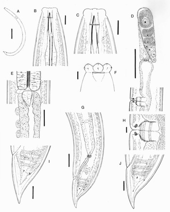
Aporcelinus abeokutaensis sp. n. (female): (a) entire body; (b, c) anterior region in lateral median view; (d) anterior genital branch; (e) pharyngo–intestinal junction; (f) lip region in lateral surface view; (g) posterior body region; (h) vagina; (i, j) caudal region. Scale bars: (a) 200 μm; (b, c, i, j) 10 μm; (d) 50 μm; (e, g) 20 μm; (f, h) 5 μm.

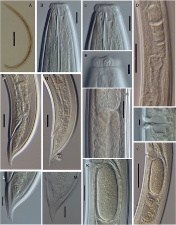
Aporcelinus abeokutaensis sp. n. (female, LM): (a) entire body; (b, c) anterior region in lateral median view; (d) anterior genital branch; (e) lip region in lateral surface view; (f, g) posterior body region; (h) pharyngo–intestinal junction; (i) vagina; (j) posterior genital branch with uterine egg; (k) uterine egg; (l, m) caudal region. Scale bars: (a) 200 μm; (b, c, l, m) 10 μm; (d, j) 50 μm; (e, i) 5 μm; (f–h, k) 20 μm.

Measurements
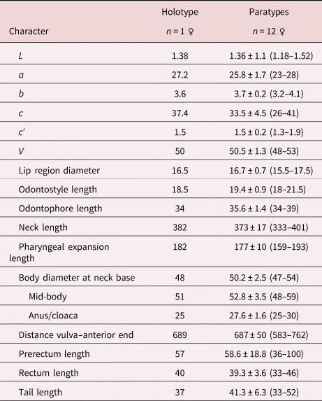
See table 1.
Morphometrics of Aporcelinus abeokutaensis sp. n. from Nigeria. Measurements in μm except L in mm and in the form average ± standard deviation (range).

Description
Female. Moderately slender nematodes of medium size, 1.18–1.52 mm long. Body cylindrical, tapering towards both ends, but more so towards the posterior extremity as the tail is conical. Habitus ventrally arcuate to a C-shape. Cuticle two-layered, 1.5–3 μm thick at anterior region, 2–4 μm in mid-body and 2–5 μm on tail; outer layer thin, nearly smooth and with nearly constant thickness throughout the body; inner layer thicker and more refractive than the outer one throughout the entire body. Lateral chord 5.5–9.5 μm broad, occupying up to one-sixth (10–18%) of mid-body diameter. Body pores indistinct. Lip region moderately angular, offset by a weak but perceptible constriction, 2.7–4.1 times as broad as high, about one-third (30–36%) of body diameter at neck base; lips moderately separate, with protruding papillae, bearing perioral liplets. Amphid fovea funnel-shaped, its opening 9.0–10 μm or 54–63% of lip region diameter. Cheilostom nearly cylindrical, with no specialization. Odontostyle strong, somewhat longer (1.0–1.3 times) than lip region diameter, 4.4–6.5 times longer than broad and 1.27–1.65% of total body length; aperture 9–10.5 μm or about one half (47–56%) of odontostyle length. Guiding ring simple but distinct, located at 8.5–11.5 μm or 0.5–0.7 times the lip region diameter from the anterior end. Odontophore rod-like, 1.7–2.1 times the odontostyle length. Pharynx consisting of a slender but muscular anterior section gradually enlarging into the basal expansion that is 5.4–6.8 times as long as wide, 3.2–4.0 times the corresponding body diameter and occupies up to one half (45–51%) of total neck length; gland nuclei located as follows: DO = 60–62, DN = 65, S1N1 = 76–77, S1N2 = 80–82, S2N = 90–91. Nerve ring located at 120–146 μm or 34–37% of total neck length from the anterior end. Pharyngo–intestinal junction surrounded by a delicate ring-like structure with a well-developed dorsal lobe; cardia conical, 16–34 × 10–20 μm. A dorsal cell mass/chord is present a short distance behind the pharyngo–intestinal junction. Intestine lacking any differentiation. Genital system diovarian, with both branches moderately developed and equally sized, 108–224 μm long or 9–15% of body length. Ovaries very variable in length (51–175 μm), often reaching and surpassing the oviduct–uterus junction. Oviduct 48–86 μm long or 1.0–1.5 times the body diameter, and consisting of a slender part made of prismatic cells and a moderately developed pars dilatata with nearly no lumen inside. A narrowing surrounded by weak sphincter separates oviduct and uterus. Uterus a tube-like structure 34–85 μm long or 0.7–1.5 times the corresponding body diameter, larger in gravid females. Uterine egg observed in two specimens, 80–88 × 34–36 μm, typical of the genus as their shell appears visibly irregular or undulate. Sperm cells not seen inside the genital tract. Vagina extending inwards 21–27 μm or up to one half (41–50%) of body diameter: pars proximalis 12–22 × 12–17 μm and with slightly convergent walls surrounded by weak circular musculature, pars refringens consisting of two visibly separate trapezoidal to oval pieces 2–5.5 × 3–5 μm and a combined width of 6.5–11.5 μm, and pars distalis 3.5–4.5 μm long. Vulva a transverse open slit. Prerectum 1.2–3.5, rectum 1.2–1.7 times the anal body diameter long. Tail conical with finely rounded tip: ventrally nearly straight or slightly convex, dorsally first convex and then bearing a variably marked dorsal concavity, with its inner core tapering at both sides and reaching up to two-thirds (56–68%) of the total tail length. Caudal pores in two pairs, situated at approximately mid-tail, one pair subdorsal, other pair lateral.
Male. Unknown.
Molecular characterization
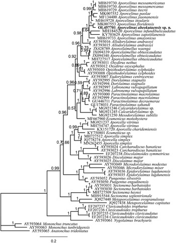
One sequence of the D2–D3 28S rDNA gene 758 bp long (OL4577701) and one sequence of 18S rDNA gene 806 bp long (OL457700) were obtained. The evolutionary relationships of the new species with other representatives of the order Dorylaimida based on the sequence of 28S rDNA are presented in fig. 3, whereas the 18S rDNA sequence did not provide any insight into the phylogenetic position of the new species.
Bayesian inferred with 50% majority of Aporcelinus abeokutaensis sp. n. based on the partial sequence of D2–D3 28S rDNA under the GTR + I + G model. The sequence of new species is indicated by bold font. Scale bar shows the number of substitutions per site.

Diagnosis
The new species is characterized by its 1.18–1.52-mm-long body, lip region offset by weak constriction, 15.5–17.5 μm broad with perioral liplets, odontostyle 18–21.5 μm at its dorsal side or 1.1–1.3 times the lip region diameter, neck 333–401 μm long, pharyngeal expansion 159–193 μm long or 45–51% of total neck length, uterus simple and 34–85 μm long or 0.7–1.5 times the corresponding body diameter long, equatorial transverse vulva (V = 48–53), tail conical with finely rounded tip (33–52 μm, c = 26–41, c′ = 1.3–1.9) and a variably distinct dorsal concavity, and male unknown.
Relationships
In having a medium-sized body up to 1.50 mm long, comparatively short odontostyle up to 22 μm long, short and simple uterus, nearly equatorial vulva (V = 48–53) and lacking males, this material resembles A. diadematus, Aporcelinus floridensis, A. granuliferus and Aporcelinus irritans.
It can be distinguished from A. diadematus in the morphology of the vagina (pars refringens consisting of separate trapezoidal vs. close together and drop-shaped sclerotized pieces, pars distalis well developed vs. nearly inexistent), vulva open (vs. close) and tail morphology (bearing an appreciably dorsal concavity vs. regularly convex at the dorsal side, inner core reaching three-fifths of tail length vs. nearly reaching the tail tip). In addition, Andrássy (Reference Andrássy2009) noted that ovarian cells of A. diadematus showed two nucleoli each, a feature that was not observed herein. For comparative purposes, the detailed description of A. diadematus by Andrássy (Reference Andrássy2009) is taken as reference inasmuch as this species has repeatedly recorded but the true identity of some of its populations remain controversial (cf. Andrássy, Reference Andrássy2009; Peña-Santiago et al., Reference Peña-Santiago, Abolafia, Nguyen, Álvarez-Ortega and Varela-Benavides2019).
Morphometrically, it is very similar to A. floridensis, only known to occur in the USA, Florida, from which it can be separated by the morphology of the vagina (pars refringens consisting of small, separate, trapezoidal vs. close together, drop-shaped sclerotized pieces; pars distalis much less developed, 3.5–4.5 vs. 4.5–6.5 μm long, and less refractive) and visibly shorter tail inner core (up to vs. more than two-thirds of tail length).
Compared to A. granuliferus, the new species can be distinguished in its less differentiated lip region (offset by weak vs. strong constriction) with (vs. without) perioral liplets, and the morphology of the vagina (pars refringens consisting of two separate trapezoidal vs. two close together drop-shaped sclerotized pieces; pars distalis not embracing vs. embracing the pars refringens). For comparative purposes, the detailed characterization of A. granuliferus by Nguyen et al. (Reference Nguyen, Abolafia, Dumack, Bonkowski and Peña-Santiago2017) is taken as reference.
From A. irritans, a deficiently characterized species originally described from Jamaica, in its less differentiated (offset by weak vs. deep constriction) and wider (15.5–17.5 vs. 14–15 μm) lip region, somewhat shorter odontostyle (1.1–1.3 vs. 1.4–1.5 times the lip region diameter) and male absent (vs. as frequent as female). In addition, the new species presents perioral liplets (vs. probably absent in A. irritans according to Thorne & Swanger's (Reference Thorne and Swanger1936) original Fig. 109) and shorter rectum (1.2–1.7 times vs. twice the anal body diameter).
Phylogenetic analysis of A. abeokutaensis sp. n. based on the partial sequence of the D2–D3 28S rDNA gene was retained and presented in fig. 3. Bayesian infrared showed that the new species as well as other species of the genus Aporcelinus grouped in a maximally supported clade, confirming the monophyly of the genus (cf. Álvarez Ortega et al., Reference Álvarez-Ortega, Subbotin and Peña-Santiago2020; Mejía-Madrid & Peña-Santiago, Reference Mejía-Madrid and Peña-Santiago2020). Interestingly, this analysis indicated that A. floridensis (MK007553) was the closest sibling of the new species (Bayesian Phylogenetics Phylogeography, BPP: 1.00), both being morphologically and morphometrically similar. Despite their close relationship and high similarity, 20 nucleotide differences in their 28S rDNA sequences explicitly shows they do not belong to the same species and verifies the proposal of the new species. Furthermore, the Aporcelinus clade was in a sister relationship with two populations of Allodorylaimus andrassyi (BPP: 0.9), proving the close relationship of Allodorylaimus and Aporcelinus, in agreement with previous findings (Mejía-Madrid & Peña-Santiago, Reference Mejía-Madrid and Peña-Santiago2020).
Type locality and habitat
South-west Nigeria, Alabata, Abeokuta (7°14′12.3″N, 3° 27′43.3″E), where the new species was collected from a watermelon (Citrullus lanatus L.) field on sandy-loam soil (sand = 68%, silt = 11%, clay = 21%, organic matter = 13.11%, pH 6.26) in an agrarian locality with history of sweet pepper (Capsicum annuum L.) and cassava (Manihot esculenta Crantz) production.
Type material
Thirteen females (holotype and 12 paratypes) deposited in the nematode collection of the University of Jaén, Spain.
Etymology
The new species is named after ‘Abeokuta’, the Nigerian locality where it was found.
Financial support
R.P.S. is grateful for the financial support received through the research program ‘PAIUJA 20121/2022: EI_RNM02_2021’, University of Jaén, Spain, for his scientific work throughout the last two years. Infrastructure and partial funding by the Nematology Unit at North-West University of South Africa is also acknowledged.
Conflicts of interest
None.
Ethical standards
The authors assert that all procedures contributing to this work comply with the ethical standards of the relevant national and institutional guides on the care and use of laboratory animals.
Author contributions
T.T.B. and M.R. contributed equally to this work and share first authorship.