Conventional congenital cardiac surgery for patients with discordant atrioventricular connections, usually associated with discordant ventricular arterial connections in the combination known as congenitally corrected transposition, leaves the morphologically right ventricle as the pump to the systemic circulation. Long-term follow-up of these patients shows progressive development of right ventricular failure in up to one-tenth of patients for each 10 years, usually in association with regurgitation across the morphologically tricuspid, or systemic atrioventricular, valve.1–2
Several instrumental diagnostic tools have been employed to study the morphology and function of the systemic right ventricle in these patients so as to evaluate its capability to sustain a systemic workload.3–7 Even if an impaired myocardial flow reserve of the right ventricle has been advocated as the possible cause for myocardial failure, the definitive and true history of the right ventricle supporting the systemic circulation in humans is not as yet known, and neither is its structural adaptation to the new workload.
To investigate these problems, we have established a rat model of pressure loading of the right ventricle by banding the pulmonary trunk. Our goals were to investigate the functional and structural changes produced by an increased workload with respect to sham-operated animals, and to evaluate whether the age of animals at the time of banding could influence the development of an adverse response.
Materials and methods
Establishment of the model
We obtained 60 male Sprague-Dawley rats from Harlan Laboratories, San Pietro al Natisone, Udine, Italy, 30 of the rats being 10 weeks old, and hence adults, while the other 30 were 3 week-old weanlings. We randomly divided these animals into experimental groups, of 20 rats each, these animals undergoing banding of the pulmonary trunk, and to control groups of 10 rats each, these animals undergoing sham operations.
The animals were housed, treated, and handled in accordance with the recommendations stated in the National Institute of Health's Guide for the Care and Use of Laboratory Animals, prepared by the Institute of Laboratory Animal Resources, National Research Council, as stated in National Institute of Health Publication number 85–23, revised 1996. The protocol was approved by the Animal Care Committee of the University of Padova Veterinary School, and by the Italian Government Committee for Animal Care.
The operative procedures were carried out under general anesthesia using 5% sevoflurane (Abbott Spa, Latina, Italy) and tiletamine chlorohydrate (Zoletil, Virbac, France) at 50 milligrams per kilogram. Rats were intubated utilizing a 16-gauge and 20-gauge polyethylene cannula in adults and in weanlings, respectively. The endo-tracheal cannula was connected to a ventilator for small animals (Rodent Ventilator 7500, Ugo Basile, Varese, Italy) and anaesthesia was maintained by a mixture of oxygen and sevoflurane. The pulmonary trunk was exposed through a limited left lateral thoracotomy in the fourth intercostal space, and banded proximal to its bifurcation using a 4.0 silk suture, which was calculated from previous pilot studies to produce a 60% decrease in the diameter, both in the adults and the weanlings. The banding was maintained for 30 days. To reduce the potential of pneumothorax, the chest was closed under positive-pressure ventilation, and a chest tube was placed during the time of the closure to evacuate all air, being removed after chest closure. Intra and post-operative pain was controlled with the non-steroidal anti-inflammatory drug tramadol, given at 2 milligrams per kilogram intramuscularly, every 12 hours, for 5 days. The animals undergoing sham procedures of both age groups had the same intra- and post-operative procedure as did the rats undergoing banding of the pulmonary trunk, except for the banding itself. Rats were subsequently weighed and inspected twice a week.
Functional tests
All rats who survived after banding had an echocardiogram on the first and thirtieth postoperatives days, performed using a Hewlett-Packard Sonos 5500 echocardiographic apparatus equipped with a 12.5 Megahertz probe. Rats were sedated by tiletamine chlorohydrate at 50 milligrams per kilogram. We measured the gradient across the band, and the thickness of the free wall of the right ventricle.
On the thirtieth post-operative day, surviving animals underwent a second operation to obtain haemodynamic measurements, and to harvest their hearts and livers. Rats were anaesthetized, placed on mechanical ventilation, and treated for pain as described above. During the operation, the animals were inspected for hydrothorax and ascites. Pressures in the systemic left ventricle and right ventricle were obtained by direct puncture of cardiac chambers with an 18-gauge needle connected to a fluid-filled polyethylene tubing to a pressure transducer and a digital monitor (Roche, Monza, Italy).
Preparation of samples for morphological analysis
Hearts were then quickly perfused with an antegrade cold cardioplegic solution to obtain a diastolic arrest and were removed from the thorax and washed with phosphate buffered saline. The liver was also explanted. We measured body weight, wet heart weight, and liver weight in all the animals. We then sectioned 30 hearts in a short axis-view plane 5 millimetres from the heart apex, sampling 12 adults after banding of the pulmonary trunk, 4 adults undergoing sham operations, 10 weanlings with banding, and 4 weanlings after sham operations, while the remaining 25 hearts were prepared by separating the free walls of the ventricles from the ventricular septum, and recording their respective weights, these measurements being undertaken in 10 banded adults, 4 adults after sham operations, 6 banded weanlings, and 6 weanlings after sham operations.
Morphometric analysis
Histology and histochemistry.
We fixed 18 heart specimens in 10% formalin-phosphate buffered saline solution and subsequently embedded these hearts in paraffin, cutting 10 micron sections, which were processed for histology, using haematoxylin and eosin staining, and histochemistry using a modified Azan-Mallory trichrome Heidenhein technique. Ventricular wall and septal thicknesses were calculated using the average of 5 measurements for each sample. The number of cardiomyocytes across the wall was defined as the number of cells along a perpendicular line to the mural axis, taking an average of 20 measurements per sample. Cardiomyocyte diameter was calculated as the mean of 80 cells sectioned equatorially. The percentage of fibrosis was calculated automatically on a virtual-colour-based system, and reported as the mean value of 10 different randomly chosen fields. Capillary density was defined as the number of capillaries per square micron within 10 different randomly chosen fields. The index of myocytes apoptosis was evaluated using the Terminal deoxynucleotidyl Transferase Biotin-2′-Deoxyuridine 5′-Triphosphate Nick End Labeling test, being stated as a proportion of the number of positive cardiomyocytes over the total number of cardiomyocytes per field.
Immunocytochemistry.
Capillaries were identified by indirect immunofluorescence using an anti-von Willebrand factor polyclonal antibody (Chemicon, Temecula, California) and anti-rabbit Immunoglobulin G coupled to fluorescein isothiocyanate as secondary antibody (Dako, Glostrup, Denmark). Cardiomyocyte apoptosis was studied by the terminal-deoxynucleotidil transferase-mediated 2′-Deoxyuridine 5′-Tri-phosphate nick-end-labelling. Sections were observed using a Zeiss Olympus BH-2 microscope equipped with a JVC 3-digital camera and Tulip Vision Line Xd 6/350, Image Pro-plus 4.1.
Immunofluorescence.
We snap-froze 25 specimens in liquid nitrogen, cutting 10 micron cryosections and processing them for expression of beta-myosin heavy chain and alpha-skeletal actin.8–9 Sections were studied using a Zeiss-Axioplan Microscope equipped with a Leica digital camera 300F and Leica Image Software IM1000.
Western blotting
Total protein extracts were obtained from about ¼ of the right ventricles. Specimens were cut, minced, and dissolved in Laemmli's buffer solution. Samples were electrophoresed either on 8% or 5% Sodium dodecyl sulfate-polyacrylamide gel electrophoresis (acrylamide/bis-acrylamide 30–0.8%) for protein separation. Proteins were transferred onto a nitrocellulose membrane (Amersham, Little Chalfont, United Kingdom) and the membranes were processed with the primary and secondary antibodies appropriately diluted in Tris-buffered saline with 0.05% tween 20, 3% milk. The secondary antibody was a peroxidase-conjugated rabbit anti-mouse immunoglobulin-G (Dako) that was revealed by enhanced chemiluminescence. Beta-myosin heavy chain expression was quantitated by densitometric analysis of the specific electrophoretic bands blotted on the paper and reacted with the specific antibody. Only values than fell within a linear range of a calibration curve were considered. This curve was obtained using a protein sample that gave a high expression level of beta-myosin heavy chain at the protein level. This sample was used as internal control in all the immunoblots and ensured for variability due to the detection method. Auto-radiographies were scanned using the Kodak Digital Science 1D program. Quantification of beta-myosin heavy chain in the various samples was performed as mean value of two different protein concentrations for each sample, analyzed in three independent experiments.
Statistics
Mean values and standard deviations of the various parameters were used for statistical analysis. Significant differences between the groups were determined by the Wilcoxon-Mann-Whitney rank sum test.10 The level of significance was set at p-value equal or less than 0.05.
Results
We lost 6 rats early after banding of the pulmonary trunk, 1 being an adult, and 4 weanlings. There were no early deaths in the animals having sham operations. The remaining 54 animals survived for the whole period of the study, and in none of them were we able to detect signs of right-sided cardiac failure, such as hydrothorax, hepatic cirrhosis, or ascites at the time of sacrifice.
Functional study
Echocardiography.
The pressure gradient across the banded pulmonary trunk increased significantly in both age group between the first and thirtieth post-operative days, from 43.94 millimetres of mercury, with standard deviation of 9.2 to 53.57 millimetres of mercury, with standard deviation of 10.90 (p-value of 0.005) in the adult rats, and from 45.81 millimetres of mercury, with standard deviation of 8.72 to 86.73 millimetres of mercury, with standard deviation of 15.02, in the weanlings (p-value less than 0.0001). The increase, at 47%, was higher in the weanlings when compared to the increase in adult rats, of 18%, probably due to the progressive narrowing of the band in relation to the increasing body weight of the animals during the period of the study. The thickness of the right ventricular free wall increased significantly in both age groups; from 0.82 millimetres, with standard deviation of 0.03 to 1.84 millimetres, with standard deviation of 0.05, in adult rats (p-value less than 0.0001), and from 0.74 millimetres, with standard deviation of 0.05 to 2.2 millimetres, with standard deviation of 0.15 millimetres, in the weanlings (p-value less than 0.0001 – Fig. 1).

Figure 1. Cross-sectional echocardiograpy (short axis view) showing the increased right ventricular mural thickness after 30 days of banding of the pulmonary trunk in a weanling rat. Postoperative day 1 (a) and 30 (b). Abbreviations: RV: right ventricle; LV: left ventricle.
Haemodynamic study.
Systolic pressure in the right ventricle was significantly higher after banding when compared to respective age-related sham operated animals, at 57.31 millimetres of mercury, with standard deviation of 15.24, versus 28.6 millimetres of mercury, with standard deviation of 3.4, in adult rats (p-value less than 0.0001), and 94 millimetres of mercury, with standard deviation of 15.17, versus 22.28 millimetres of mercury, with standard deviation of 1.49) in weanlings (p-value less than 0.0001). No difference was found in systolic and diastolic pressures in the left ventricle between the banded rats and those undergoing sham operations in either age group. The ratio of systolic pressures between the right and left ventricles was 0.58 in the adult rats, and 0.95 in the weanlings, after banding of the pulmonary trunk. The right ventricular diastolic pressure was increased only in the adults after banding when compared to adults undergoing sham operations, at 3.4 millimetres of mercury, with standard deviation of 0.83, versus 1.5 millimetres of mercury, with standard deviation of 0.7 (p-value of 0.009).
Morphometric study
After 30 days, the weight of the adult rats subsequent to banding increased from 335.26 grams, with standard deviation of 16.18 to 416.94 grams, with standard deviation of 30.46, whilst the weight of the banded weanlings increased from 40.06 grams, with standard deviation of 0.96 to 246.33 grams, with standard deviation of 27.59. No significant differences in body weight were found between the animals undergoing banding and their sham operated controls.
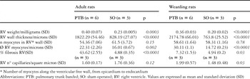
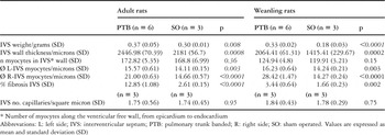
The wet weight of the heart was significantly increased in banded rats compared to their controls, from 1.03 grams, with standard deviation of 0.04 to 1.33 grams, with standard deviation of 0.14, in adults (p-value less than 0.0001), and from 0.84 grams, with standard deviation of 0.05 to 1.2 grams, with standard deviation of 0.2, in weanlings (p-value equal to 0.0002). This increase was likely to be due to the increased weight of the right ventricle, at 42.5% and 44.9%, respectively, in adult and weanling rats, and the ventricular septum, at 20% and 44.1%, respectively, in the two groups (Fig. 2, Tables 1–2). A significant increase was also found in right ventricular mural thickness, mainly caused by an increase in cardiomyocyte diameters, from 14.72 microns, with standard deviation of 0.23 to 30.11 microns, with standard deviation of 1.1, in banded weanlings, and from 16.01 microns, with standard deviation of 0.67 to 22.31 microns, with standard deviation of 2.26, in adults after banding, and extra-cellular fibrosis, from 4.94% with standard deviation of 0.2 to 7.32% with standard deviation of 0.53 in weanlings after banding, and from 4.88%, with standard deviation of 0.35 to 43.62% with standard deviation of 2.55 in banded adults (see Figs. 3–4, and Table 1). A similar response of cardiomyocytes and interstitial tissue is also shown for the ventricular septum (see Table 2). The number of cardiomyocytes across the transverse section of the right ventricular free wall was decreased in banded adults when compared to adults undergoing sham operations, albeit that the difference was not significant. Regarding the density of capillaries in the right ventricular myocardium, this was significantly increased in banded weanlings, at 1.99 capillaries per square micron, with standard deviation of 0.57, if compared to the sham-operated weanlings, when values were 1.48 capillaries per square micron, with standard deviation of 0.48. In contrast, the density of capillaries in the right ventricle of banded adult rats, at 1.60 capillaries per square micron, with standard deviation of 0.17, was mildly decreased if compared to the values for adults undergoing the sham operation, at 1.76 capillaries per square micron, with standard deviation of 0.36 (Table 1). The weight of the systemic left ventricular component of the heart, and the weight of the liver, was not increased subsequent to banding in either group of animals (data not shown). The apoptotic index was less than 1% in both adult and weaning rats after banding if compared to their controls.

Figure 2. Haematoxylin-eosin staining of cardiac sections cut showing the increase of right ventricular mural thickness consequent to banding of the pulmonary trunk (black arrowhead). Abbreviations: PTB: pulmonary trunk banded; SO: sham operated; LV: left ventricle.
Table 1. Morphometric data – Right ventricle.

Table 2. Morphometric data – Interventricular septum.


Figure 3. Haematoxilin-eosin staining of cardiac section of right ventricle taken from weanling and adult rats with banding of the pulmonary trunk and sham operations. Note the significant increase of myocyte diameter of rats with banding (both age groups). Note also the complex three-dimensional mesh arrangement of the right ventricular mass.

Figure 4. Azan-Mallory trichrome Heidenhein-modified staining of cardiac sections of right ventricle taken from weanling and adult rats with banding of the pulmonary trunk and sham operations. Note the significant increase of extracellular fibrosis in adult rats with banding.
Expression of developing markers in hypertrophic cardiomyocytes
The effect of the banding of the pulmonary trunk on the profile of cardiomyocytic differentiation was assayed by testing the expression of beta-myosin heavy chain and alpha–skeletal actin at the cellular and molecular level. The expression of these two proteins is markedly enhanced in hypertrophic myo-cardium in models of both left and right ventricular hypertrophic myocardium other than banding of the pulmonary trunk. Immunostaining of cryosections from the cardiac muscles of the four groups of animals is shown in Figure 5. Beta-myosin heavy chain is distributed according to a mosaic of positive and negative fibres in the heart of normal adult rat (not shown). As shown in Figure 5A, B, adult and weanlings rats show a minor proportion of strongly reactive cells after the sham operation, while both groups display high levels of reactivity and homogeneous staining in the right ventricle after banding (Figs. 5C, D). The left ventricle, on the other hand, maintained the pattern for the sham operation subsequent to banding (Figs. 5C, E). Similar results were obtained by analysis of expression of alpha-skeletal actin (Fig. 5F, I). In contrast to the pattern of expression of beta-myosin heavy chain, however, the immunostaining pattern of alpha-skeletal actin in banded rats was a mosaic of strongly and more weakly stained reactive fibres.

Figure 5. Expression of molecular markers of cardiomyocyte hypertrophy in rats with banding of the pulmonary trunk. Cardiac cryosections from adult and weanling rats with banding and sham operations were process by anti-beta-myosin heavy chain (A through E) and anti-alpha-skeletal actin (F through I) antibodies. The arrow in (F) indicates a coronary artery surrounded by cardiomyocytes strongly reactive with the anti alpha-skeletal actin antibody (scale bar, 100 microns).
The increase in expression of beta-myosin heavy chain was investigated further by immunoblotting (Fig. 6). In comparison with sham operated rats, the level of accumulation increased three-fold in adult rats after banding, and more than sixteen-fold in banded weanlings (Fig. 6A and Table 3). Significantly higher expression, though not quantified, was also found for alpha-skeletal actin when comparing the rats undergoing banding with those submitted to the sham operations (Fig. 6B).

Figure 6. Western blots used for the quantitative evaluation of markers of car-diomyocyte hypertrophy (8% Sodium dodecyl sulfate-polyacry-lamide gel electrophoresis gels with calibrated samples and the corresponding immunoblots are shown). Panel A: beta-myosin heavy chain expression in the blotted antigens as revealed after incubation with the respective antibody. Panel B: alpha-skeletal actin expression in the blotted antigens as revealed after incubation with the respective antibody. Only the area corresponding to the reactive bands is visible.
Table 3. Levels of expression of beta-myosin heavy chain at right ventricular level.

Discussion
We have created a reproducible model of acute increased afterload of the low-pressure morphologically right ventricle by banding the pulmonary trunk. The limitation in involvement of the ventricular septum to its right side is in agreement with previous observations.3, 11
All animals tolerated well the increased workload, and all were in good clinical conditions after 30 days of banding, showing no signs of cardiac failure, such as loss of weight, hepatic congestion, hydrothorax, or ascites. Marked echocardiographic and morphometric changes are seen for both adult and weanling rats in our banded model of compensated right ventricular hypertrophy, specifically a significant increase of the thickness of its wall and its weight. Whilst these data seem to suggest a good adaptation of the right ventricle to pressure overload, a more thorough structural investigation reveals a different situation. The thickening of the right ventricular wall is predominantly due to hypertrophy of the cardiomyocytes and development of interstitial fibrosis.
Hypertrophy of the cardiomyocytes is seen when banding is undertaken at both ages, but it is significantly greater in weanlings. Conversely, hyperplasia is not evident in our model, despite this now being considered as an additional important component of the response of the myocardial cell to increased workload.3–6, 12 Since the ability of the cardiomyocyte to proliferate is lost soon after birth, it may be that the time window we used for weaning the rats is such that remodelling of the ventricular wall is made possible solely by hypertrophy of preexisting cardiomyocytes.13
The other player involved in the process of structural and functional remodelling of right ventricular myocardium is the interstitial fibrosis. The ventricular mass is known to be arranged as a complex three-dimensional mesh of tangential and diagonally intruding fibres,14, 15 supported by a collagenous fibrous matrix, the latter being better represented in right ventricular myocardium of rats that underwent increased overload (Figs. 3 and 4).
In comparison to the animals undergoing sham operations, the percentage of ventricular area in which fibrosis develops is significantly higher in banded rats from both age groups, but is more pronounced in adult rats (p-value less than 0.0001) than in the weanlings. There is a complete absence, however, of any condensations of fibrous tissue within the supporting matrix that might have permitted the distinction of any tertiary arrangement of reproducible myocardial bundles.15–16 The density of capillaries, nonetheless, is dramatically different in the adults compared to the weanlings subsequent to banding, and this could suggest that the neo-angiogenetic potential, and hence the adaptive response of the vascular network to an increased workload, is weaker in adults that in weanlings. Since apoptosis was low in both groups after banding, it is reasonable to assume that most of the observed fibrosis is not of reparative origin, in other words, it does not represent a disproportionate increase in synthesis and/or inhibition of degradation of extra-cellular matrix as consequence of death of cardiomyocytes.17 Obviously, we cannot exclude that a small part of the fibrosis develops as scarring replacement of dead cardiomyocytes due to inadequate vascular supply or sustained stress. Likewise, we cannot say whether apoptosis is more evident during the first phase of the increased afterload,17, 18 since we examined only a single time point subsequent to banding.
It was noticeably that the minimal fibrosis but good capillary response seen in weanlings after banding are coupled to higher levels of myocytic hypertrophy. This event is accompanied by the increased expression of alpha–skeletal actin and beta-myosin heavy chain, well known members of the “fetal gene programme” which is reactivated coincidentally with cellular hypertrophy. The mechanical stretch of the myocardium, which inherently characterized the model of banding of the pulmonary trunk, can up regulate, among others, the renin-angiotensin-aldosterone system that, via the type 1a angiotensin receptor, can cause both hypertrophy and fibrosis. The role played by this autocrine-paracrine cross-talk between cardiomyocytes and interstitial fibroblasts is now under scrutiny in our laboratory.19
The expression of alpha–skeletal actin and beta-myosin heavy chain is higher in the weanling rats than in their adult counterparts, implying that, in younger animals, the actomyosin contractile machinery has been shifted to a functional condition in which the myocardium displays a decreased shorting velocity, and therefore a decreased power output, inasmuch as beta-myosin heavy chain is associated with slower rates of contraction.8 This might potentially reflect a decreased systolic performance in the weanlings after banding in comparison with their sham operated controls, but this is not the case. Thus, changes of beta-myosin heavy chain expression in the banded weanlings do not attain a functional significance. Conversely, interstitial fibrosis could play a key role in influencing the diastolic function, inasmuch as the mean diastolic pressure in the right ventricle of banded adults was significantly higher than in sham operated animals, and this could represent an initial sign of compromise of diastolic function at this age. In fact, the increase of non-contractile elements could, if sufficiently large, result in an increase in cross-sectional area and, thereby, in a decrease of the force-generating ability. This can, in turn, interfere with the maximum velocity of shortening of the insulted myocardium.20
Our model provided data about the structural and physiologic adaptation of a low-pressure right ventricle to an increased workload. The age of the animals at the time of banding seems to be a risk factor for adverse changes in such rats exposed to increased pressure load. It remains to be established what is the molecular and cellular base for the weak responsiveness of younger animals for interstitial fibrosis in a compensated model of right ventricular hypertrophy.21
This animal model of right ventricular overload simulates the clinical condition of patients with congenital heart disease characterized by a systemic right ventricle; and our findings are in agreement with our previous unpublished data about the post-mortem histologic evaluation of the morphologically systemic right ventricle in 4 adult patients who had congenitally corrected transposition, and died with right ventricular failure. In all these 4 patients the percentage of extra-cellular fibrosis was increased in the systemic morphologically right ventricle if compared to that of normal right ventricle in age-matched controls, at 21.8% versus 10.6% (Fig. 7). Furthermore, the density of capillaries was also found to be significantly lower in the myocardium of the systemic morphologically right ventricle myocardium if compared to controls.

Figure 7. Azan-Mallory trichrome Heidenhein-modified staining of cardiac sections from the systemic morphologically right ventricle taken from a 54-year-old patient with congenitally corrected transposition (A) compared to that of a normal low-pressure ventricle (B). Note the significant increase of extra-cellular fibrosis.
Future animal models relating to the development of a pharmacological modulation of myocardial angiogenesis, in addition to the possible repopulation of ventricular myocardium with stem cells, could conceivably play a key role in the myocardial response to an increased workload. Such findings could be possibly transferred to the clinical arena to improve the myocardial response of patients with failing systemic morphologically right ventricles, postponing other complex surgical options and eventually orthotropic heart transplantation.
Limitations
There are limitations to our study. We have not determined the expression of alpha–skeletal actin and beta-myosin heavy chain in cardiomyocytes at the beginning of the study period in either of our two groups of animals. Thus, we cannot rule out the possibility that part of the observed changes in the levels of expression for these two markers is attributable to physiological variations. In Sprague-Dawley rats, however, these changes are known to be modest.21 The banding of the pulmonary trunk in the weanling rats produced progressive narrowing in relation to the increasing body weight of the animals during the period of the study. As a result, at the end of the study the afterload in the banded weanlings was greater compared to banded adults, despite the initial imposition of 60% decrease in the diameter of the pulmonary trunk in both age groups.
Ackowledgements
This work was supported in part by a grant from Consortium Agreement European Community “Heart failure and cardiac repair” IP 018630. We thank Dr Sophie Clement for providing the alpha–skeletal actin antibody. We also thank Dr Aldo Castaneda for reviewing the manuscript.