

In 1784 anatomist and accoucheur William Hunter wrote,
Large collections [of anatomical specimens] which modern Anatomists are striving, almost every where to procure, are of infinite service to the art; especially in the hands of teachers. They give students clear ideas about many things, which it is very essential to know, and yet which it is impossible that a teacher should be able to shew otherwise, were he ever so well supplied with fresh subjects.Footnote 1
The statement, which comes from Hunter's introductory lectures on anatomy, suggests that collections of specimens were of crucial significance to the art of anatomy. They served anatomical science itself, but were primarily of use to teachers, serving to impart some ideas more clearly than bodies themselves. And they functioned in groups, in collections. Knowledge in late eighteenth- and early nineteenth-century anatomy was made, embodied and spread through what this article argues was a system of visual display, of which these collections of specimens were only one part. That system was used in pedagogical contexts, contexts that were also the sites of anatomical research. Anatomical museums functioned alongside books and bodies to form a set of pedagogical tools and objects of study. Individual elements of display, such as atlases or dissected bodies, were understood to function together as parts of a system that was created and deployed in a particular setting and for a particular purpose.
Wax models; preserved specimens in jars, housed in collections; schematic chalk drawings from the classroom; elaborate engravings and less elaborate etchings found in books; dead bodies; paintings and sculptures; and living bodies – each of these was, to some extent, the object of study in anatomy. Together, they were taken to form several things: Nature, a representation of Nature, and a representation of a particular argument about how the natural world was constituted. They were material embodiments of scientific knowledge at the same time as they were its objects of study. As a constellation of interrelated tools, along with the texts that framed them, they were understood to function together to serve pedagogical and research functions, endeavours that often coalesced in a science that was rooted in the classroom.
In 1814, Charles Bell wrote to his brother that the ‘whole system must be kept in full operation – preparations, drawings, models, cases, lectures, clinical lectures, &c’.Footnote 2 Bell's ‘system’ was both visual and pedagogical. It was visual in the sense that all parts of the system were visual and that seeing was privileged, even for anatomical parts like the hand that we usually associate with touch; but not all elements of the system were simply visual. The system was pedagogical because knowledge in Enlightenment and early nineteenth-century anatomy was produced and disseminated in a pedagogical setting. Students who entered Bell's classrooms had been witness to bodies both living and dead long before they ever began professional training. They watched Bell and his house pupil dissect a cadaver at the front of the room, over a full course, stretching weeks on end. They heard him narrate such a dissection, clarifying the relationships between parts and systems of anatomy in a messy anatomical field, and they watched him impose order on the blackboard, with rough sketches showing function or how parts came together. These same students could walk a short hallway to the museum, full of collections of dry and wet specimens collected in multiple to give a sense of anatomical variation considered normal and that which could be classified as pathological. Such specimens were sometimes brought into the classroom for lectures, where students would pass minute and detailed forms of anatomy not visible in the performance of dissection, or rarer specimens, preserved because they were hard to come by. A few decades earlier, William Hunter had even described the way in which they were to be passed around the room, one student describing to the next what was to be seen. And students learned by doing – training their hand and eye together, by preparing their own specimens and preservations, using models made of wax, drawing from the specimens and from atlas illustrations. The hand and eye were seen as analogous organs, their training interrelated and simultaneous: doing was a part of seeing.Footnote 3 Reference books for students also served as manuals for practitioners. And grand atlases, meant to impress, illustrated nature and also the order their authors perceived in nature.
This paper treats as its central historiographical problems notions of realism and naturalism in imagery as self-evident, the use of atlases as the privileged sources of visual representations in science, and dichotomies of representation and nature and of image and knowledge. This puts it in dialogue with a variety of work on art and anatomy, including Lorraine Daston and Peter Galison's important and widely cited work Objectivity. Footnote 4 One can recontextualize and extend their work on atlases by focusing on the uses of visualization and display. A focus on use demonstrates that illustrated books served as only a portion of a system of visual display that was taken to function in its entirety, of which anatomical atlases were just one element.
If we decentralize these expensive texts to include three-dimensional objects and more ephemeral visual displays, we get a broader sense of what constituted science and its audiences. To do so shifts the focus from the products of science to their uses. Understood that way, it becomes clear that the visual epistemologies that Daston and Galison group together under the heading of ‘truth-to-nature’ reflect notions of the knowability of Nature herself and of the indistinguishability of object and representation better than they do notions of subjectivity and objectivity. We also find that pedagogical functions united most of these forms of knowledge.
In his ‘History of science and its sociological reconstructions’, Steven Shapin wrote that knowledge ‘is always tailored to doing things. It is in the course of doing things with knowledge that its meaning is produced; thus the notions of use and meaning are intertwined’.Footnote 5 The assertion, so crucial to much work in the history of science, should be applied equally to visual representations of (or as) knowledge – they were made for doing things; we ought to look to those contexts of use to determine the meaning of the visual. Visual displays were used to systematize pedagogical training and to root it in educational theories of people like Johann Pestalozzi, who believed that observation of objects themselves formed the best mode of teaching.Footnote 6 Pedagogy – as carried out in the private schools of London – was at the centre of British medical science in the Enlightenment and the Age of Reform. It created the space, the audience and the support for anatomical research.Footnote 7 If Daston and Galison and others do not see atlases situated in their broader context, it may be because they do not take pedagogy seriously as the space in which science was developed in this period.Footnote 8
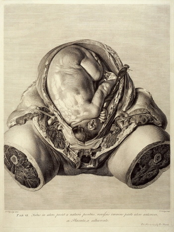
How, then, do that basing of anatomical science on pedagogy and a focus on context of use accord with the type of visualization supposedly at work in anatomical atlases? According to Daston and Galison, ‘not only do images make the atlas; atlas images make the science’.Footnote 9 However, atlases such as William Hunter's Anatomia uteri humani gravidi tabulis illustrata (The Anatomy of the Human Gravid Uterus, 1774) were large and expensive.Footnote 10 Hunter's was an elephant folio printed by John Baskerville, a printer known for his high-quality papers and inks. Baskerville published no other scientific books and Hunter's atlas was the most expensive book Baskerville published in his career.Footnote 11 This expense was not peculiar to Hunter's atlas – atlases were, by necessity, expensive books that were not particularly portable. It would be an odd understanding of the scientific endeavour, then, that saw them as the essence of science – these books that were rarely bought and used, limited primarily to the classrooms of their authors. To understand their place better within a pedagogical endeavour, alongside other objects and images, we ought to examine the contexts of use of the other elements in the system of display, looking at the intervention or creative work involved in fashioning them, at their uses once created, and at their intended audiences.Footnote 12 Each of these is important to understanding what is sometimes termed ‘visual culture’,Footnote 13 to understanding how visual displays, when combined in a system, stood in both for nature and for science, or for knowledge about nature.
Anatomy in Britain: the Hunters, Baillie and the Bells
William Hunter, Charles Bell, John Hunter, John Bell and Matthew Baillie were all recognized in their own time as pre-eminent British anatomists, known for developing their science, but also for their contributions, in some fashion, to what I call visual display. William Hunter, born and trained in Scotland, moved to London to seek fortune and professional accomplishment. He found both. He established the Great Windmill Street School of Anatomy in 1769 and became physician to Queen Charlotte. He was elected a fellow of the Royal Society in 1767 and was appointed professor of anatomy to the Royal Academy in 1768.Footnote 14 William's younger brother, John, was a surgeon and an enthusiastic collector of anatomical specimens who ran an anatomy museum in Leicester Square. Matthew Baillie, best known for his work on ‘morbid anatomy’ (now called pathology), was a nephew of the Hunters and was trained, to a great extent, under William at Great Windmill Street. There he gained proficiency in making preparations, and, when William Hunter died in 1783, Baillie took over the Great Windmill Street School with William Cruikshank.Footnote 15
John and Charles Bell were also Scottish, trained in Edinburgh. Charles received some of his training from John, his older brother, who ran a successful anatomy school on Surgeon's Square in Edinburgh, but while, like the Hunters, Charles left Edinburgh for the open medical marketplace of London, John stayed in Edinburgh, eventually closing his school after engaging in disputes with powerful professors at Edinburgh University and being blocked from bringing students into the dispensary.Footnote 16 Charles, who left Edinburgh because of his brother's unpopularity, built his career on teaching, using it to publicize his discovery of the separate roots of motor and sensory nerves. Charles taught both artists and anatomists at the Great Windmill Street School from 1812 onwards.Footnote 17
Though they are a group of Scotsmen, the group is otherwise one that is representative of British anatomy as a whole between 1750 and 1850. John and Charles Bell both produced their own anatomical illustrations; William Hunter taught at the Royal Academy. All owned considerable collections of specimens, practised anatomy as a science and published illustrated treatises. And, following a dominant model of what constituted learning, all wrote in strikingly similar terms about the uses of the visual in teaching anatomy, talking about knowledge ‘more distinctly impressed upon the mind by figures … being exhibited to the eye’ (Baillie),Footnote 18 or about an image giving ‘clearer ideas of most natural objects … [which] makes stronger impressions on the mind’ (William Hunter),Footnote 19 or about a book of engraved plates depicting anatomical structures that would ‘fix them in his [the student's] memory in a way which no description can accomplish’ (Charles Bell)Footnote 20 – that is to say, talking about visual display as an important pedagogical tool.
Seeing the big picture in its complexity and its order: dead bodies, dissection on display and schematic sketches to learn by
Hunter begins his posthumously published Two Introductory Lectures, Delivered by Dr. William Hunter to His Last Course of Anatomical Lectures at His Theatre in Windmill Street (1784) with a discussion of the origins of anatomy, saying that
the observance of bodies killed by violence, attention to wounded men, and to many diseases, the various ways of putting criminals to death, the funeral ceremonies, and a variety of such things … have shewn men, every day, more and more of themselves; especially as curiosity and self-love would urge them powerfully to observation and reflection.Footnote 21
The sort of casual and unstructured, informal experience of death as well as of living bodies formed one end of the spectrum of visual displays that were understood to constitute the experience of anatomy students and medical students. Witnessing funerals, executions, deaths and illness, bodies were not foreign or distant things to medical men in Georgian Britain. In such encounters – visual displays of death, a theatre of dead bodies – the bodies themselves were not representations, nor were they even objects of formal study, but, as Hunter himself articulates, they functioned as a backdrop, a set of common experiences that would underlie formal anatomical study. Such bodies, unlike anatomical corpses, the term I will use for bodies that have been anatomized, were not especially the objects of scientific intervention or the subject of a particular scientific aesthetic; their uses were not understood to be scientific and their audiences were multiple, not confined to the professional or the man of science.Footnote 22
Such casual experiences with dead and diseased bodies were complemented by formal dissections taking place in lecture theatres. John Bell said of dissection that it ‘is the first and last business of the student’.Footnote 23 William Hunter's theatre was set up with the care befitting such important pedagogical business:
You may observe that this theatre is particularly well constructed, both for seeing and hearing; a strong sky-light is thrown upon the table, and the glass being ground, that is, made rough upon one surface, the glare of sun-shine is not admitted: the circular seats are brought as near the table, as ease in sitting would admit of; and, as they go back, they are a good deal raised, which is a considerable advantage both in seeing and hearing …Footnote 24
The interventions of anatomical science in this setting were both active (the dissection of the body by the demonstrator) and descriptive, categories that should not be seen as dichotomous and were rarely uncoupled. Hunter described the elaborate construction of anatomy theatres to promote hearing and seeing because the narration of dissection was crucial to situating its display. Charles Bell, some thirty years later, would say of his dissection course in the same theatre that ‘regular and full Demonstrations of the Parts dissected are given; where the Application of Anatomy to Surgery is explained, and the Methods of operating shown on the Dead Body’.Footnote 25 Things were demonstrated and shown at the same time as they were explained.
The audience for such dissections was made up almost exclusively of students. Hunter described the way in which dissection demonstrations functioned within a medical education, saying that the student would ‘see the preparatory dissection for every lecture; which will make the lecture itself much more intelligible, and fix it deeper in the mind; he will see all the principal parts dissected and demonstrated over and over again [on] a number of bodies dissected in succession’.Footnote 26 Dissection was good for seeing the ‘principal parts’, for seeing gross figures. Even with an abundant source of corpses for dissection, as Hunter clearly assumed when he wrote about ‘a number of bodies’, however, cadavers too were only a part of a system, not an object of study that could only be approximated through other sorts of display.
The complicated mess of a decaying corpse, dissected by a demonstrator, was good for seeing some things – for a sense of scale and overall relationship of the parts – but it was less good for others. Even as a representative of the anatomy of a whole body it was complemented by other kinds of visual display. All of these forms of display were meant to work together in a sort of interrelated whole. At the other end of a spectrum of displays from overly complicated bodies sat a type of display that left little evidence for the historian – the rough schematics and chalk drawings of the classroom. These too required explanation and expert narration, but visually they simplified the complexity of a body and diagrammatically showed its functions. They too helped to explain gross anatomy and relationships between parts. They were solely the product of the anatomist and not of nature. But they were also the element of the system of display with the narrowest audience, existing only fleetingly in the classroom for a professional audience of men taking a course, and only occasionally reproduced in professional medical journals. They were ephemeral, but also the true representations of anatomical science, representing rather than depicting their subjects.
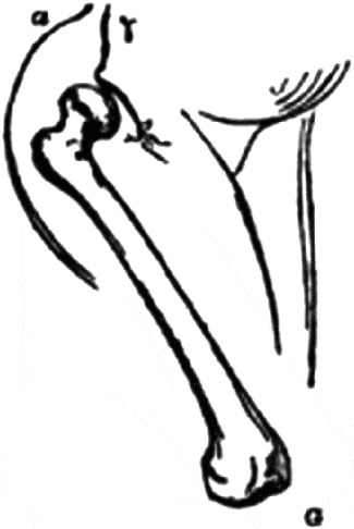
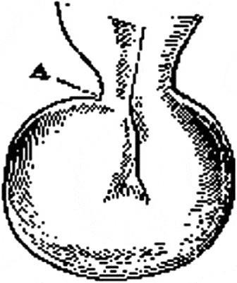
We have little access to these drawings, often made on a board with chalk and erased at the end of a lecture, but when weekly medical journals first appeared in the 1820s they began to publish accounts of lectures given around London, including, on some occasions, replications of drawings. Some such drawings, such as those by Bell in Figures 1 and 2, were published in issues of the London Medical Gazette. It is clear that they were an integral part of lectures. Bell's wife wrote after his death, ‘By constant practice he became an attractive lecturer … I have been told that his rapid and effective sketches on the black-board were a great aid’.Footnote 27 And Bell often wrote in his letters to his brother about making drawings for his class.Footnote 28
A rough schematic of a femur from the London Medical Gazette article. It acts as a representation of an anatomist's idea, and would have been an ephemeral product of the classroom. It was clearly significant enough within the lecture itself, however, to warrant a place in its subsequent publication. Charles Bell, ‘Diseases and accidents to which the hip-joint is liable’, London Medical Gazette (1828) 1(6), p. 137 (see Footnote footnote 29).

Image of a herniary sac, irreducibly distended by intestinal contents. The triangle and line indicate the narrowed neck of the sac which prevents the intestine from being forced back into the abdomen. Charles Bell, ‘Clinical lecture upon hernia’, London Medical Gazette (20 December 1829) 3, pp. 105–108, 105.

In the cases of both Figure 1 and its description, it is clear that the ephemeral classroom drawings that were very much a part of lectures helped to convey the ideas of their maker, what John Bell termed ‘plans’. Bell's lecture associated with the drawing was on causes of repeated dislocation and the image appears to depict the angle of the femur in the case of dislocation.Footnote 29 It conveys a relationship between parts, a rough approximation, an idea. Similarly, the descriptions of Bell's drawings of nervous systems of leeches and man suggest that Bell was after a set of relationships, an idea about an anatomical system supported by comparative visual illustrations of a rough and approximate nature. While these whole bodies and diagrammatic sketches gave a good, rough sense of the whole and of relationships of parts, of the plan of the human body, other visual tools were necessary in order to display, and indeed to create knowledge about, finer structures.
Seeing the invisible, holding Nature still: the minute and the rare on display in collections of specimens, preparations and models
The function of dissection of whole bodies within the system is clear in the way Hunter speaks about passing another element of that system, specimens and preparations, around the classroom. He offers detailed instructions to students, saying:
the preparations, must be sent round the company; that every student may examine them in his own hand … [P]reparations are to go round from right to left; in the second bench, from left to right; and so alternately, to the farthest seat of all. To prevent loss of time, when you give a preparation to your neighbour, be so good as to point out the part, or circumstance which is then to be examined; as I shall do, when it is first handed round…Footnote 30
Dissection demonstrations were good for showing large parts, principal parts, of the body and for showing relationships within the body as a whole, but Hunter himself recognized dissection as functioning directly alongside anatomical preparations that were prepared for, and used in, the classroom. Preparations revealed the minute parts of anatomy.
Hunter moved from his discussion of the origins of a science of anatomy in casual exposure to bodies to a discussion of the most recent progress in anatomy, associating two kinds of display as constitutive of that progress. ‘Were the great Harvey to rise from his grave, to examine what has been done since his time, I imagine that nothing would give him more pleasure, than to view with attention, the cabinets of some of the Anatomists of the present times’.Footnote 31 Those specimens and preparations have often been seen as mere curiosities, public spectacles, strange and macabre evidence of a widespread collecting culture that extended to human remains, or as evidence of a fascination with monstrosities.Footnote 32 But the progress Hunter attributed to these displays reveals that they were far more than a general sort of culture of collecting: ‘In the latter part of the last century Anatomy made two great steps, by the invention of injections, and the method of making what we commonly call preparations. These two modern arts have really been of infinite use to Anatomy’.Footnote 33 It is significant that Hunter framed the greatest advances in anatomy as having been useful ones; not therapeutically useful, but scientifically so – useful in generating knowledge. Preservations, injections, models, dessicated specimens, were all used as objects of study; as a mode of training; as a way of ‘seeing’ systems of barely visible anatomical parts with clarity, away from the messiness of the body; and of maintaining for pedagogy and for research bodies that otherwise too quickly decayed.
When grouping visual displays by their context of use within the system, two anatomy displays that could be seen as very different – wax models and jarred organs – actually end up occupying the same place. If we focus on their use and meaning to historical actors, rather than on the material products themselves or the means by which they were produced, we find that these two objects shared a purpose, an audience and often a physical space.
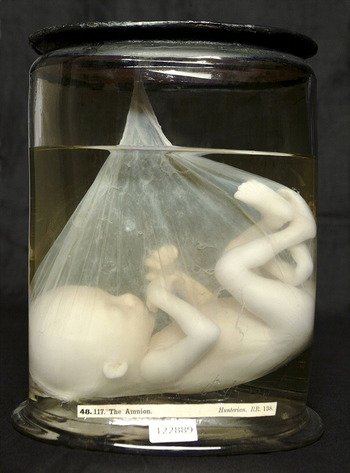
How, then, were injections and preservations, such as the eighteenth-century foetus in Figure 3, used? Hunter described the ways in which they made nature's delicately tiny contents visible in a predictable and consistent way, one that did not depend on significant labor and more significant luck, placing them alongside corpses as displays to be used in conjunction:
Besides dead bodies, we said, that a professor of Anatomy should have a competent stock of preparations … Preparations serve two purposes chiefly, to wit, the preservation of uncommon things, and the preservation of such things as required considerable labour to anatomize them, so as to shew their structure distinctly. Of the first sort are, the pregnant uterus, diseases, parts of singular conformation, &c. Of the second class are, preparations of the ear, the eye, and, in general, such as shew the very fine and delicate parts of the body, which we call the minutiæ of Anatomy.Footnote 34
‘The Amnion’, the Hunterian Museum, RR138, 122889, 48.117. According to Hunter's catalogue, this is a foetus ‘about the sixth month … The connecting medium between the amnion and the choiron…is so tender that the least force or rough handling separates these two membranes’. All preservations were catalogued and many, as in the case of this foetus, displayed parts of anatomy not commonly seen. © The Hunterian, University of Glasgow 2012.

John Hunter, John Bell and Charles Bell, like William Hunter, all kept museums or large collections of such injected, dried or jarred specimens;Footnote 35 Roy Porter has noted that the specimens themselves were greatly responsible for Hunter's ability to attract students.Footnote 36 Simon Chaplin has argued compellingly that such specimens functioned alongside dissection as didactic tools, but that the specimens themselves, in the context of the museum, also helped to construct notions of anatomical displays as a ‘form of “natural” spectacle’, simultaneously naturalizing the art of dissection as a way of learning about living bodies and conferring ‘upon dissection a degree of epistemological legitimacy (as a valid way of knowing about living things) in the eyes of “expert” spectators drawn from [John] Hunter's medical and scientific peers’.Footnote 37 The specimens were the tools both of students and of experts. They allowed students to see what would otherwise have been invisible in a crowded lecture theater as anatomy was demonstrated through dissection by the instructor. They captured the possible variations of normal and pathological in a systematic way. And, by creating them, advanced students both contributed materially to classroom research and learned mechanical and visual skills.
In his Two Introductory Lectures, Hunter moves from his description of injected vessels to casts and wax models, sometimes also called by anatomists in this period ‘specimens’ or ‘preparations’. Hunter said of casts, ‘The proper, or principal use of this art, is, to preserve a very perfect likeness of such subjects as we but seldom can meet with, or cannot well preserve in a natural state; a subject in pregnancy, for example’.Footnote 38 Casts functioned, in other words, much like actual specimens. The point of these three-dimensional visual displays was not to act as representations of nature, but to freeze nature herself so that she could be studied. Wax casts were meant to be a ‘perfect likeness’ of the real thing. In the continuum of visual displays, they served as an intermediate between nature and representation. Though they were wholly crafted by men of science, they were not understood to act themselves as creations or representations. The intervention of science, the labour of anatomists and their craftsmen, was not meant to make an argument or to idealize or to reveal a hidden truth: that intervention was merely made to prevent decay and to hold nature still. In so doing, it rendered accessible those things which were not everyday, those that were not the normal or typical or ideal. Hunter said of preparations, ‘the object is ready to be seen at any time. And, in the same manner [the anatomist] can preserve anatomical curiosities, or rarities of every kind; such as, parts that are uncommonly formed; parts that are diseased; the parts of the pregnant uterus and its contents’.Footnote 39 Even the names ‘preservation’ and ‘specimen’ indicate that these displays themselves were the object of study: these objects were taken to be Nature for purposes of anatomical observation.
This view was not particular to Hunter. As Charles Bell's wife recalled, Bell ‘had discovered a method of modelling morbid appearances in wax retaining their colour in its original freshness, so as to perpetuate for the student much that was lost to them in the usual manner of preserving them’.Footnote 40‘Colour’ and ‘freshness’ were valued in Bell's models because they were things lost quickly in dissections. Again, they offered the opportunity to prevent decay. These man-made wax models were preserving nature.
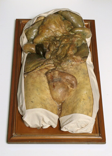
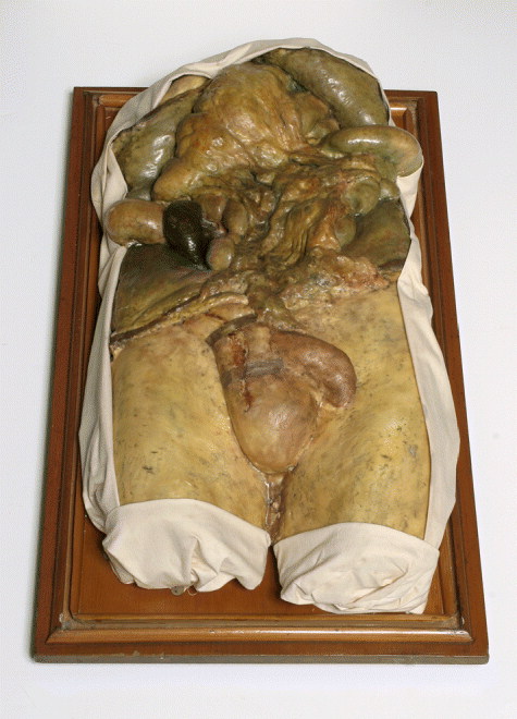
Clear evidence of this can be found in Bell's accompanying museum catalogue. Of the wax model in Figure 4, he notes, ‘From an adult male who survived the operation of herniotomy during several days but without alleviation of symptoms … Though successfully reduced by operation the strangulated loop of intestine was black and gangrenous’.Footnote 41 Casts and models were made from specific bodies, taken again as ways of preserving such bodies. If jarred specimens were for the body's minute parts, casts and models situated those parts or preserved the not-often-seen anatomies of a slightly larger scale. One could not ‘jar’ a torso or dessicate it, so instead one modelled it. William Hunter made casts of each of the subjects contained in his atlas on the gravid uterus and displayed them alongside the books, a three-dimensional object preserving nature.Footnote 42 Preservations and models were both the products of a great deal of scientific labour – sometimes not even intervention, but creation. But when made successfully, that labour was obscured, such that both preserved specimens and models, or casts, could appear to be direct products of fresh corpses, anatomized at the moment of death, or sometimes even of living bodies with lifelike colour – the natural objects of study for anatomists.Footnote 43
Wax and plaster cast of torso. Royal College of Surgeons of Edinburgh, Bell Collection, GC 1.43.04. Such casts, taken from individual bodies, provided common ways of preserving the anatomy of larger parts and their relations. This one was taken ‘From an adult male who survived the operation of herniotomy during several days but without alleviation of symptoms’. Reprinted with the permission of the Royal College of Surgeons of Edinburgh.
The audience for these anatomical objects was wide and varied, as the specimens were often housed in museums that were open to the public, but whose primary intended audience was medical in nature (the public element becomes curious and significant only when these three-dimensional objects were removed from a system of display). These anatomical objects were teaching tools, at a time when objects were, according to both Bell and Hunter, the best ways of ‘making an impression on the brain’. Specimens and models are often seen as imperfect stand-ins for the gold-standard of anatomy teaching tools – corpses – at a time when dissection was taboo, but in fact, even if students had had fresh corpses in abundance to dissect for themselves, that would not have taught them to see properly. Hunter says as much: preparations
are of infinite service to the art; especially in the hands of teachers. They give students clear ideas about many things, which it is very essential to know, and yet which it is impossible that a teacher should be able to shew otherwise, were he ever so well supplied with fresh subjects.Footnote 44
These three-dimensional objects, thought to be a sort of ‘Nature-unchanging’, must be understood as functioning within collections and not as individual pieces. A visit to John Hunter's museum at the Royal College of Surgeons in London reveals the scope of Hunter's seven thousand museum items – multiple specimens of individual body parts are housed with drawings and paintings, skeletons and stuffed animals. Collections like Hunter's, combining both specimens of the same body part in series to show variation of that part across a species and specimens of different body parts to represent the total organism, represent varieties of normal and pathological tissue preserved by various means. Thus, in his System of Operative Surgery (1807), Charles Bell wrote, ‘The cast of this subject and the dissected bladder completes the series of preparations of fistula in perineo, to be seen in my Collection’.Footnote 45 The preparation was a part of a system of bladders with similar pathologies as well as normal varieties. Bell said of his collections, housed in a museum, that the museum
is a room admired for its proportions of great size, with a handsome gallery running round; the class room door opens from the gallery. It would require a month to go round the museum with a book in your hand. I knew that this was a thing to me above all value, and already, by good arrangement, and by the addition of my own preparations, I have filled the room.Footnote 46
If we need evidence that these natural philosophers of the late Enlightenment did not rely on an idealizing sort of vision or style of representation, that they meant to take in and also to depict the whole of nature in their scientific displays, we need only appreciate the vast numbers of specimens they collected and displayed together.
Bell strongly advocated training hand and eye together – doing was seeing in anatomy. Anatomical objects provided vision to the hands that held and described, and eventually replicated, those objects. While the hand was taught, it was taught to see, rather than to feel, in order to know. Though these classes trained surgeons and artists, and tactility was important to both, these displays were tools that primarily aided a natural-philosophical vision. Bell taught advanced students by having them dissect out and preserve their own specimens, in effect having them conduct their own research. But seeing required the system. Bodies could not stand alone intelligibly. Students learned to see by creating and preserving anatomical objects and by drawing, using their hands. At the same time, Bell says, they looked at drawings. While nature unedited was the object of vision and of study, it still required explanation. Texts helped to situate and define visual displays, as did systematic arrangements within a collection.
Part of the system of visual display, and one that helped to define the context for the three-dimensional objects as disciplinary and pedagogical, included not only the arrangement of specimens together, but a textual as well as a visual context for these displays. Scanty but significant text surrounding atlas images, as well as the catalogues that almost always accompanied museums, both situated individual objects and designated a particular way of seeing for a particular audience. A public audience could admire museum displays or atlas images in a casual fashion, but these objects were used as tools for examining nature by the audience of anatomists and medical men whose attention addressed the system of visual display as a whole and perceived the interrelation of its parts. Bell's description of the wax model as having been taken ‘from’ a patient offers one such example of a catalogue entry. In another, describing the dried specimen in Figure 5, Bell wrote,
the Patient lay long in the Middlesex Hospital being kept very low, and occasionally bled, his sufferings were by no means so acute, as we would imagine must necessarily result from such extensive disease … Tumour has burst through to the back part, where it formed a very large Tumour during life, notwithstanding the distance of this posterior sac, from the Heart, the pulsation of the Tumour was at all Times very distinct … he died exhausted from weakness.Footnote 47
This catalogue entry offered a description of the patient's symptoms before death, of the situation of the diseased organ within the individual's body upon dissection, and of how this case compared to others. The catalogue, an integral part of the display itself, offered a context for an individual pathological specimen, specifying what in it was normal and what pathological. Most museums would have had a similar sort of contextualizing catalogue, filled with the history of the specimens being displayed.Footnote 48 Captions attached to atlas images played a similar role within the system of visual display, binding together and situating its elements.
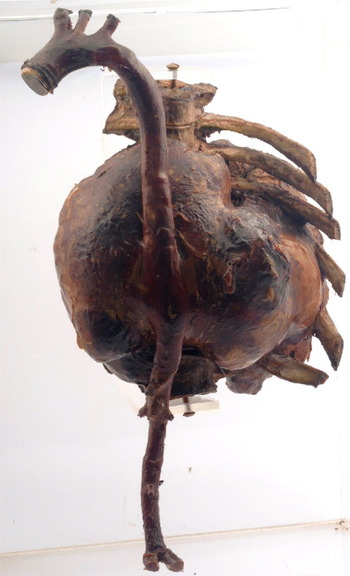
Thoracic aorta with aneurysm. Royal College of Surgeons of Edinburgh, Bell Collection, BC.xii.2.M.57. GC 11006. This is clearly a dry preparation, though such anatomical specimens were often ‘wet’ and kept in a preservative spirit in jars, as in Figure 3. There was a variety of preservation techniques that attempted to preserve a life-like quality as well as the structure of the organ. Reprinted with the permission of the Royal College of Surgeons of Edinburgh.

Breaking dichotomies through books: uniting the minute and the whole, seeing anatomy and the plan
Books, the most familiar subjects of study for historians, are the objects within systems of display to which we most often turn when we seek to understand the role that visual representations played in science, and indeed they do deserve a prominent place, but a place alongside other materials of science. Illustrated books fulfilled more than one role within a system of visual display, depending on the style of book and the style of illustration – both of which determined the price, and therefore use, of the book.
Both etching and engraving could be used to reproduce images. Etching was a cheaper technique that Charles Bell used frequently in books that were designed to be affordable for students and practising medical men. Those books were meant to be used in conjunction with dissection and other forms of display and might more accurately be termed ‘reference books’, rather than textbooks, as they were not meant to stand alone and were often designed for, and used by, those who had taken on only what unsystematic training they could afford and continued to try to learn after beginning to practise. John Bell said of texts designed for the student, ‘when drawings are made for his use, the body should be laid out, as he is to order it in dissection’.Footnote 49 Dissections were ordered based on how quickly parts and systems putrefied, so texts that were designed to follow this order were designed to do so purely to match the exigencies of dissection and not for reasons of anatomical logic. The book seemed to bring together in two dimensions those elements gross and minute covered by the two separate three-dimensional forms of display, dissections and specimens, taking both as the books' objects. John Bell explained that plates should portray ‘first all the individual parts one by one, and then join them, showing how the whole is composed, without which regular form of demonstration, nothing could be clearly understood of parts so very intricate and difficult, and having so long a catalogue of hard names connected with them’.Footnote 50 Thus the drawings of reference books would unite the detailed work of preservations and specimens with the plan represented in dissection. Elsewhere Bell described another attempt at such a union, saying, ‘The ingenious Mr. Cruikshank, with the design of explaining all that he or Dr Hunter had injected of the lymphatic system, in one consistent view, took a delicate and elegant drawing of the human body, and laid his lymphatics upon it’.Footnote 51 In this attempt to combine elements of a system of visual display, the interrelatedness is made clear.
The texts were didactic. Authors tended to be explicit about both advantages and disadvantages of the costs of production, sometimes apologizing for the economizing nature of small volumes and less detailed etchings, sometimes accentuating those elements as evidence that the text could be widely used. Such quarto texts often sacrificed the ability to literally stand in for Nature, the object of inquiry in anatomical science, for the sake of portability, affordability and the capacity to present a plan of anatomy to students. Given those constraints, anatomists then attempted the kind of naturalism and detail that the text's size and etching technique would allow, but always with the aspiration of achieving the same sort of faithful reproduction of individual, observed subjects as were depicted by folio-sized, engraved atlases. When his brother mistook some drawings for engravings, Bell responded in an 1809 letter, ‘My bones engraved! Not a touch of them … besides they will not cost me one pound a-piece. Engraving would have been at the rate of six guineas; though a splendid book it will be cheap and circulate wide’.Footnote 52
Why, then, given their expense and the general classroom context of use for systems of visual display, produce atlases full of costly engravings at all? The reasons seem to have been threefold, and speak to the inappropriateness of dichotomies such as ‘nature’ versus ‘representation’, or knowledge as opposed to its object.Footnote 53 First, and most straightforwardly, atlases could serve as expensive and elaborate showpieces for patrons and wealthy buyers, the atlases having been endowed with a sort of cultural capital that Simon Chaplin describes as having motivated often the accumulation of specimens and preparations beyond what was useful or by those not teaching anatomy.Footnote 54 Atlases were indeed a way to convey a discovery or to impress, to demonstrate the anatomist's success. In order to afford to have one printed, one had to have ‘made it’ already. That function, book as asset, required no system of display.
Atlases taken within the context of the system of display, however, could both embody the nature about which knowledge was made and simultaneously articulate discovery or scientific excellence – make a knowledge claim both to peers and to potential students. Historians have forced these images into one role or the other – nature or knowledge – where their creators saw no such opposition. Atlases could act as any of these three things (asset, nature, discovery), or as all of them at once, reflecting different but related forms of knowledge claims and ideas about how knowledge is produced depending on the anatomist and the audience. That multiplicity provided both their assumed value and a seeming ambivalence that sometimes appears to produce confusion.
William Hunter spoke quite self-consciously about the decision to publish his study of the gravid uterus as an elephant folio rather than a less expensive set of etchings in a small book. Some might think
that a great part of the expense might have been spared, and the work thereby rendered of more general use, if the figures had been made to a smaller scale, if the engraving had been less finished, and if some of the figures, which are very similar to others, had been omitted.
But the size of the book allowed Hunter to show
the peculiar habit and composition of parts, as well as the outward form, situation and connection of them … if the natural size of the object be tolerably fit for an engraving, that must be of all others the very best, as it has the advantage of shewing such an important circumstance. Upon these considerations, all the figures in this work were made of the natural size …Footnote 55
The elephant folio was chosen as the best way of achieving naturalism in the images. John Bell, who did not have the money at his disposal for such a large-scale project, apologized for his ‘little plates’ for reasons that mirror Hunter's for preferring an elephant folio, saying,
The [etchings] want that size which gives splendor to a grander work, and of course that proportion, which gives the full idea of the human body; they want that elegant drawing, and careful engraving, which should do any idea justice, which is so necessary in delivering the minuter parts with character and truth, all is wanting that belongs to the idea of a grander work …Footnote 56
Each anatomist desired to faithfully embody nature in his atlas images.
Charles Bell, in a collaboration with his brother in 1801, observed,
Of [any] twenty bodies not one will be found fit for drawing; but still I conceive that we are not to work out a drawing by piecing and adding from notes and preparations; we are to select carefully from a variety of bodies, that [one body] which gives largeness of parts, where the varieties of parts are well marked, and where there is the most natural distribution of vessels.Footnote 57
Bell's unwavering commitment to depicting Nature exactly as she was found – to selecting well, but drawing the individual body in front of him – that is expressed here required that he not create some sort of anatomical composite of the ‘ideal’ or ‘normal’.Footnote 58 When seeking a body to draw, he looked, as an anatomist would, for ‘normal’ distribution of the parts, but he also kept in mind the requirements of the artist, and looked for a body in which the anatomical parts he was drawing were ‘well-marked’ and large. To those who would do otherwise or who objected to the peculiarity of individual bodies, Bell offered text as an antidote, saying, ‘let us allow ourselves no license but copy accurately. By noting in the description any little deviation every necessary end is answered’.Footnote 59 Thus the text served to provide indications of what could be universalized.
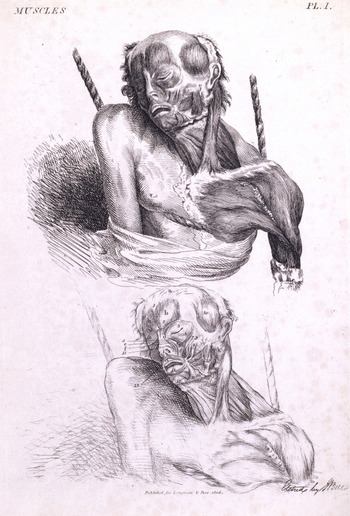
John Bell, like his brother, adopted the practice of depicting individual corpses and using text to situate them. Describing the plate from Figure 6, which depicts muscles of the face, neck, throat, shoulder and breast, he said,
It was drawn from a subject that had been hanged, and the neck being broken, the head lies flatter upon one shoulder, than it should do even in the dead body, for the Atlas and Dentatus, the two first Vertebrae of the Neck, were fairly broken loose from each other. – The Muscles are more distinctly seen on the left side, on the right side they are thrown into shadow, and are but faintly indicated …Footnote 60
The caption clearly demonstrates that text, even minimal text, was hardly incidental. It allowed for the depiction of the very particular – a corpse made the subject of dissection by hanging – in order to create general knowledge about the normal anatomy of the human body, describing ways in which a hanging victim's neck muscles would be like and unlike those of individuals that a surgeon–anatomist would be likely to encounter in practice.
John Bell, Engravings of the Bones, Muscles, and Joints, London: Longman and Rees, and Cadell and Davies, 1804, Book 2, Plate II, p. 93. John Bell wrote of this image, ‘This plate belongs chiefly to the Throat … This Plate explains first all the individual parts one by one, and then joins them, showing how the whole is composed, without which regular form of demonstration, nothing could be clearly understood of parts so very intricate and difficult, and having so long a catalogue of hard names connected with them.’ Reprinted with the permission of the Royal Academy of Arts, London.

Hunter too espoused direct representation of what was directly seen – a depiction of the natural that preserved the immediate sensory experience and observation of an individual body by the anatomist – and instructed his artists to copy directly from dissected corpses, warts and all (Figures 7 and 8). He famously engaged in a debate with the artist Joshua Reynolds about whether the copying of nature could itself constitute art, or whether mere imitation without either embellishment or essentializing constituted only a craft.Footnote 61 Hunter's verdict, after responding to Reynolds by considering the merits of both the artist's tendency to idealize or universalize and the anatomist's desire for strict accuracy in representing the individual, was,
The one [a faithful copy of nature] may have the elegance and harmony of the natural object; the other [an artist's synthetic rendering] has commonly the hardness of a geometrical diagram: the one shews the object, or gives perception; the other only describes or gives an idea of it. A very essential advantage of the first is, that it represents what was actually seen, it carries the mark of truth, and becomes almost as infallible as the object itself.Footnote 62
By representing the object as it was seen, it becomes almost as infallible as nature, and also ‘gives perception’, whereas the idealized image gives only a description or idea of an object – the sort of thing that unaccompanied text could provide. Hunter was sometimes in conflict on this point with his own artist, Jan van Rymsdyk, but said of his illustrated atlases that they were done with ‘not so much as a joint of a finger having been moved to shew any part more distinctly, or to give a more picturesque effect’.Footnote 63 Like Hunter, John Bell described ‘a continual struggle between the anatomist and the painter; one [the artist] striving for elegance of form, the other [the anatomist] insisting upon accuracy of representation’.Footnote 64
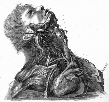
Charles Bell, ‘Nerves of the neck’, from Bell, A Series of Engravings Explaining the Course of the Nerves, London: Longman and Rees, 1803, Plate II. As is evident here, Bell included individual features that were meant to enhance both the beauty and credibility of the drawings, demonstrating a commitment to representation of the particular. © The Wellcome Library, London.

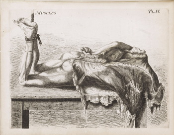
John Bell, Engravings of the Bones, Muscles, and Joints, London: Longman and Rees, and Cadell and Davies, 1804, Book 2, Plate IV, p. 109. This plate ‘explains’ muscles belonging to the scapula and back, which are, according to Bell, ‘very remarkable in beautiful statues’. John Bell's drawings are, in fact, known for being ugly to the point of being grotesque – a way of demonstrating their realism. © The Wellcome Library, London.

It is worth returning to Daston and Galison at this point. They ask us to distrust Hunter's words, his own claims to naturalism, saying, ‘It would be a mistake to take Hunter entirely at his word – to believe that his figures did indeed represent the object “exactly as it was seen” ’. Their argument has the implication of dismissing or ignoring the considerable debate between artists and anatomists about how to depict nature, eliding the groups by reading only the images themselves. They argue that Hunter ‘considered it part of truth-to-nature to inject the womb with “some spirits to raise it up to the figure it had when the abdomen was first opened” ’.Footnote 65 But in fact Hunter was not trying to perfect Nature, to instruct her in truth, but instead to preserve nature in a state that quickly disappeared and decayed in death, and he was doing so in an entirely routine manner. His explicit purpose was to make a working object of study to be used. Hunter was doing what Bell would do when he said that a wax model was taken from a corpse – making nature, the nature of a living person's anatomy, usable and reusable for research and teaching. Naturalism and realism need to be historicized like any other set of values or ambitions in science. And Hunter clearly thought of his images (see Figure 9 below) as fitting a naturalistic aesthetic and as representing the particular or individual.
William Hunter, The Anatomy of the Human Gravid Uterus, ‘The gravid uterus at full term’, Plate IV. The individual uterus is represented in isolation, severed from the rest of the body. © The Wellcome Library, London.

These atlas images-as-nature were used in the classroom of their author, alongside casts and other figures. As Bell described his museum, which featured two-dimensional displays alongside preparations, ‘My little collection begins now to look well, and, as you may conceive, peculiar, having the paintings placed in the interstices of the preparations’.Footnote 66 And William Hunter framed the drawings of his atlas in the context of other visual displays on which he was working:
The first ten plates are represented in the museum by a number of plaster of Paris casts. These were taken actually from the same subject, and show the same stages of the dissection as certain of the drawings; they were subsequently coloured after nature … The whole of them are exactly nature herself, and almost as good as the fresh subject. We have a good many of them to help us on; they are most useful, especially where it is so difficult to get a subject of this kind to explain upon in a course of lectures.Footnote 67
These atlases sat in museums along with preparations, some depicting the same bodies as were jarred on the shelves, revealing other structures, other vantage points, and giving permanence to other oft-invisible subjects. They aided lecturers. That was their context of use. When representing nature (and Hunter's casts and drawings were depicting the same subjects because they were, in fact, meant to act as nature, as object of study), the plates were objects for pedagogical use and for pedagogical audience. They supplemented three-dimensional displays, highlighting minute details that were necessarily obscured in converting the dissections into permanent preparations.
Selections producing knowledge of Nature and the pathological: seeing Nature and her order in the same frame
Unlike the preparations and specimens, where the labour involved in crafting the display was obscured in order to render the specimens natural in appearance, in the case of atlas images the intervention of science in their creation was not only obvious, but highlighted. These books were objects that could not simulate the colour and texture of a body, its three-dimensionality, which were aspects contributed by and therefore valued in wax models and specimens, but they could unite the gross and the minute, showing together the complexity of nature and its inherent structure, relationships and order. Anatomists like John Bell often wrote in their prefaces about the labour that went into such books: ‘a few words concerning the mechanical labour. I have drawn the plates in my own hand. I have engraved some of these plates, and etched almost the whole of them. Which I mention only to show, that they have their chance of being correct in the anatomy’.Footnote 68 Seeing, even seeing in such a way as to produce a faithful reproduction of an anatomical object, was made possible by an individual's expertise.
Anatomists used atlases, products of their craftsmanship, to claim expertise for the sake of increasing lecture enrolment, to enlist patrons, or to secure a discovery. They intended to highlight their own role in the production of this most expensive element of visual display because to do so would increase their standing among one of the audiences for such atlases. Hence while all three considered their work governed by a sort of naturalism, the drawings of John Bell, Charles Bell and William Hunter all had different styles, styles that would have been recognizable as theirs to patrons or to students. Selections, and not idealizing alterations, defined those styles, whose existence is not at odds with the notion that all three authors considered their depictions faithful to nature.
Atlases became a worthwhile investment for both producer and subscriber, because they brought together multiple meanings in a single object. They were the ‘nature’ under study, and in that sense they served as both working and pedagogical objects. Students looked at them, but also drew from them, and they sat in museums alongside casts, supplementing gross anatomy with fine detail. But they were simultaneously a way of seeing, organizing and making sense of the world. They could constitute a claim of knowledge or claim to discovery. Thus Charles Bell said that he would reveal his greatest contribution, his discovery of a system of the nerves, ‘by magnificent engravings of the whole nervous system’.Footnote 69 His work would culminate in a set of engravings, the finest expression of his work, because these engravings were meant to show Nature and her relationships – her plan – and also because the objects themselves were expensive evidence of an anatomist's standing, affordable only to the wealthy. But such an atlas would not stand alone.
The images from William Hunter's and Charles Bell's atlases are not, according to their authors, full of idealized images, though they are selections. The knowledge itself was a matter of selection and vision. As Charles Bell explained, they are images of bodies selected for ‘normal’ anatomy and for distinct, large, easily drawn parts. Images were always selected for something. And they always served a particular context of use, or in this case multiple uses. There was reasoning built into the drawings themselves. Hunter described them as a ‘universal language, [one that] conveys clearer ideas of most natural objects than words can express … and gives an immediate comprehension of what it represents’.Footnote 70 In this case the drawings represented both human bodies and anatomical knowledge. But when these selected images are understood to have functioned within the context of the classroom of their author alongside descriptive texts, whole bodies, schematic drawings and collections of jarred specimens and wax models (objects that were often deliberately selected as representations of the pathological or of oddities), it becomes clear that drawings of ‘normal bodies’ were a part of a broader system. Visual displays were selected because, taken together, they acted as tools to allow the discipline to ‘see’ a nature that was both finite and ordered in its variation and therefore displayable. They were the tools of visualization in a pedagogical philosophy that valued sensory experience as the primary means of learning. They were accumulated and wrangled into order by instructor–anatomists as a way of recruiting students in a competitive medical marketplace. If knowledge was enabled by all and distributed across the system, the system was only made meaningful by the anatomist himself, who provided the text and narration that brought the system together, situated its parts and showed the student what he was seeing.
Part of the reason why a system of visual display provided such an important component of anatomical study was that during the period in question there was an understanding of Nature herself as systematic, even in her pathological state.Footnote 71 Pathological specimens and references books serve as good reminders of why bodies depicted in visual displays were selected for being representative and typical. Representations and other visual displays were not idealizing, but they did select for ‘normal’ and were informed by underlying classificatory ideals. But again, if we return to context of use, it would be unlikely to suppose that those disciplines most reliant upon visual display – anatomy, botany, natural history – did not require some form of ‘idealizing’. All of these disciplines require categorization. It would be impossible to sort plants or specimens without a ‘normal’ version of each species or object, because without such a typical model, as well as its predicted variations, one could not determine what would be far enough off the ‘normal’ to place in a different pile. That may be even more the case in anatomy, where categorization was made into normal and pathological, and the two categories were defined in relation to each other, rather than absolutely. Visions of the normal were necessary in order to determine the pathological against which they were defined, the pathological themselves often falling into supposedly known and predictable patterns of deviation.
Matthew Baillie, William Hunter's nephew, whose 1793 text on morbid anatomy was the first systematic treatment of pathology in English,Footnote 72 wrote about presenting pathological deviations from normal anatomy in his A Series of Engravings, tending to illustrate the Morbid Anatomy of some of the most Important Parts of the Human Body (1799): ‘It seemed to me, therefore, to be an important desideratum in Anatomy, to comprehend in one work, upon some regular plan, Engravings of the chief Morbid Changes of Structure in the most essential parts of the human body’.Footnote 73 Pathology itself, the abnormal, could be depicted in an atlas because it took familiar and predictable, rather than monstrous or random, forms. This can also be seen in the careful attempts to develop comprehensive collections of pathological anatomical specimens, housed together and systematically alongside collections of normal anatomy.
The deviations from normal anatomy were depicted in much the same fashion as in the usual atlases of human anatomy. They were drawn from collections of preserved specimens or from individual bodies.Footnote 74 Again, text provided context, situating the individual on the page within a system of natural pathological anomalies. In one place Baillie talks in terms of ‘rare’ and ‘common’ pathological variations.Footnote 75
Nature had an order. That system could be displayed through individual objects and subjects, taken together and situated by texts that gave a sense of the individual's relation to a whole class of like objects or parts. Displays were chosen for reasons to do with clarity of the depiction and also an idea of what was normal or typical. The latter was significant to a discipline like medicine, in which atypical often meant pathological, though presumably some idea of normal or typical was also essential to any subject that involved classification. When selections of individual objects for display were made, they were not averaged or even improved in their representation. Instead they revealed an orderly world in which selections of ‘normal’ and of a range of divergences from typical could be made on the basis of assumptions about form, function, viability and non-random deviation. Such an assumed regularity or pattern in Nature herself would later be provided differently – through statistics rather than natural philosophy, a mathematical sense of a random world rather than philosophical treatment of its internal logic.Footnote 76 Anatomists' images were nature itself. As Charles Bell put it in one of his advertisements, students, when studying anatomy in clinical lectures that involved observation of patients, would be ‘under the Correction of Nature herself’.Footnote 77 Nature was the highest authority, faithfully and systematically revealed through a system of display.
Conclusion
What the world will speak of is my drawings. I have often been troubled with the perverseness of people attaching merit to the drawings of my book, and closing their eyes altogether on the reasoning … You know that this subject cannot have due importance given to it by etchings on the margin of a book, – that it requires a great establishment of casts and models, – that is what I regret. I am quite conscious of possessing a talent in the way of modeling [sic] superior to those of many I have visited, – and this is buried.Footnote 78
Charles Bell's words make evident a particular context of use for anatomical drawings. They existed as part of a didactic system of display, alongside casts and models, as a subject of study and claim to knowledge and understanding. Atlas images alone were not the embodiment of representational practices; we need to look beyond texts and books to look at objects of visual culture not in isolation but in an integrated context of use. Context of use here defines a system of visual display. Students flocked to the halls of those professors who had the best specimens and preparations, cadavers and drawings; in other words, the best systems of visual display. They trained their hands and eyes first by looking and then by crafting visual displays. This system of display cultivated a quintessentially British gaze, rooted in an amalgamation of natural philosophy and clinical practice and therapeutics. ‘Seeing’ anatomy in Britain was to see a body full of anatomical systems, understood in detail and in entirety, by the system of display. It involved freezing a living body in death, restoring its color, its vivacity and its forms through displays that did not perfect nature; they perfected vision, providing tools to render anatomy perceptible to the senses rather than just to the mind.
For late eighteenth- and early nineteenth-century anatomists, displays constituted a form of reasoning and a way of teaching. According to Hunter, ‘The whole of them are exactly nature herself, and almost as good as the fresh subject.’Footnote 79 The classrooms of medical men were filled with displays that taught future doctors and surgeons to see, to know and to remember. Elements of the system of display, some ephemeral and some valued for their permanence, taught students to see and learn in slightly different ways – at a slightly different level or with a different lens – such that when taken together, they formed a sort of comprehensive three-dimensional anatomy of a living person. Anatomists did not mistake displays for Nature, nor did they consider them mere representations of Nature; they deliberately allowed such displays, in combination, to stand in for Nature, holding Nature still in order to study her. Atlas images embodied not merely what the anatomists were looking at but also what they were seeing: Nature and its order, not in opposition, but in the same frame.
Both Bell and Hunter wrote explicitly about the beauty and elegance of nature. Their natural world had order. Pathologies, anomalies and deviations from the ideal took a finite set of forms, and the predictable variations could be embodied in a system of visual display because of this assumption of an underlying order. This view and similar such systems surely prevailed in other sciences of the period besides anatomy, especially in natural history. We might characterize the overall relationship between image, nature and science as ‘a system of nature on display’, or as embodied systems of nature. Enlightenment and early Victorian anatomy preserved nature, held nature still, and ordered those distilled visions of nature. They saw in nature a teachable beauty only to be revealed through a system of complementary visual displays, a system of displays that together were ‘exactly nature herself’.