Introduction
The herring Clupea harengus L. has been the subject of parasitological attention for many years. This attention, however, focused mainly on adult herring and, with few exceptions (Rosenthal, Reference Rosenthal1967; Arthur & Arai, Reference Arthur and Arai1980a; Ivanchenko & Grozdilova, Reference Ivanchenko and Grozdilova1985; Heath & Nicoll, Reference Heath and Nicoll1991), and then mostly regarding Pacific herring, juveniles have been given little attention. In a checklist of parasites of Pacific and Atlantic herring, Arthur & Arai (Reference Arthur and Arai1984) listed 82 parasite taxa, mostly identified to species, comprising 9 Protozoa, 13 Monogenea, 21 Digenea, 8 Cestoda, 10 Nematoda, 12 Acanthocephala, 2 Arguloidea, 6 Copepoda and 1 Agnatha. In a paper on relationships between herring and its parasites, MacKenzie (Reference MacKenzie1987a) listed 87 species of parasites; 11 Protozoa, 17 Monogenea, 18 Digenea, 9 Cestoda, 12 Nematoda, 10 Acanthocephala, 1 Hirudinea, 2 Branchiura and 7 Copepoda. Later, another digenean, Aponurus laguncula, was reported from the English Channel (Bray & MacKenzie, Reference Bray and MacKenzie1990), and should thus be added to these lists. At least 13 species have their distributions restricted to the Atlantic only; they are Kudoa clupeidae, Pseudoterranova decipiens, Mazocraes harengi, Cercaria pythionike, C. doricha, Hemiurus luehei, Lepeophtheirus pollachius, Lernaeenicus sprattaes, Pleistophora sp., Mazocraeoides georgei, Calliobdella vivida and Argulus alosae (MacKenzie, Reference MacKenzie1987a). Of these, six species have been reported only from the north-east (NE) Atlantic and six species from the north-west (NW) Atlantic alone. The former includes M. harengi, C. pythionike, C. doricha, H. luehei, L. pollachius and L. sprattae, for which herring may serve as the intermediate or definitive host. Parasites covered by studies of Arthur & Arai (Reference Arthur and Arai1984) and MacKenzie (Reference MacKenzie1987a), however, were recovered mainly from adult herring. The aims of the present study were to add to our knowledge of parasite fauna of young-of-the-year Atlantic herring and also to add some details to earlier descriptions, especially regarding the external morphology, and some aspects of the ecology of herring parasites.
Materials and methods
During summer and autumn 1994–97, 771 specimens of 0-ring herring (ranging from 40 to 110 mm in length) were collected live from Gullmarsfjord and Brofjorden off the Swedish west coast. Fish from each haul were transferred to and kept in tanks supplied by filtered sea water. Examination of fish took place 1–10 days after they had been transferred to the tanks. Some fish from each haul were deep frozen after being kept for 24 h in the tanks, and were examined later.
During examination, the total length of each fish was measured to the nearest millimetre, and each fish was weighed and sexed. The skin and all gill arches of each fish were screened for ectoparasites, and the body cavity was opened and examined for endoparasites. Squashed preparations of visceral organs, including heart, liver, spleen, kidney, gall bladder, swim bladder and gonads, were made and examined under a light microscope. The digestive tract was removed and divided into sections, which were separately screened under stereo- and light microscopes.
Living parasites were studied under a light microscope and photographed. Drawings were made with the aid of a drawing tube, and measurements were made with calibrated eyepieces. Whole mounts of digeneans, nematodes and cestodes were fixed in either: (1) Berland's fluid, transferred to 70% ethanol and then to lactic acid and stained with ‘Mexican Red’, Dylon, textile stain (Berland, 1995) and mounted in glycerol jelly; or (2) in Bouin's fluid and stained with acetic carmine or haematoxylin and mounted. Specimens for serial sectioning were fixed in Bouin's, embedded in paraffin and sectioned into 5–8 μm thick sections, which were stained with haematoxylin–eosin. Monogeneans were fixed with ammonium picrate–glycerine (Malmberg's fixative), transferred to a slide, covered with a coverslip and sealed with nail polish (Malmberg, Reference Malmberg1957). Material for scanning electron microscopy (SEM) studies was washed several times in physiological saline in the case of endoparasites, or in filtered sea water in the case of ectoparasites, fixed for 3–4 h in 2.5% glutaraldehyde buffered with cacodylate (pH 7.4, 4°C), washed in buffer and post-fixed for 3 h in cacodylate-buffered OsO4 (4°C), dehydrated in ethanol and acetone and finally by critical-point dryer. Specimens were coated with gold–palladium spatter and examined in a DSM 950, Zeiss SEM and photographed using Ilford FP4 (125 ASA) black and white films. Negative films were later scanned for digital recording.
All measurements are given in micrometres, unless otherwise stated, and are mostly in mean (minimum–maximum) order. Digeneans in this study were arbitrarily divided into three groups according to their stage of maturity: without eggs, with fewer than 10 eggs and with more than 10 eggs.
Results and discussion
During the study, 15 parasitic species were found on and in fingerling herring. These were: five Protozoa, one species of uncertain affinity, two Monogenea, five Digenea, three plerocercoids of Cestoda, one Nematoda larvae and one Copepoda.
Protozoa
Trichodina sp. Ehrenberg, 1838 (Ciliophora: Trichodinidae)
The parasite, mean prevalence 2%, was not only found on the gills but also in the lateral line system of the head, and even as a hyperparasite on another gill parasite, the monogenean Pseudanthocotyloides heterocotyle. Adhesive disc: 35 (33–38) in diameter, denticulate ring, 12 (10–14) in diameter, bearing 18 denticles with well-developed thorns and blades. The intensity of infection was low and no more than 20 specimens per fish were recorded.
This genus has been reported from gills of Clupea pallasi in the north-west Atlantic (Lom & Laird, Reference Lom and Laird1969). Due to the large number of similar and highly variable species in the genus Trichodina, the species could not be identified with certainty at this stage. However, this parasite does not look like any of the previously described Trichodina that the author could access. No gross signs of pathological effects were observed, probably due to the low intensity of infection.
Ceratomyxa auerbachi Kabata, Reference Kabata1962 (Myxosporidia: Myxosporea)
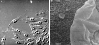
Spores of C. auerbachi were found floating in the gall-bladder fluid of 15% of fishes. Developed spores, straight to slightly curved (fig. 1a), length 10.5 (10.0–11.5), thickness 75.0 (73.0–80.0), diameter of polar capsule 4.5 (4.1–5.0). All trophozoite stages, as well as deformed spores, as described by Kabata (Reference Kabata1962), were also observed in this study. In SEM examination, the spore body was smooth and two polar capsule openings were seen (fig. 1b).
(a) A mass of fresh Ceratomyxa auerbachi from the gall bladder of herring; scale bar = 20 μm. (b) SEM of spore of Ceratomyxa auerbachi with two polar opening (arrowheads); scale bar = 1 μm.

This parasite has been reported from herring in the North Sea (Kabata, Reference Kabata1962), the Norwegian Sea (Tolonen & Karlsbakk, 2003) and north-east Pacific (Arthur, Reference Arthur1978; Arthur & Arai, Reference Arthur and Arai1980a). Ceratomyxa acadiensis from the gall bladder of herring of the north-west Atlantic, as reported by Ellis (Reference Ellis1930) and Sindermann (Reference Sindermann1961) was also considered to be C. auerbachi by Arthur & Arai (Reference Arthur and Arai1984).
Experiments carried out to determine the transmission mode of different species have yielded different, and sometimes contradictory, results. While direct experimental transmission has been successful for Myxosoma cerebralis (Prihoda, 1983), Ceratomyxa shasta (Ibarra et al., Reference Ibarra, Gall and Hedrick1991) and Sphaerospora renicola in fresh water, there have been some unsuccessful attempts, sometimes involving the same species, e.g. M. cerebralis (Markiw & Wolf, Reference Markiw and Wolf1983), or different species, e.g. Myxobolus insidiosus (Wyatt, Reference Wyatt1978), Myxobolus cotti (El-Matbouli & Hofmann, Reference El-Matbouli and Hofmann1989), Myxidium oviforme and S. renicola (Grossheider & Körting, Reference Grossheider and Körting1993). Wolf & Markiw (Reference Wolf and Markiw1984) were the first to claim involvement of a species of freshwater tubificid worms in the life cycle of Myxobolus cerebralis, in which ingested spores entered yet another sexual process and sporogenesis. The indirect life cycle and involvement of an intermediate host harbouring an actinosporean stage of the life cycle were later demonstrated experimentally for several other freshwater species (Kent et al., Reference Kent, Andree, Bartholomew, El-Matbouli, Desser, Devlin, Feist, Hallet, Hedrick, Hoffmann, Khattra, Lester, Longshaw, Palenzuela and Xiao2001; Morris & Adams, Reference Morris and Adams2006), and confirmed by comparative studies of rDNA sequence data of actinosporean and myxosporean phases of the life cycle (Andree et al., Reference Andree, Gresoviac and Hedrick1997; Bartholomew et al., Reference Bartholomew, Whipple, Stevens and Fryer1997; Longshaw et al., Reference Longshaw, Fiest, Canning and Okamura1999). The same indirect life-cycle path has also been described for a few marine species involving marine sipunculids, oligochaetes and polychaetes (Ikeda, Reference Ikeda1912; Hallett et al., Reference Hallett, Erseus and Lester1999; Køie, Reference Køie2000, Reference Køie2002, Reference Køie2005). While some attempts to induce direct fish-to-fish infections of Sphaerospora spp. and Kudoa thyrsites to marine fish species have been unsuccessful (Moran et al., Reference Moran, Whitaker and Kent1999), there have been successful direct transmission experiments of K. thyrsites, Kudoa ovivora, Myxidium leei and Enteromyxum leei (Diamant, Reference Diamant1997; Moran et al., Reference Moran, Whitaker and Kent1999; Swearer & Robertson, Reference Swearer and Robertson1999; Golomazou et al., Reference Golomazou, Athanassopoulou, Karagouni, Tsagozis, Tsantilas and Vagianou2006). Despite all those contradictory results, the involvement of a benthic invertebrate as intermediate host has been considered by Tolonen & Karlsbakk (2003) to probably be true for Ceratomyxa auerbachi. But, examination of herring kept in tanks for more than 24 months, during the present study, showed that the prevalence of infection increased to 98% after 2 weeks and remained at this level for the entire period. Golomazou et al. (Reference Golomazou, Athanassopoulou, Karagouni, Tsagozis, Tsantilas and Vagianou2006) observed almost the same increase of prevalence in 19 days, when they were studying the transmission of Enteromyxum leei in sea bream, Diplodus puntazzo, by cohabitation. These observations strongly suggest a direct life cycle for at least some myxozoan species, including C. auerbachi. Even very heavy infections by the parasite in this study did not seem to cause any mortality, or any obvious damage, to the host.
Species of uncertain taxonomic status
Ichthyophonus hoferi Plehn and Mulsow, 1911
The prevalence of I. hoferi among fingerling herring was 2% in summer 1992, and 0% during the following years.
I. hoferi has long been assigned to different families of Protozoa and Fungi (Lauckner, Reference Lauckner and Kinne1984). However, based on genetic analysis, the organism is now recognized as belonging to a novel, as yet un-named, clade of protistan (Ragan et al., Reference Ragan, Goggin, Richard, Cerenius, Jamieson, Plourde, Rand and Söderhäll1996; Spanggaard et al., Reference Spanggaard, Skouboe and Rossen1996).
Morphology, pathology and epizootiology of I. hoferi, as observed in the present study area, are discussed elsewhere (Rahimian & Thulin, Reference Rahimian and Thulin1996; Rahimian, Reference Rahimian1998). I. hoferi has a cosmopolitan distribution and has been reported from more than 100 species of fish from both freshwater and marine environments (Rand, Reference Rand1990). The parasite causes death of its host between 30 and 180 days after infection (Sindermann & Scattergood, Reference Sindermann and Scattergood1954; Sindermann, Reference Sindermann1990; Rahimian & Thulin, Reference Rahimian and Thulin1996) and death of the host seems to occur earlier in younger herrings (Rahimian & Thulin, Reference Rahimian and Thulin1996). The infection among young-of-the-year herring was observed only during summer 1992. I. hoferi has, probably, a direct life cycle and it seems likely that young herring acquire the infection from spores that had remained infective from the epizootic and consequent mass mortality that occurred during 1991 among herring populations (Rahimian & Thulin, Reference Rahimian and Thulin1996).
Monogenea
Gyrodactylus harengi Malmberg, Reference Malmberg1957 (Monogenea: Monopisthocotylea, Gyrodactylidae)
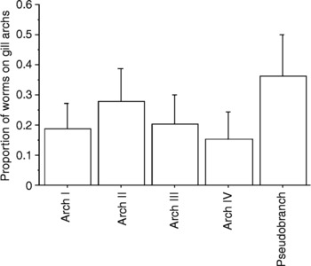
The parasite was found on the gill arches as well as on the pseudobranchs of 22.5% of fish. No infection was detected at the beginning of each season, with the first infection appearing in late June and reaching a peak of prevalence in August. The mean of intensity was 2.5 with a maximum intensity of 12. Regarding the site selection of the parasite, differences between infection on different gill arches were significant (P = 0.017, α = 0.5), with the highest intensity on the pseudobranch and second gill arch (fig. 2).
Interaction bar plot for occurrence of Gyrodactylus harengi on different gill arches of herring.

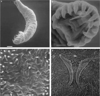
Body elongate, length 417 (390–440), width 96 (78–130) at mid body. Adhesive organs, two, at anterior terminal end, mouth at first quarter of body. Whole body, including haptor, covered by microvilli (fig. 3a–c).
(a) SEM of Gyrodactylus harengi; scale bar = 20 μm. The whole body, including the haptor (b), is covered by intensive microvilli (c). (b) SEM of the haptor of G. harengi; scale bar = 10 μm. (c) SEM shows details of the microvilli coverage of the body of G. harengi; scale bar = 500 nm. (d) Light microscopy of hamuli and its dorsal and ventral transverse bars of G. harengi.

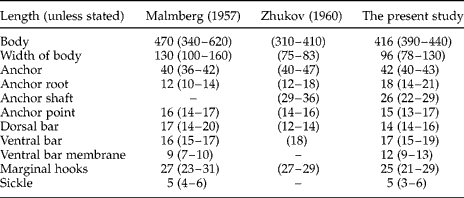
Distinction between the body proper and the opisthaptor is obvious when using SEM, while it is not so pronounced in traditional preparations (fig. 3a). In the opisthaptor, 16 marginal hooks (fig. 3b), length 25.1 (21.0–28.6), distributed evenly. Hamuli (fig. 3d), one pair, single rooted, supported by one dorsal and one ventral transverse bar; measurements are given in table 1.
Comparative measurements (in micrometres) of Gyrodactylus harengi specimens found in the present study with those reported by Malmberg (Reference Malmberg1957) and Zhukov (Reference Zhukov1960).
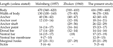
Malmberg (Reference Malmberg1957) proposed the name Gyrodactylus harengi for specimens collected from C. harengus membras from the Baltic Sea. The species was later reported from C. harengus pallasi and Ammodytes hexapterus hexapterus from the Bering Sea (Zhukov, Reference Zhukov1960). Malmberg (Reference Malmberg1970), having examined those specimens, stated that worms from the latter fish belonged to a separate species of Gyrodactylus. He also noticed slight differences between specimens and proposed that specimens from C. harengus pallasi belonged to a different subspecies than did those described from C. harengus membras.
Measurements of the specimens found in this study overlap most of the measurements of previous descriptions (Malmberg, Reference Malmberg1957; Zhukov, Reference Zhukov1960). However, some differences exist, not only in sizes of hard parts of the opisthaptor (table 1), but also in the general morphology of these organs, i.e. distance between dorsal and ventral bars, which is greater in the specimens studied in the present work. Malmberg (Reference Malmberg1970) suggested that three subspecies of Gyrodactylus are infecting three subspecies of C. harengus. However, characters of hard parts of Gyrodactylus can be affected by environmental factors, e.g. water temperature (Malmberg, personal communication), that might have resulted in these differences.
Gyrodactylus has a pair of strong hamuli and 16 marginal hooks supporting the attachment of the worm on the host surface. Consequently, G. harengi could potentially infect all gill arches equally. Results of this study, however, show transverse partitioning, but no preference for the left or right side of the body was observed. Polyansky & Shulman (Reference Polyansky and Shulman1956) have distinguished two basic types in the age dynamics of parasite fauna of fish. In their categorization, Gyrodactylus, with a direct life cycle, falls into the ‘freshwater type’ in which initial infection of young fish occurs after association of young and adult fishes. Fish examined during the present study had not yet associated with adults. Consequently, it is most probable that infection of young herring comes about as a result of their association with other fish species, notably sprat Sprattus sprattus – Gyrodactylus is viviparous and the possibility of infection by overwintering eggs (as can happen in some oviparous monogeneans) is ruled out, and the gill arches of older sprats have the same size and morphology as those of young herrings. It should also be noted that G. harengus is the only Gyrodactylus infecting these two fish species.
Pseudanthocotyloides heterocotyle (van Beneden, Reference van Beneden1871) Euzet & Prost, Reference Euzet and Prost1969 (Monogenea: Polyopisthocotylea: Mazocraeidae)
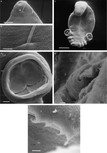
Pseudanthocotyloides heterocotyle were found on the gill arches with a mean intensity of 1.5 and maximum intensity of 5, on 8% of fish. The general morphology of this worm has been described elsewhere (Euzet & Prost, Reference Euzet and Prost1969; Rahimian et al., Reference Rahimian, Longshaw, MacKenzie and Thulin1999). The body surface of the worm is naked, with abundant and irregularly distributed pores (fig. 4a, b), diameter 0.25–1.00. These pores seem to be involved in secretion of mucus. The haptor is not distinct from the body proper and has four pairs of clamps, with the fourth pair much larger than the others (fig. 4c). Inside each clamp, there are two sliding plates (fig. 4d) which, by their movements, acting on sclerites, open or close the clamp. Clamps do not seem to have a sucking function. Instead, the worm obviously uses them as a ‘grasping’ apparatus by which it grasps the gill lamella of the host (fig. 4e). Of one pair of hamuli and two pairs of marginal hooks, located at the most posterior terminal of the haptor, only the small hamuli and one pair of the marginal hooks (fig. 4f) seem to contribute, a little, to maintaining the hold of the worm.
Pseudanthocotyloides heterocotyle. (a) SEM of anterior part; note the genital pore (arrowhead) from which an egg's polar filament is extruding; scale bar = 20 μm. (b) Higher magnification of the area around the genital pore, note numerous small pores on the body surface; scale bar = 2 μm. (c) SEM of the body proper of P. heterocotyle, note relative sizes of different clamps, ∧ shows the fourth pair of the clamps; scale bar = 200 μm. (d) SEM of an open clamp with two sliding plates; scale bar = 20 μm. (e) SEM of clamp of P. heterocotyle grasping gill lamella of herring; scale bar = 10 μm. (f) SEM of the most posterior part of opisthaptor of P. heterocotyle, showing hamuli and one pair of marginal hooks; scale bar = 5 μm.

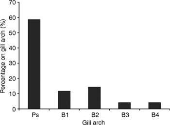
Distribution of P. heterocotyle was significantly different on different gill arches of herring (fig. 5). More than 60% of worms were found on the pseudobranchs, while gill arches I and II harboured about 12% and 15%, respectively, and arches III and IV housed only about 5% of worms.
Distribution of Pseudanthocotyloides heterocotyle on different gill arches of herring. Ps, pseudobranch; B1–4: gill arches I–IV.

P. heterocotyle was first described by van Beneden (Reference van Beneden1871) as Octostoma heterocotyle from sprat from the Mediterranean Sea. Van Beneden & Hesse (Reference van Beneden and Hesse1863) had earlier described Octocotyle harengi from herring. Descriptions of both species give very few valid characteristics. Sproston (Reference Sproston1946) included both species in the genus Mazocraes Herman 1782, as M. heterocotyle and M. harengi. M. heterocotyle from sprat was redescribed and placed in the new genus Pseudanthocotyloides by Euzet & Prost (Reference Euzet and Prost1969). In the present study only P. heterocotyle was found from this genus. Rahimian et al. (Reference Rahimian, Longshaw, MacKenzie and Thulin1999) speculated that M. harengi is synonymous with M. alosae. Except for a report of P. heterocotyle from wild herring in captivity (Longshaw, Reference Longshaw1996) there has been no report of this parasite from herring, or from any fish species from the area of the present study.
In their study, Rahimian et al. (Reference Rahimian, Longshaw, MacKenzie and Thulin1999) found P. heterocotyle only on small herring; no adult herring was infected. It seems that the mechanisms by which the parasite remains attached to or moves on the host is the main factor that determines the size of the host to which the parasite can attach and/or hold on. As hamuli and marginal hooks are very small in comparison with the worm size, they seem to have a minor role in holding the worm on to the host. Therefore, it seems that clamps are the organs playing the major role in the positioning of the worm, and the size, or thickness, of the gill lamella is apparently the factor that determines whether or not clamps can grasp and hold on. As fish grow, so do the gill lamellae, and the bigger the lamellae, the less clamps are able to hold on to them. Differences between the size of the lamellae of the pseudobranch and those of the gills are not significant, and it seems likely that the difference observed in occurrence of P. heterocotyle on different gill arches is mostly due to the speed and amount of water passing between arches. On the other hand, the pattern of worms' distribution on different gill arches might be an evolutionary adaptation for the parasite, since polyopisthocotyleans feed primarily on blood (Cheng, Reference Cheng1986) and the pseudobranch is supplied with blood that has already been oxygenated in the first branchial arch (Hughes, 1984); the parasite therefore receives blood rich in oxygen. Furthermore, in many cases, the pseudobranch is considered to be non-respiratory and hence settling on this site by a relatively large parasite would cause minimal disturbance to respiratory function of the gills. This idea is also supported by an overcrowded situation, when P. heterocotyle occurred in high densities on wild herring in captivity, causing mortality. In that instance worms were found to be distributed over all gill arches (Longshaw, Reference Longshaw1996), possibly due to population pressure. The observed distribution pattern, therefore, seems to be caused by the worms' preferences and not by physical limitations.
Digenea
Metacercariae Cryptocotyle lingua (Creplin, 1825) Fischoeder 1903 (Digenea: Opisthorchioidea, Heterophyidae)
Metacercariae of Cryptocotyle lingua were found encapsulated on the body surfaces, including skin, gills, eyes, and even in the intestine wall, of 70.2% of fish, with mean intensity of 26.3 and maximum intensity of 160. Sixty-eight per cent of capsules were found in areas above the middle line of the body, where they were distributed randomly. Capsules were spherical to ovoid, 600 × 460 in diameter, and each completely filled with one metacercaria. On the skin and in the flesh, capsules were pigmented. The species is adequately described in its original descriptions and by Sindermann (Reference Sindermann1966) and Køie (Reference Køie1977).
Metacercariae of C. lingua have been reported from herring of the north-west Atlantic (Sindermann & Rosenfield, Reference Sindermann and Rosenfield1954; Sindermann, Reference Sindermann1966; McGladdery & Burt, Reference McGladdery and Burt1985), the North Sea (MacKenzie, Reference MacKenzie1987a) and the Baltic Sea (Reimer, Reference Reimer1970). With finding this parasite on herring in the present study area, it is now known on both sides of the North Atlantic and its adjacent waters. The life cycle of C. lingua includes two intermediate hosts, and adults occur in the intestine of piscivorous birds (Køie, Reference Køie1977). Herring, among many other fish species, serves as the second intermediate host and the lifespan of the metacercaria in the host is about 1 year (MacKenzie, Reference MacKenzie1987a). The first intermediate host, Littorina littorea, occurs in coastal areas and cercariae are released when the water temperature is above 10°C (Sindermann, Reference Sindermann1966). Consequently, due to their choice of habitat, young herring will be infected, while none or few metacercariae of C. lingua will have the chance to infect adult herring.
Metacercariae Renicola sp. (Digenea: Renicolidae)
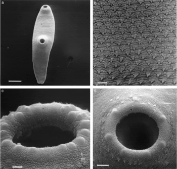
The hyaline, thick-walled, oval capsules, 300 (280–340) by 220, enclosing the metacercariae of Renicola were found on the outside of the pyloric caeca of 6.8% of fishes, with a maximum intensity of 3; in most cases only one capsule per fish was observed. Through the capsule wall a pink pigmentation could be seen. Body elongated, narrow and thin in post-ventral-sucker region (fig. 6a), 850 (800–950) length, 230 (200–260) width. Body surface covered by obliquely arranged pyramidal spines (fig. 6b). Oral sucker, terminal, 94 (90–96) in diameter, prepharynx not evident, pharynx globular, 30 μm in diameter, caeca bifurcated shortly after pharynx, terminating blindly near the posterior part of the body. Ventral sucker, 95 (90–96), equatorial, equal to slightly larger than the oral sucker (fig. 6a). Sensory structures, possibly touch receptors, prominent around both oral and ventral suckers (fig. 6c, d), though their numbers and arrangement were different. These structures were also distributed on the body surface (fig. 6a).
Metacercaria of Renicola sp. (a) SEM of metacercaria of Renicola sp.; scale bar = 100 μm. (b) SEM of body surface of metacercaria of Renicola sp.; scale bar = 5 μm. (c) SEM of oral sucker of metacercaria of Renicola sp., note semi-hemispherical sensory organs, possibly touch receptors; scale bar = 5 μm. (d) SEM of ventral sucker of metacercaria of Renicola sp.; scale bar = 10 μm.

Metacercariae of Renicola have so far been reported exclusively from the north-east Atlantic and mainly from clupeoid fishes; from sprat (Wright, Reference Wright1956), sardines (Timon-David, Reference Timon-David1953) and herring (MacKenzie, Reference MacKenzie1975). Rothschild & Sproston (Reference Rothschild and Sproston1941), however, reported the parasite from the gadoid fish bib, Trisopterus luscus, and whiting Merlangius merlangus. Wright (Reference Wright1956) recognized two distinct types of metacercariae in sprat, ‘pythionike’ and ‘doricha’, and MacKenzie (Reference MacKenzie1975) set criteria to distinguish the two types. Using those criteria, the morphology of metacercariae found in herring of the present study were of the ‘pythionike’ type, while MacKenzie (Reference MacKenzie1975) found both types of metacercariae in juvenile herring. In his study, he also found that more than 70% of some juvenile herring populations off the east coast of Scotland were infected with up to 130 metacercariae. In the present study, however, the maximum intensity was only 3.
Results from the present study expand the range of Renicola sp. in herring from the North Sea into the Skagerrak. Adult Renicolidae are parasites of renal ducts of various piscivorous birds, and the first intermediate host is the gastropod mollusc Turritella communis (Wright, Reference Wright1956), both being present in the area of the present study.
Hemiurus luehei Odhner, 1905 (Digenea: Hemiuridae)
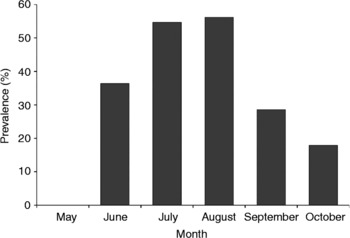
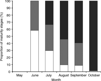
Hemiurus luehei was mostly found in the stomach (70%), occasionally in the pyloric caeca (25%) and rarely in the intestine (5%). The mean prevalence of infection was 47.1%, with a maximum intensity of 133 and mean intensity of 12.3. The prevalence rose from 0 in May to 54.7 in July, remained almost the same during August, and decreased toward the autumn (fig. 7). The proportion of flukes in different maturity stages during the sampling months are shown in fig. 8. The proportions of those in stages I and II continuously decreased from the peak in June to October. At the same time the proportion of flukes in stage III increased to 99% of total recovered flukes in October.
The mean prevalence of Hemiurus luehei infection during May–October, from 1994 to 1997.

Proportions of different maturity stages of Hemiurus luehei infecting juvenile herring during May–October, from 1994 to 1997; □ Stage I,  Stage II and ■ Stage III.

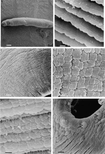
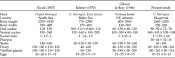
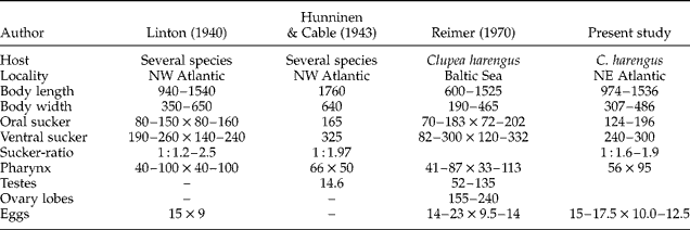
H. luehei has been described in detail by Gibson & Bray (Reference Gibson and Bray1986). The body of the fluke is cylindrical and covered with a plate-like tegument. The shape and arrangements of these plates are different in different parts of the body. On most parts, the plates are arranged in imbricate ring form (fig. 9a, b), which is interrupted in regions around the anterior and posterior suckers, where they become scale-like imbricate. On the posterior part of the soma, the tegument is scale-like and in juxtaposed arrangement (fig. 9c, d). The change between these two extremes on the body surface is gradual, and intermediates can be seen between the ends of the body (fig. 9e). Tegument of the ecsoma is smooth. Oral sucker subterminal, surrounded by some hemispherical papillae (fig. 9f). The ventral sucker is larger than, and situated close to, the oral sucker (sucker ratio 1 : 1.55–1.60). The measurements of H. luehei found in the present work in comparison to those found in previous studies are presented in table 2.
SEM of Hemiurus luehei: (a) SEM of the body covered by imbricate ring form, interrupted in the sucker regions; scale bar = 200 μm. (b) High magnification, SEM of imbricate rings on area anterior to the ventral sucker; scale bar = 1 μm. (c) SEM of the most posterior part, note that the telescopic naked ecsoma is not exposed; scale bar = 5 μm. (d) High magnification SEM of the posterior part of soma with scale-like tegument in juxtaposed arrangement; scale bar = 2 μm. (e) High magnification SEM of the tegument between ventral sucker and the most posterior part of the body; scale bar = 1 μm. (f) SEM of the oral sucker, note the hemispherical papillae (>) on the outer edge of the sucker; scale bar = 5 μm.

Comparative measurements (in micrometres) of Hemiurus luehei specimens found in the present study with those reported by Nicoll (Reference Nicoll1907), Reimer (Reference Reimer1970) and Gibson & Bray (Reference Gibson and Bray1986).
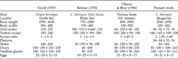
Clupeid fish, especially herring, are among the most important hosts for H. luehei, while other fishes, e.g. salmonids and gadids, are probably infected by preying on these plankton feeders (Gibson & Bray, Reference Gibson and Bray1986). H. luehei has been reported from herring of the north-east Atlantic and its adjacent waters (see Arthur & Arai, Reference Arthur and Arai1984). Furthermore, the species reported as H. raabei from herring of the Baltic Sea (Rokicki, Reference Rokicki1973; Gaevskaya, Reference Gaevskaya1977) may also be a synonym of H. luehei (Arthur & Arai, Reference Arthur and Arai1984).
The life cycle of H. luehei has been studied by Køie (Reference Køie1990). Her studies show that the tectibranch opistobranch, Philine denticulata, is the first intermediate host, calanoid copepods (under experimental conditions) are the second intermediate hosts, and Sagitta sp. is a paratenic host.
The prevalence of infection in herring of the Baltic Sea has been found to be more or less constant (58%) throughout the year (Reimer, Reference Reimer1970). In the North Sea, on the other hand, the prevalence was found to fluctuate dramatically, between 2% during winter and up to 70% during summer (Hansen, Reference Hansen1955). These data led Køie (Reference Køie1990) to speculate that either the short lifespan of H. luehei or the migration pattern of herring populations might have caused those differences. In the present study, the 0-ring herring first became infected in May–June and showed the highest prevalence in July, then the prevalence decreased during the autumn. Consequently, these results strongly indicate that such changes in the prevalence, at least in the study area, are mainly due to the short lifespan of this fluke. The copepods hosting metacercariae of H. luehei usually die within 3 weeks post infection, but Sagitta, as paratenic host, can prolong the lifetime of the metacercariae (Køie, Reference Køie1990). Small herring do not feed upon Sagitta and mostly prey on small copepods; this indicates that the observed decrease in the prevalence is due to unavailability of metacercariae in high numbers, combined with dying out of flukes in fish in a matter of a few months.
Differences in the tegumental structures of different regions of the body of H. luehei are reported here for the first time. Hemiuridae are unusual amongst digeneans, since adults usually occur in the stomach of their marine fish hosts, where they are exposed to extreme mechanical and chemical stresses due to the presence of food items (that mostly include the hard parts of plankton), acid and proteolytic enzymes (MacKenzie & Gibson, Reference MacKenzie and Gibson1970; Gibson & Bray, Reference Gibson and Bray1979). It has been proposed that the special tegument of Hemiuridae is an adaptation allowing them to tolerate those extreme conditions of the stomach, and the ‘naked’ ecsoma acts as a ‘feeding organ’, extending from the body when circumstances are most tolerable (Gibson & Bray, Reference Gibson and Bray1979). This idea was supported by experimental and transmission electron microscopy studies by Matthews & Matthews (Reference Matthews and Matthews1988). They showed that the ecsomal tegument, unlike the somal tegument, is adapted for transference of materials between the parasite and its host. The idea can also be supported by the observed differences between the tegument of H. luehei and that of L. confusus, another Hemiuridae. The differences in the tegumental structure of the most posterior part of body, however, may facilitate retraction and extension of the telescopic ecsoma. On the other hand, with the venetian blind-like arrangement of the tegumental elements (fig. 9b), at least some part of the soma may be exposed to the environment, when conditions are good, by raising the tegumental ‘slats’, and be protected from the environment when the ‘slats’ overlap.
Lecithaster confusus Odhner, 1905 (Digenea: Hemiuridae)
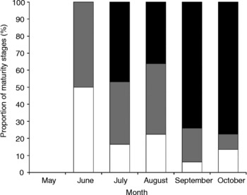
Adults of Lecithaster confusus were found both in the intestine and pyloric caeca of 58.8% of fish, with a mean intensity of 5.1 and maximum intensity of 49. Prevalence of infection, after appearance of infection in June (27.3%) increased to 44.2% in July and 76.8% in August and then decreased to 47.9% in September and was almost constant in October (53.8%). The proportion of worms in different maturity stages, however, changed, with the highest proportion of stage I in June, stage II in July and stage III in October. The proportions of stages I and II were the same in June and generally decreased during the following months, while the proportion of stage III increased (fig. 10).
Proportions of different maturity stages of Lecithaster confusus infecting juvenile herring during May–October, from 1994 to 1997; □ Stage I, Stage II and Stage III.

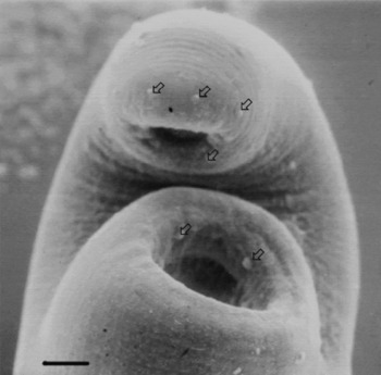
Body naked, fusiform, tapering towards posterior end, with greatest diameter at posterior end of the ventral sucker (fig. 11). Sensory structures, possibly touch receptors, prominent around both the oral and ventral suckers (fig. 11). Measurements of L. confusus from the present study overlap those of previous studies (table 3).
SEM of Lecithaster confusus: anterior part of the body, note receptors ( ), possibly touch receptors, on the anterior and ventral suckers; scale bar = 20 μm.

Comparative measurements (in micrometres) of Lecithaster confusus specimens found in the present study with those reported by Linton (Reference Linton1940), Hunninen & Cable (Reference Hunninen and Cable1943) and Reimer (Reference Reimer1970).
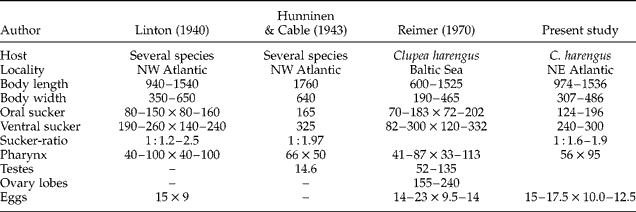
L. confusus has been reported from the intestine of herring from the Baltic Sea (Reimer, Reference Reimer1970; Odening, Reference Odening1978), north-west Atlantic (Linton, Reference Linton1940), White Sea (Shulman, Reference Shulman1956) and Barents Sea (Polyansky, 1955). Naidenova (Reference Naidenova1974) proposed that L. gibbosus and L. confusus be considered as subspecies with northern and southern distributions. But both species have been reported from herring at a wide range of latitudes, with the exception of L. confusus from the Pacific herring (MacKenzie, Reference MacKenzie1987a). L. gibbosus has been reported from herring of both sides of the Atlantic and its adjacent waters, as well as from the Pacific (Arthur & Arai, Reference Arthur and Arai1984). L. confusus and L. gibbosus are difficult to distinguish but, according to Gibson (Reference Gibson1996), the vitelline lobes in L. confusus are short, a little longer than wide, and eggs are usually < 19 μm. In L. gibbosus, on the other hand, the vitelline lobes are longer than wide and eggs are normally >19 μm. Considering these criteria, specimens found in the present study are L. confusus.
After the initial infection of herring, the prevalence of infection did not change dramatically. This suggests either a relatively long lifespan of adults and short exposure period to metacercariae, or a continuous exposure of host to metacercariae and relatively short lifespan of adults. The proportions of the parasites at different maturity stages, however, indicated that the maximum exposure to metacercariae was in June with lower, but constant, exposures during the rest of study period.
The parasitic snail Odostomia trifida serves as the first intermediate host of L. confusus and metacercariae live, un-encysted, in the haemocoele of the copepods Acartia spp. (Hunninen & Cable, Reference Hunninen and Cable1943). The only species of that snail genus occurring in this area is O. rissoides, which parasitizes Mytilus edulis. Across the Kattegat and along the Danish coasts, Køie (Reference Køie1983) reported L. gibbosus as the ‘only’ Lecithaster species occurring in several fish species, including clupeoids. If this is true, then the genus Lecithaster can be used as a biological tag to separate herring spawning in the eastern from those spawning in the western Skagerrak–Kattegat. However, it seems questionable that two different species with such diverse prevalence occur in the same host species at such close localities, especially since the first intermediate host occurs on both sides of the Skagerrak–Kattegat.
Pseudobacciger harengulae (Yamaguti, Reference Yamaguti1938) (Digenea: Fellodistomidae)
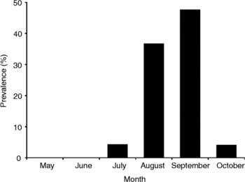
Pseudobacciger harengulae occurred in the pyloric caeca and intestine, of which the pyloric caeca was the main site of infection. The prevalence of infection was 21.5% with the mean intensity of 4.8 and maximum intensity of 52. The highest prevalence was observed in September. The pattern of prevalence changes is shown in fig. 12.
Changes in the mean prevalence of Pseudobacciger harengulae in fingerling herring, during May–October, from 1994 to 1997.

P. harengulae has been adequately described (Yamaguti, Reference Yamaguti1938; Dimitrov et al., Reference Dimitrov, Bray and Gibson1999; Rahimian & Thulin, Reference Rahimian and Thulin2003). Occurrence of P. harengulae in the cold Swedish waters is surprising since previously it has been reported from tropical, subtropical and generally warmer areas (Yamaguti, Reference Yamaguti1938; Manter, Reference Manter1947; Madhavi, Reference Madhavi1975; Kim & Chun, Reference Kim and Chun1984; Gaevskaya, Reference Gaevskaya1996; Dimitrov et al., Reference Dimitrov, Bray and Gibson1999). The life cycle of P. harengulae was studied in Korea (Kim & Chun, Reference Kim and Chun1984) (under the incorrect name of Bacciger harengulae). According to that study Solen strictus and Meretrix lusoria were among the molluscs serving as the first intermediate host of the parasite. None of those species occurs in the area of present investigation. However, the two families, Veneridae and Solenidae, to which these molluscs belong, have some representatives here. Particularly, the family Solenidae is of special interest. This family has two species represented in the area, Ensis ensis and Cultellus pellucidus. In addition, a new, alien species, Ensis directus, was introduced to the German North Sea coast in 1978 (von Cosel et al., Reference von Cosel, Doerjes and Muehlenhardt-Siegel1982) and spread from Gammelgab (Denmark) to the Balgzang (The Netherlands) soon after. By the late 1980s and early 1990s E. directus appeared in large numbers in the Gullmarsfjord, the area of study, but later suffered heavy loses and now can only occasionally be found in the fjord. Considering the coincidence of E. directus and P. harengulae, it is very likely that this digenean has been introduced to the area in its sporocyst form, together with the clam. Still, study of the life cycle of this fluke in the present study area in comparison with the source area could be very interesting and fruitful.
Cestoda
Plerocercoids of Bothriocephalus sp. (Cestoda: Bothriocephalidae)
Plerocercoids of the cestode family Bothriocephalidae were found free in the intestine of 7.6% of young herring, with mean and maximum intensities of 1.8 and 4, respectively. This parasite first appeared in June, but then infestation continued without any significant changes in either prevalence or intensity. Individual fish with more than one parasite, however, were found more often towards the autumn.
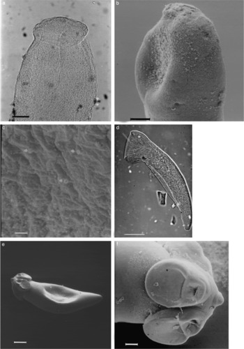
The specimens were sub-cylindrical, 800 (400–1500) long, 280 (100–300) in width at the most posterior part of scolex. Scolex elongated, unarmed, with apical disc, bothria shallow, covered with microvilli (fig. 13a, b). On the body surface there were numerous minute cylindrical, open-ended tubes (fig. 13c), which were particularly abundant on the neck area. The nature of these tubes is unknown. However, considering the absence of microtriches, it seems that these tubes are secretion openings of some glands (Bray, personal communication).
Different cestodes found in fingerling herring. (a) Light micrograph of scolex of Bothriocephalus sp. plerocercoid with the apical disc; scale bar = 50 μm. (b) SEM scolex of Bothriocephalus sp. plerocercoid with the apical disc and shallow bothria; scale bar = 50 μm. (c) SEM of the body surface of Bothriocephalus sp. plerocercoid, note minute tubes (arrows); scale bar = 2 μm. (d) Plerocercoid of acrobothriid gen. sp. with a typical cup-like scolex; scale bar = 50 μm. (e) SEM of plerocercoid of Tetraphyllidea spp.; scale bar = 200 μm. (f) SEM of scolex of plerocercoid of Tetraphyllidea spp.; scale bar = 50 μm.

Specimens found in this study are similar to those reported as plerocercoids of Bothriocephalus scorpii from herring of the Baltic Sea by Reimer (Reference Reimer1970) and Rokicki (Reference Rokicki1973); however, there was no information on what criteria those authors used for their identification of the plerocercoids to the specific level. Furthermore, there are reports of the occurrence of B. scorpii in herring from the north-west Atlantic (Linton, Reference Linton1941) and of Pseudophyllidea gen. sp. in herring from the Bering Sea (Zhukov, Reference Zhukov1963), the Pacific, north-west Atlantic and White Sea (Arthur & Arai, Reference Arthur and Arai1984), but this is the first report of this parasite from the north-east Atlantic.
At present, it is not possible to establish a link between the plerocercoids found in the present study and adults of B. scorpii, based on the available morphological information. Having said that, however, the larvae found in the present study may be defined as Bothriocephalus sp. Adults of B. scorpii have been reported from more than 50 species of teleost (Williams & Jones, Reference Williams and Jones1994). Members of the order Pseudophyllidea generally use copepods as their first, and sometimes the only, intermediate host, in which the hexacanth larva penetrates into the haemocoel and develops to the procercoid. If a second intermediate host, a fish, is involved, the plerocercoid develops in the musculature or in the body cavity (Williams & Jones, Reference Williams and Jones1994). However, in this study, as in other studies on herring, the hosts harboured the parasite only in their intestines.
Plerocercoids of Acrobothriidae Olsson, 1872 (Cestoda: Spathebothriidea)
Plerocercoids of acrobothriids were found free, or attached to the wall, in the intestine of 5.8% of the fish. The maximum intensity was 3 with a mean intensity of 1.2. The worms were elongated–funnel shaped, length 250–370, width at the mid-body 55–60, with a cup-like scolex (fig. 13d), 75–80 in diameter, and 50 in depth. The parasite was first observed in July and both the intensity and prevalence of infestation remained the same throughout the study period. However, due to low prevalence and intensity, the evidence is not conclusive.
The specimens are similar to those described as and called Scolex bothriosimplex by Reimer (Reference Reimer1970) from herring and other hosts in the Baltic Sea. In their checklist of parasites of herring, Arthur & Arai (Reference Arthur and Arai1984) included ‘Cestoscolex’ of van Beneden (Reference van Beneden1871) and S. bothriosimplex as unidentified Cestoda. As far as I am aware, there are two orders of Cestoda having members with a single cup-like scolex: Nippotaeniidea Yamaguti, 1937 and Spathebothriidea Wardle & McLeod, 1952. All six species of Nippotaeniidea have been reported from freshwater fishes in Japan, China, the former Soviet Union and New Zealand (Bray, Reference Bray, Kalil, Jones and Bray1994). Members of Spathebothriidea, on the other hand, are parasites of freshwater and euryhaline chondrostean and teleost fishes (Gibson, Reference Gibson, Khalil, Jones and Bray1994). This order comprises two families, Spatheobothriidae Yamaguti, 1934 and Acrobothriidae Olsson, 1872, the former of which has no distinctly differentiated scolex. Acrobothriidae have a differentiated scolex with one or two sucker-like attachment organs and occur especially in salmonids, pleuronectids and soleids, all of them abundant in the study area. There is also an alternative possibility that the scolex of plerocercoids found in this study are not yet developed, consequently all plerocercoids mentioned may belong to a different order.
Plerocercoid of Tetraphyllidea, Carus, 1863 (Cestoda)
Plerocercoids of Tetraphyllidea were found either free in, or attached to the wall of, the intestine of 2.2% of young herring. No fish were found infected with more than one worm. The worm (fig. 13e) is fusiform when relaxed, 930 (700–1000) long and 280 (200–300) wide. Scolex (fig. 13f) with four sub-elliptical, unarmed bothria, each having two loculi that could appear or disappear with scolex movements in live specimens. A hemispherical apical organ, with numerous cilia-like structures at its base, was also present. Body covered with microvilli. Live specimens had a pink ring at the base of each bothrium. The low prevalence and intensity of infection did not allow any analysis.
Specimens found in this study correspond to those found by McGladdery & Burt (Reference McGladdery and Burt1985). They reported the plerocercoid as Scolex pleuronectis Müller, 1788, and noted two forms: ‘microscopic’ with four unilocular, and ‘macroscopic’ with four bilocular bothria. These two forms might have been two stages in development of the same parasite or, alternatively, could have been due to different fixation of the same animals resulting in different shapes of the scolex, as was observed in the present study. S. pleuronectis and S. polymorphus are collective names for plerocercoids of various tetraphyllidean species and have been reported from herring from all of its geographical range (Arthur & Arai, Reference Arthur and Arai1984). Considering the fact that the scolex of specimens found in the present study would develop even further, assignment of the specimens to family and species is not possible; but, nevertheless, they are similar to those described as Spiniloculus spp. by Avdeeva (1989).
Nematoda
Larval Hysterothylacium aduncum Rudolphi, 1802 (Nematoda: Anisakidae)
Larvae of Hysterothylacium aduncum were found free in the intestine of 3.5% of examined fish, with a mean intensity of 1.1 and maximum intensity of 2. The low prevalence and intensity of infection did not allow any seasonal interpretation of the infection. Worms were all in their fourth larval stage, elongate, cylindrical, between 1.4 and 5.0 mm long. The anterior part had four semi-spherical papillae around the boring tooth. The cuticle was segmented, segmentation being most conspicuous on the post-anal region.
The occurrence of H. aduncum in herring is worldwide and has been reported from the Atlantic and Pacific and their adjacent waters (Arthur & Arai, Reference Arthur and Arai1984). MacKenzie (Reference MacKenzie1987a) found that the prevalence and intensity of H. aduncum in herring are consistently lower than those in most other fish species.
The morphology and life cycle of H. aduncum have been investigated and described by Smith (Reference Smith1983) and Køie (Reference Køie1993). In the North Sea, larvae of H. aduncum were found to naturally infect the euphausiid species Thysanoessa raschii, T. couchii and T. inermis (Smith, Reference Smith1983). In an experimental study on the life cycle of H. aduncum, Køie (Reference Køie1993) was able to infect the calanoid copepod Acartia tonsa and harpacticoid copepods by eggs containing the third larval stage of H. aduncum. In her experiments, mysids, amphipods and isopods were also infected when exposed to a large number of eggs. Furthermore, upon examination of wild copepods of the Sound (between Sweden and Denmark) she found infections in Acartia sp. and Pseudocalanus elongatus. Køie (Reference Køie1993) also found that larval H. aduncum must reach a certain length, of at least 1.5–2.0 mm, in their copepod host, before they can survive in fish host. In the present study, however, fish were found to be infected with larvae of half that supposed critical size. Herring seems to serve as the second intermediate host for H. aduncum.
Copepoda
Caligus elongatus von Nordmann, 1832 (Copepoda: Caligidae)
The parasitic copepod Caligus elongatus was found on the body surface, especially around the anus and on the anal fin of 2% of examined young herring. On live fish kept in tanks, however, the parasites were observed to move around on the body surface. Most infected fishes had only one copepod, and only two specimens infected with two copepods were found. Fish infected with C. elongatus were found only during September–October, and all parasites were adult females.
Two specimens of C. elongatus were heavily infected with up to 45 Udonella sp., a monogenean hyperparasite.
C. elongatus, sometimes under other names, has been reported from more than 80 species of both teleosts and elasmobranchs, worldwide (Kabata, Reference Kabata1979). On herring, C. elongatus has been reported from both sides of the north Atlantic (Dollfus, Reference Dollfus1956; Sindermann, Reference Sindermann1957; Rosenthal, Reference Rosenthal1967 (experimental); Kabata, Reference Kabata1979). The morphology of adult and larval stages of this copepod has been described by Kabata (Reference Kabata1979) and Piasecki (Reference Piasecki1996).
Conclusions
The present study focused on the parasite fauna of 0-ring herring from a limited geographical area. During the study 15 parasitic species, including 2 Protozoa, 1 species of uncertain taxonomic status, 2 Monogenea, 5 Digenea, 3 Cestoda, 1 Nematoda, and 1 Copepoda were found. Records of Trichodina sp., Ceratomyxa auerbachi, metacercariae of Renicola sp., plerocercoids of Bothriocephalus sp., acrobothriid and tetraphyllid and the copepod Caligus elongatus from herring of the study area are all new geographical records. Observation on mixed-age adult herring from the Kattegat and Skagerrak (unpublished personal observations) revealed the presence of following parasites: Goussia clupearum, Eimeria sardinae, Ichthyophonus hoferi, Gyrodactylus harengi, Cryptocotyle lingua, Renicola sp., Hemiurus luehei, Lecithaster confusus, Pseudobacciger harengulae, Bothriocephalus sp., Tetraphyllidea, Anisakis simplex and Hysterothylacium aduncum.
MacKenzie (Reference MacKenzie1987a) categorized parasites of herring according to their host specificity, into five groups: (1) herring as abnormal host; (2) herring as the most important host; (3) herring as one of several equally important hosts; (4) herring as a host with minor importance; and (5) other. Considering his categories, none of the parasites found in this study can be assigned to the first category; Ceratomyxa auerbachi and Gyrodactylus harengi can be placed in the category of parasites to which herring is probably the most important host; Pseudanthocotyloides heterocotyle, Pseudobacciger harengulae, metacercariae of Renicola sp., Hemiurus luehei and Caligus elongatus fall into the third category; and metacercariae of Cryptocotyle lingua, Lecithaster confusus, all metacestodes and Hysterothylacium aduncum fall into the category of parasites into which herring has a minor importance as a host. Trichodina sp. requires more investigations before its position in these categories can be determined, but for now it should be placed in the fifth category.
MacKenzie (Reference MacKenzie1987a) stated that the parasite faunas of juvenile and adult herring are not very different, since herring remain plankton-feeders throughout their lives. While this is true for parasites with indirect life cycles, it does not seem to be the case for parasites with direct life cycles. A study of parasites of juvenile herring can give an index of parasites that adult herring may be infected with while in the area of present study. Adult herring enter and spawn in the study area in May and leave soon after. Hatched larvae remain and feed in the area, before leaving in the autumn. These fish then come back to the area, after approximately 2 years. Since fish examined in this study, most probably, have been born and grown in the area, their parasite fauna reflects the parasites of herring present in this area. This can provide a tool to be used in future biological tagging studies. Some parasites of herring have been used as biological tags for fish and fisheries research. Parasites of herring have, for example, been used for stock separation (Arthur & Arai, Reference Arthur and Arai1980a, Reference Arthur and Araib; Kulachkova, Reference Kulachkova1989; Moser, Reference Moser1991), recognition of recruitment migration (Heath, Reference Heath1984; McGladdery & Burt, Reference McGladdery and Burt1985; MacKenzie, Reference MacKenzie1985) and studies on herring diet and feeding behaviours (MacKenzie, Reference MacKenzie1987b). Furthermore, intestinal helminths may be used for further seasonal migration studies (MacKenzie, Reference MacKenzie1987a). In these types of studies, digeneans are especially important since they are usually highly specific to their first intermediate host, usually a mollusc. From parasites reported in the present study, metacercariae of Renicola sp. and Hysterothylacium sp. have been shown to be useful for population studies of herring (MacKenzie, Reference MacKenzie1985; Moser, Reference Moser1991). Except for Ichthyophonus hoferi, which is highly pathogenic to its host, and Caligus elongatus, which can easily drop off the host, all other parasites may be potentially suitable for fish and fisheries research of herring populations in the Skagerrak–Kattegat.
Acknowledgements
I wish to thank sincerely Professor Jan Thulin for reading and commenting on early drafts of the paper. Also my thanks go to Dr Rodney Bray for his comments on the manuscript. The present work was carried out at Kristineberg Marine Research Station, Fisheries Research Laboratory, Lysekil, Zoology Institute, Gothenburg (Sweden), and School of Biology, University of Tehran. So, I wish to thank all the staff who helped me during the course of this study. This study was part of a larger-scale study on parasites and diseases of herring, carried out with financial support from Fiskbranschen Abba, Orvar & Gertrud Nybelin's Found, Skandiakonserv, Rahmns Stiftelse, Swedish National Board of Fisheries, The Royal Swedish Academy of Science, Adlebertska hosipitiefonden and Hvidtfeldska fonden.