Introduction
Chronic cough lasting 8 weeks or more is often seen as an intractable problem in childhood (Irwin et al., Reference Irwin, Baumann, Bolser, Boulet, Braman, Brightling, Brown, Canning, Chang, Dicpinigaitis, Eccles, Glomb, Goldstein, Graham, Hargreave, Kvale, Lewis, McCool, McCrory, Prakash, Pratter, Rosen, Schulman, Shannon, Smith Hammond and Tarlo2006). One of the most common causes of chronic cough is bronchial asthma (Dicpinigaitis, Reference Dicpinigaitis2006). The prevalence of bronchial asthma has increased during the past 20–30 years in developed countries, but there is a considerably lower prevalence in developing countries (Cookson & Moffatt, 1997). There are numerous environmental risk factors, such as pollution, increased exposure to allergens, changes in eating habits, and a decreased intake of antioxidants, which can account for atopic disorders, including bronchial asthma (Britton et al., Reference Britton, Pavord and Richards1994; Hill et al., 1997; Soutar et al., Reference Soutar, Seaton and Brown1997; Vormann, Reference Vormann2006).
There are two newly recognized entities in the background of chronic cough: cough variant asthma (CVA) and non-asthmatic eosinophilic bronchitis (NAEB). The prevalence of CVA varies by race and geographically, occurring more frequently in Asia than in Europe (Todokoro et al., Reference Todokoro, Mochizuki, Tokuyama and Morikawa2003; Lai et al., Reference Lai, Chen, Liu, Luo, Zhong, He, Li and Zhong2006). It may be the precursor of bronchial asthma in almost 30% of cases (Fujimura et al., Reference Fujimura, Abo, Ogawa, Nishi, Kibe, Hirose, Nakatsumi and Iwasa2005). The diagnosis of CVA is based on chronic cough without wheezing, with a normal chest X-ray and normal spirometry, but with bronchial hyperresponsiveness (BHR) to non-specific agents such as methacholine and histamine, and cough relief with the use of bronchodilator drugs (Irwin et al., Reference Irwin, Baumann, Bolser, Boulet, Braman, Brightling, Brown, Canning, Chang, Dicpinigaitis, Eccles, Glomb, Goldstein, Graham, Hargreave, Kvale, Lewis, McCool, McCrory, Prakash, Pratter, Rosen, Schulman, Shannon, Smith Hammond and Tarlo2006). Patients with eosinophilic bronchitis have a bronchodilator-resistant chronic cough in 13% of cases (Brightling et al., Reference Brightling, Ward, Goh, Wardlaw and Pavord1999), a normal lung function, atopy and no BHR to methacholine (Dicpinigaitis, Reference Dicpinigaitis2006).
Atopy can be characterized by an elevated IgE level, and increased numbers of eosinophilic cells produced in the inflammatory process, resulting from stimulation of the Th2-dependent immune response (Holt et al., Reference Holt, Macaubas, Stumbles and Sly1999).
Toxocara infection is associated with an increased prevalence of airway symptoms and may be a possible aetiological agent of asthma (Buijs et al., Reference Buijs, Borsboom, van Gemund, Hazebroek, van Dougen, van Knapen and Neijens1994). Toxocara canis is a nematode, a euryxenic parasite which can infect humans and canines and whose life cycle is based on transmission between animals. The frequency of Toxocara seropositivity in children with bronchial asthma varies between 6 and 68% (Oteifa et al., Reference Oteifa, Moustafa and Elgozamy1998; Minvielle et al., Reference Minvielle, Niedfeld, Ciarmela, De Falco, Ghiani and Basualdo1999; Zacharasiewicz et al., Reference Zacharasiewicz, Auer, Brath, Stohlhofer, Frank, Aspöck and Zwick2000; Chan et al., Reference Chan, Anuar, Fong, Debruyne and Ibrahim2001).
The presence of migrating larvae in the paratenic host contributes to the pathology, depending on the site of the larvae and the intensity of infection (Kerr-Muir, Reference Kerr-Muir1994; Magnaval et al., Reference Magnaval, Glickman, Dorchies and Morassin2001). Allergic diseases are common in childhood, but their causes, in most cases, remain unknown.
The aim of the present study was to examine the potential role of T. canis infection in our outpatients with chronic cough.
Materials and methods
Four hundred and twenty-five consecutive and unrelated outpatients aged from 2 to 17 years who attended the Department of Pediatrics between January 2000 and September 2006 with a chronic cough (lasting 8 weeks or more) were enrolled into this study. In all cases, skin prick tests (SPTs), lung function and chest X-ray examinations were performed, and measurements were made of the peripheral blood eosinophil count, the serum total IgE level, and specific IgG antibodies against excretory and secretory (E/S) T. canis antigen at presentation.
Questionnaires for patients and their families
Detailed questionnaires were completed by the family, to ascertain the history of the child from the perinatal period, exposure to infections, the occurrence or not of smoking at home, risk factors for T. canis infection, the severity and nature of the cough, eating habits, the medication used, the character of the atopy, environmental factors and the family history.
Skin prick tests and lung function
Twelve common inhalative and 10 common nutritive allergens (Haarlems Allergens Laboratory, The Netherlands) were used in the SPTs, which were performed in each subject on the volar side of the forearm according to the guidelines of the Subcommittee on Skin Tests of the European Academy of Allergology & Clinical Immunology (1989).
Spirometry and bronchial reversibility tests were also performed. The latter were considered positive if the improvement in the forced expiratory volume in 1 second (FEV1) after the administration of 200 μg of a short-term β2-agonist was >15%. Non-specific bronchus provocation tests with a provocative dose of methacholine, histamine or adenosine and exercise producing a 20% decrease in FEV1 were carried out in patients who could cooperate in these tests (Global Initiative for Asthma, 1998).
Laboratory measurements
Blood eosinophil counts were measured with a haematologic analyser (Pentra 80 RAB108EA, ABX Horiba Diagnostics, Montpellier, France). The serum total IgE concentration was measured with ImmunoCAP™ Technology equipment (Pharmacia Diagnostics, Uppsala, Sweden). Results over 120 U ml− 1 were considered to be elevated.
Toxocara canis serology
The serological examination for T. canis, based on the enzyme-linked immunosorbent assay of IgG (EIA Toxocara canis IgG, Test-Line, Clinical Diagnostics, Czech Republic; TLEIA) and more sensitive and specific Western blotting methods (Toxocara Western Blot IgG, LDBIO Diagnostics, France; LWB), was performed by the National Reference Laboratory for Helminthozoonoses at the National Center for Epidemiology in Budapest. The TLEIA was carried out according to the manufacturer's instructions. For the semiquantitative characterization of TLEIA results, the ratio method was used: the sample OD/cut-off ratio was multiplied by 10 and was rounded to the closest integer. With this index of positivity (IP), the sample is negative if IP is in the interval 0–8, borderline if IP is 9–11 and positive if IP ≥ 12. The LWB was carried out and interpreted in accordance with the manufacturer's instructions. The result was interpreted as positive if there were at least two visible bands on the strip in the range 24–35 kDa (Danka et al., Reference Danka, Kucsera, Orosz, Horváth and Szénási2003).
The seroprevalences of symptomatic children were compared with those of 1605 asymptomatic children aged from 1 to 17 years, obtained in the frame of a national survey, especially from rural areas, where the presence of dogs, cats and other animals at home was more frequent than in towns. All symptomatic patients with positive serology for T. canis were referred for ophthalmological examination and were treated with albendazole in a dose of 10 mg kg− 1 day− 1 for 7 days. Patients weighing >40 kg received 400 mg albendazole a day for 7 days. All patients with bronchial asthma and CVA were treated with inhaled corticosteroid (ICS), as were patients whose histories revealed a beneficial effect of previously occasionally administered ICS. Further, short-term β2-mimetics were sometimes administered as needed in cases of dyspnoea.
Blood samples were collected from each child to determine the larva-specific IgG antibodies and blood chemical parameters at the first presentation and after a 12-month follow-up period. At 2–3, 6 and 12 months their physical status, lung function, and demands for ICS and other medication were evaluated.
Informed consent to participation was obtained from parents and children aged 5 years and upward. All procedures are well documented in our patient record files. This study was approved by the Local Human Investigation Review Board of Albert Szent-Györgyi Medical and Pharmaceutical Center.
Statistical analysis
GraphPad Prism version 4.00 for Windows (GraphPad Software, San Diego, California, USA) was used for statistical evaluation. Data are expressed as means ± standard error of the mean. Parametric data such as the blood eosinophil and absolute eosinophil counts, the total IgE level in the serum, the levels of specific IgG antibodies against T. canis antigen and the requirements for ICS at the beginning and end of the 12-month period, were calculated by using variance analysis and the paired or unpaired t-test. The chi-squared test was used to establish differences in BHR within the groups at the beginning and at the end of the study and in SPT results at the beginning of the study. P < 0.05 was considered significant. Non-parametric data, such as ages, were calculated by using the Mann–Whitney test between genders.
Results
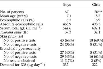
Of the overall 425 children, 136 (32%) exhibited seropositivity for T. canis antigens, 43 (21 treated and 22 not treated against T. canis) of the 136 were unavailable for follow-up because they did not attend the control visits, but the remaining 93 were adequately assessed, diagnosed and followed up during 1 year. Table 1 shows the characteristics of these patients and the demand for ICS at the beginning of the study period. The seropositivity for T. canis was more prevalent among boys than among girls. The girls and boys did not differ significantly in terms of the peripheral eosinophil count, the absolute eosinophil count, Toxocara serology, the SPT findings as concerns either inhalative or nutritive allergens, non-specific bronchial provocations, the serum total IgE level or the demand for ICS. Since there was no significant gender difference, we decided to analyse the data on boys and girls together.
Characteristics of patients with seropositivity for Toxocara canis. Values are given as means; IP, index of positivity; IP >12 was considered positive; ICS, inhaled corticosteroid. Percentages in brackets indicate prevalence of negative and positive results of skin prick tests and bronchial hyperresponsiveness among boys and girls.
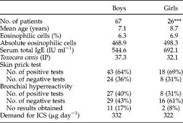
There was a significant difference in the numbers of boys and girls; ***P < 0.001.
Seroprevalence for Toxocara canis and chronic cough
The overall prevalence of T. canis seropositivity in the study population was 32%, as compared with 17% seropositivity in asymptomatic children (P < 0.01).
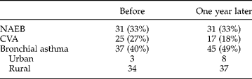
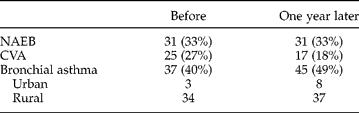
The diagnosis of bronchial asthma, NAEB or CVA was based on a history of attacks of dyspnoea and wheeze, the lung function, the BHR, the response to the bronchodilator and the susceptibility to atopy. Overall, 31 (33%) children were diagnosed with NAEB, 25 (27%) with CVA and 37 (40%) with bronchial asthma at the beginning of the study, as shown in table 2.
Distributions (number of patients (%)) of bronchial asthma, non-asthmatic eosinophilic bronchitis (NAEB) and cough variant asthma (CVA) at the beginning and at the end of the study.
Eosinophil count, total IgE level and T. canis IgG level
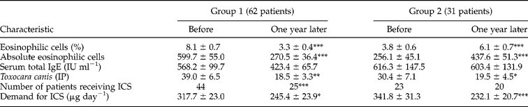
The eosinophil count in the peripheral blood formed the basis of the patient classification. The 93 children were divided into two groups: 62 (group 1) with a decreased, and 31 (group 2) with an increased peripheral eosinophilic cell count at the end of the 12-month period. Table 3 shows their laboratory characteristics and the demand for ICS at the end of the study period. There were significant decreases in the levels of antibodies against T. canis and in the demand for ICS in both groups, and in the number of patients who received ICS in group 1. No significant difference was found in either group as concerns serum total IgE levels by the end of the 12-month period.
Laboratory characteristics of patients with seropositivity for Toxocara canis in group 1 (patients with a decreased eosinophil count) and group 2 (patients with an increased eosinophil count) before and at the end of the study. IP, index of positivity; IP >12 was considered positive; ICS, inhaled corticosteroid.

Values are given as means ± SEM; significant differences between results before and at the end of the study in both groups are indicated as *P < 0.05; **P < 0.01; ***P < 0.001.
Efficacy of anthelminthics and ICS
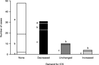
Figure 1 shows the changes in the demand for ICS by the end of the 12-month period. At the beginning of the study, ICS was regularly administered to all patients with CVA and bronchial asthma and to five patients with NAEB whose histories revealed a beneficial effect of previously occasionally administered ICS. From the end of the second or third month of the study, ICS administration was required in only two of those with NAEB and in 17 of the 25 with CVA.
Demand for inhaled corticosteroid (ICS) by patients with non-asthmatic eosinophilic bronchitis (a), cough variant asthma (b) and bronchial asthma (c) at the end of the 12-month period.

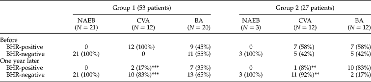
At the first presentation, spirometry, bronchial reversibility tests and non-specific bronchus provocation tests could not be performed in 13 children aged < 5 years because of a lack of cooperation. BHR was detected in 35 (44%) patients. By the end of the study period, the BHR was significantly improved in the cohort overall. A significant improvement in BHR was observed in those with CVA, in contrast with those with bronchial asthma in both groups (table 4).
Changes in bronchial hyperresponsiveness (BHR) during the 1-year period in patients with chronic cough (N=80). NAEB, non-asthmatic eosinophilic bronchitis; CVA, cough variant asthma; BA, bronchial asthma; number of patients and distribution (%) of positive and negative results are indicated in each group (group 1: patients with a decreased eosinophil count; group 2: patients with an increased eosinophil count).
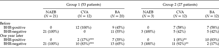
Significant differences between results before and at the end of the study in both groups are indicated by **P < 0.01; ***P < 0.001.
Ophthalmological and chest X-ray examinations of seropositive children gave negative results.
Discussion
The purpose of the present study was to investigate the possible role of Toxocara infection and the prevalence of bronchial asthma, CVA and NAEB among patients with chronic cough. Laboratory parameters such as the eosinophilic cell count, the Toxocara serology and the serum total IgE level were assessed for their value in the setting-up of the diagnosis and at the end of the 1-year period. We examined the efficacy of early treatment with ICS and anthelminthics in children with CVA and bronchial asthma, and only with anthelminthics in those with NAEB.
Seroprevalence for Toxocara and chronic cough
In 93 patients with chronic cough who were observed during 1 year, bronchial asthma was diagnosed in 40%, CVA in 27% and NAEB in 33%. Toxocara canis seropositivity was more frequent in boys than girls. Great differences are observed worldwide in the overall prevalence of Toxocara infections. Sharghi et al. (Reference Sharghi, Schantz, Caramico, Ballas, Teague and Hotez2001) found that the overall prevalence of toxocariasis in children living in urban areas was 10.2%, whereas a Brazilian study indicated a prevalence of 54.8% among children living in poor circumstances (Figueiredo et al., Reference Figueiredo, Taddei and Menezes2005). The seroprevalence in developing countries may be 86% (Thompson et al., Reference Thompson, Bundy, Cooper and Schantz1986). A Hungarian national survey (unpublished data) indicated that the seroprevalence for Toxocara was significantly lower in healthy children than in those with chronic cough enrolled in the present study.
Some authors have reported a close association between seropositivity for T. canis and bronchial asthma (Buijs et al., Reference Buijs, Borsboom, van Gemund, Hazebroek, van Dougen, van Knapen and Neijens1994; Oteifa et al., Reference Oteifa, Moustafa and Elgozamy1998; Figueiredo et al., Reference Figueiredo, Taddei and Menezes2005). The majority of our patients came from rural areas, which appeared to be an acceptable explanation for the 40% prevalence of bronchial asthma among seropositive children with chronic cough.
The published prevalence of NAEB varies between 10 and 30% (Brightling, Reference Brightling2006). Fujimara et al. (Reference Fujimara, Ogawa, Nishizawa and Nishi2003) concluded that NAEB is not a precursor of classic asthma. NAEB was diagnosed in 33% of our seropositive patients, and no members of this group became asthmatic.
Dicpinigaitis (Reference Dicpinigaitis2006) found that the one of the most important causes of cough is CVA. Lai et al. (Reference Lai, Chen, Liu, Luo, Zhong, He, Li and Zhong2006) and Todokoro et al. (Reference Todokoro, Mochizuki, Tokuyama and Morikawa2003) reported CVA in 13.6% and 75%, respectively, of their cases with chronic cough. Although CVA seems to be more prevalent in Far Eastern countries, it is also an important entity in Europe, especially as regards the differential diagnosis of children with chronic cough (Bateman et al., Reference Bateman, Hurd, Barnes, Bousquet, Drazen, FitzGerald, Gibson, Ohta, O'Byrne, Pedersen, Pizzichini, Sullivan, Wenzel and Zar2008). Recent data suggest that CVA is part of a continuum in the development of asthma symptoms and in the asthmatic inflammatory process (Abouzgheib et al., Reference Abouzgheib, Pratter and Bartter2007). Thirty to 54% of those with CVA develop typical asthma within several years (Todokoro et al., Reference Todokoro, Mochizuki, Tokuyama and Morikawa2003; Fujimara et al., Reference Fujimara, Nishizawa, Nishitsuji, Nomura, Abo and Ogawa2005). Nakajima et al. (Reference Nakajima, Nishimura, Nishiuma, Kotani, Nakata and Yokoyama2006) concluded that the majority of patients with CVA develop asthmatic symptoms after 1 year. We found that 8 of 25 patients with CVA (32%) presented asthmatic symptoms at the end of the 1-year period.
Eosinophil count, and total IgE and T. canis IgG levels
The basis of the patient classification was the eosinophil count in the peripheral blood. Sixty-two children (group 1) displayed a decreased, and 31 (group 2) an increased peripheral eosinophilic cell count. Some authors consider that there is a close positive association between toxocariasis and eosinophils and elevated IgE levels (Glickman et al., Reference Glickman, Magnaval, Domanski, Shofer, Lauria, Gottstein and Brochier1987; Figueiredo et al., Reference Figueiredo, Taddei and Menezes2005). The lower the eosinophilic cell count, the lower the T. canis IgG level and the lower the total IgE level observed in the serum in group 1. These three findings and symptoms are likely to be indicators of an improvement in the condition. The higher the eosinophilic cell count, the higher the total IgE level in those in group 2. These parameters and symptoms are likely to be predictors of a decline in the condition.
Efficacy of anthelminthics and ICS
A high helminth burden may alter the immune response and modify the prevalence of bronchial asthma. Exposure to mild or chronic Toxocara spp., leading to seropositivity, is associated with an increased prevalence of asthmatic symptoms, inducing a higher Th2 response with no activity of the regulatory system. In these cases, airway symptoms are alleviated after anthelminth treatment (Lynch et al., Reference Lynch, Lopez, Di Prisco-Fuenmayor, Hagel, Medouze, Viana, Ortega and Prato1987). The early administration of ICS is recommended for the effective treatment of CVA, with a view to restoration of the normal responsiveness of the bronchi and prevention of the progression of CVA to classic asthma (Shirahata et al., Reference Shirahata, Fujimoto, Arioka, Shouda, Kudo and Ikeda2005; Matsumoto et al., Reference Matsumoto, Niimi, Takemura, Tabuena, Yamaguchi, Matsuoka, Hirai, Muro, Ito, Mio, Chin, Nishiyama and Mishima2006). Those patients who do not take ICS for a prolonged period develop typical asthma (Fujimara et al., Reference Fujimara, Ogawa, Nishizawa and Nishi2003, Reference Fujimara, Nishizawa, Nishitsuji, Nomura, Abo and Ogawa2005). Presuming the aetiopathogenetic role of T. canis in the inflammatory process of chronic cough, we decided to treat the children not only with ICS, but also with a 1-week course of anthelminthics. Figure 1 shows that ICS therapy could be stopped after 2–3 months in 17 (68%) of the 25 with CVA. The other eight children might be candidates for bronchial asthma. Six of them needed only a decreased, and two an unchanged, or increased, dose of ICS at the end of the 1-year period. This may be explained by the fact that these children were exposed to contaminated soil in parks and playgrounds, either in urban areas or in rural communities, where infections had been acquired or re-acquired. These circumstances can cause a decline in the condition. Our experience and results support the observation that deworming therapy supplemented with ICS may prevent the transformation of CVA into asthma in the majority of seropositive patients with chronic cough.
Toxocara canis may stimulate polyclonal IgE antibodies (Buijs et al., Reference Buijs, Borsboom, van Gemund, Hazebroek, van Dougen, van Knapen and Neijens1994). IgE can bind to the membrane of bronchiolar and alveolar mast cells and release mediators that lead to inflammation of the bronchial mucosa and to asthma-like symptoms (Minvielle et al., Reference Minvielle, Niedfeld, Ciarmela, De Falco, Ghiani and Basualdo1999), which can be alleviated by the use of effective anthelminthics. The patients with bronchial asthma required the long-term use of ICS; only two children were able to stop using it within 1 year. The dose of ICS was decreased significantly in 23 of the 37 (62%) children, while 12 (32%) needed an unchanged or an increased dose of ICS at the end of the 1-year period.
Brightling et al. (Reference Brightling, Ward, Goh, Wardlaw and Pavord1999) regarded NAEB as a common cause of chronic cough. Since the aetiopathogenetic role of toxocariasis was assumed in the background of NAEB, only those patients who displayed no BHR and corresponded to the diagnostic criteria of NAEB participated in deworming treatment without ICS. Toxocara canis was the sensitizing agent identified in our cohort. It seemed that this therapy and optimal avoidance of sensitizers were sufficient; no-one needed ICS.
Toxocariasis in Hungary is mild or sporadic. Deworming therapy alleviates airway symptoms without exacerbation in patients with bronchial asthma and can be of definitive help for children with NAEB and the majority of those with CVA.