

Introduction
In recent years, thyroid cancer has become a type of malignant solid tumour with the fastest increasing incidence among many malignant tumours (Wang and Ganly, Reference Wang and Ganly2018). In the USA, thyroid cancer has become the highest incidence of cancer among people over the age of 65 years. In China, the incidence of thyroid cancer had increased approximately five-fold in the past 30 years, and ranks as the fifth leading cause of female cancer death (Janjua and Wreesmann, Reference Janjua and Wreesmann2018). At present, the mechanism of thyroid cancer is still not clear and may involve the regulation of multiple signalling pathways and relate to some specific molecules (Han and Kim, Reference Han and Kim2018; Nicholson and Yip, Reference Nicholson and Yip2018). Therefore, it is of great practical significance to clarify the pathogenesis of thyroid cancer and find effective biomarkers for the diagnosis and treatment of thyroid cancer.
Long non-coding RNAs (lncRNAs) are a type of non-coding RNA (ncRNA) with lengths of more than 200 bp. They can regulate gene expression through epigenetic regulation, transcriptional regulation and post-transcriptional regulation, and therefore participate in many biological processes such as metastasis, invasion, autophagy, proliferation and apoptosis of tumour cells (Quinn and Chang, Reference Quinn and Chang2016; Lin and Yang, Reference Lin and Yang2018). Current studies have shown that lncRNAs can be used as biomarkers for many human cancers, including thyroid cancer. For example, Zhang H et al. (2018) reported that BISPR silencing inhibited the proliferation and invasion of papillary thyroid cancer (PTC) cells, which can be used as a biomarker of tumorigenesis. In addition, lncRNA NR_036575.1 was upregulated in PTC tissues and cells, and had a certain significance for PTC diagnosis (Sun W et al., 2016). Small nucleolar RNA host gene14 (SNHG14) is a recently discovered tumour-promoting gene that is found in various tumours. Previous studies have demonstrated that SNHG14 promoted the proliferation and metastasis of rectal cancer by targeting EZH2 (Di et al., Reference Di, Weinan, Xin, Zhiwei, Xinyue, Jinxue and Mingqi2019), promoted the progression of gastric cancer through downregulating miR-145, and promoted the development of ovarian cancer through sponging miR-219a-5p (Li et al., Reference Li, Zhang and Li2020). In addition, SNHG14 can function as an oncogene in cervical and pancreatic cancers (Deng et al., Reference Deng, Chen, Cai, An, Wu, Chen, Sun, Yang, Shi and Yang2019; Zhang YY et al., Reference Zhang, Li, Xu and Shang2019a;). Overexpression of SNHG14 significantly promoted clear cell renal cell carcinoma cell migration and invasion (Liu G et al., 2017). SNHG14 may play a carcinogenic role by sponging miR-340 in non-small-cell lung carcinomas (Zhang Z et al., 2019b). SNHG14 could promote cervical cancer progression (Ji et al., Reference Ji, Wang, Bao, Yan and Ji2019). lncRNA SNHG14 promotes hepatocellular carcinoma progression by H3K27 acetylation of activated PABPC1 by PTEN signalling (Zhang H et al., 2020a). However, the expression pattern and regulatory mechanism of SNHG14 in thyroid cancer are still unknown, and there is still much room for research.
MicroRNAs (miRNAs), as a new generation molecular marker, have been shown to have clear expression specificity in many kinds of tumours and are closely related to the occurrence and development of various tumours (Kwok and Tay, Reference Kwok and Tay2017). miRNAs are a class of endogenous non-coding small molecule RNAs with a length of approximately 21–25 nucleotides and can combine with multiple target genes to influence tumour progression (Liu and Olson, Reference Liu and Olson2010; Fang et al., Reference Fang, Zhu, Dong, Zhou, Wang, Liu, Fan, Miao, Liu, Xu and Li2012). miR-93-5p is located on chromosome 11q22.1 and participates in the regulation of various tumour progressions. Sun XY et al. (Reference Sun, Han, Zhao, Cheng and Zhang2019) found that miR-93-5p significantly promoted the occurrence and development of cervical carcinoma by targeting the THBS2/MMPS signal pathway. In lung cancer, miR-93-5p could enhance the proliferation of non-small-cell lung cancer cells, and the expression level of miR-93-5p was negatively correlated with poor prognosis for patients with lung cancer (Yang W et al., 2018). Although miR-93-5p has been studied in many tumours, its biological function and regulatory mechanism in thyroid cancer have not been explored.
Our study explored, for the first time, the expression changes and regulatory mechanism of SNHG14 in thyroid cancer and revealed that SNHG14 could promote the proliferation, invasion and migration of thyroid tumour cells by targeting miR-93-5p in vivo and in vitro, providing a new strategy for the diagnosis and treatment of thyroid cancer.
Materials and methods
Cell culture
Thyroid normal follicular epithelial cells Nthy-ori3–1 and thyroid tumour cells (TPC-1, B-CPAP, SW579 and 8505C) were purchased from the Shanghai Cell Bank of the Chinese Academy of Science (Shanghai, China). Nthy-ori3-1 and 8505C cells were cultured in RPMI-1640 medium containing 10% fetal bovine serum (FBS) (Sigma), 100 U/ml of penicillin and 100 U/ml of streptomycin. The remaining cells were cultured in Dulbecco’s modified Eagle’s medium (DMEM) containing 10% FBS. All cells were cultured in an incubator containing 5% CO2 in air at 37°C. Subculture was carried out when the cell growth density reached 90% confluency. Cells in logarithmic phase were used for the follow-up experimental study.
Cell transfection
Short hairpin RNA (shRNA) against SNHG14 (sh-SNHG14-1 and sh-SNHG14-2), negative scrambled shRNA (sh-NC), miR-93-5p mimic and the corresponding NC (miR-NC) were obtained from Guangzhou RiboBio Co., Ltd. The SNHG14 overexpression vector pcDNA-SNHG14 and empty control vector pcDNA (pcDNA-NC) were constructed at Shanghai GenePharma Co., Ltd. Cells in logarithmic phase were inoculated in a 6-well plate at a density of 2 × 10⁵ per well and cultured for 24 h. These above plasmids were transfected into cells using Lipofectamine 2000 (Qiagen) strictly according to the manufacturer’s instructions. At 48 h after transfection, transfection efficiency was detected using qRT-PCR. The sequences of the shRNAs were as follows: sh-SNHG14-1: Sense 5´-GCAAAUGAAAGCUACCAAU-3´; Anti-sense 5´-AUUGGUAGCUUUCAUUUGC-3´; sh-SNHG14-2: Sense 5´-GCACAAUAUCUUUGAACUA-3´; Anti-sense 5´-UAGUUCAAAGAUAUUGUGC-3´.
Relative quantitative real-time PCR (qRT-PCR) analysis
TRIzol reagent (Sigma) was used to extract total RNA from cells according to the manufacturer’s instructions. Then, RNA was transcribed into cDNA using the Verso™ cDNA kit (ABgene, UK). qRT-PCR was performed on an ABI 7500 system using the SYBR Prime Script kit (TaKaRa Bio Inc., USA). The relative expression levels of SNHG14 and miR-93-5p were calculated using the 2−ΔΔCt method. Glyceraldehyde 3-phosphate dehydrogenase (GAPDH) or U6 were used as an internal reference. The sequences of primers were: SNHG14-F: 5´-GGGTGTTTACGTAGACCAGAACC-3´, SNHG14-R: 5´-CTTCCAAAAGCCTTCTGCCTTAG-3´; GAPDH-F: 5´-GACAAGCTTCCCGTTCTCAG-3´, GAPDH-R: 5´-GAGTCAACGGATTTGGTCGT-3´; miR-93-5p-F: 5´-GGCAGCAAAGTTCTGAGACAC-3´, miR-93-5p-R: 5´-GTGCAGGGTCCGAGGTATTC-3´; U6-F: 5´-CTCGCTTCGGCAGCACA-3´, U6-R: 5´-AACGCTTCACGAATTTGCGT-3´. Thermocycling conditions were 95°C for 10 min, followed by 40 cycles of 95°C for 15 s and 60°C for 1 min.
CCK-8 assay
Cell proliferation was detected using the CCK-8 assay. Transfected cells were seeded onto 96-well plates at a density of 2 × 10⁴ cells/well. After conventional culture for 24, 48 or 72 h, 10 μl CCK-8 solution was added to each well and the plates were incubated for 2 h. The absorbance of each well at 450 nm wavelength was detected using an Automatic Microplate Reader (Syngene, USA).
Immunofluorescence assay
The expression levels of E-cadherin (ab15148; Abcam), N-cadherin (ab12221; Abcam) and vimentin (ab45939; Abcam) were detected using an immunofluorescence assay. Transfected cells distributed in Petri dishes were washed with phosphate-buffered saline (PBS) and fixed for 10 min with 4% paraformaldehyde. Next, the cells were permeated for 10 min with 0.1% Triton X-100 and sealed for 30 min with 10% FBS. After completing the above steps, the cells were incubated with primary antibodies E-cadherin, N-cadherin and vimentin at 4°C for 24 h. Then cells were incubated with the corresponding secondary antibody against goat-anti-rabbit Cy3 (Beyotime Institute of Biotechnology, China) for 2 h. Finally, samples were incubated with 4′,6-diamidino-2-phenylindole (DAPI) for 5 min and washed twice with PBS. Cells were observed using a Bio-Rad 400 confocal microscope and the state of cells was recorded.
Immunohistochemistry assay
The tumour tissue of mice was fixed in 4% paraformaldehyde for 24 h and then dehydrated. The paraffin-embedded tissue sections were cut into 4 μm. Tissues were stained with Ki67 antibody (ab15580; Abcam). The stained tissue was observed and photographed under an optical microscope (Eclipse 80i, Nikon, Japan).
Wound-healing assay
The ability to cell migrate was detected using a wound-healing assay. Cells were seeded into 12-well plates at a density of 6 × 10⁴ per well and cultured in DMEM without FBS. When the degree of cell fusion reached 80%, a sterile 200 μl liquid transfer gun was moved vertically across the surface of the culture plate. After 24 h of routine culture, the scratches were photographed and analyzed using an optical microscope.
Transwell assay
Cells were seeded into the upper chamber of transwell inserts (Corning, Inc.) containing Matrigel (BD Biosciences) at a density of 3 × 10⁴ cells/well. DMEM containing 10% FBS was added to the lower chamber. After 24 h of routine incubation, the cells were washed with methanol and stained with 0.1% crystal violet. The state of cell invasion was photographed using an optical microscope.
Luciferase reporter assay
Binding sites for SNHG14 and miR-93-5p were predicted using starBase v.2.0 software (http://starbase.sysu.edu.cn/). In total, 4 × 10⁴ cells were seeded into 24-well plates and cultured for 24 h in a humidified atmosphere of 5% CO2 in air at 37˚C prior to dual-luciferase reporter assay. Cells were plated into 60-mm cell culture dishes and cultured for 24 h until reaching 60–80% confluency. The plasmids containing wild-type (WT) and mutant 3′UTR were obtained from Promega. SNHG14 WT or SNHG14 Mut and miR-93-5p mimic or miR-NC were transfected into cells using Lipofectamine® 2000 (Invitrogen; Thermo Fisher Scientific, Inc.) and incubated for 24 h. Following the incubation, relative firefly and Renilla luciferase activities were detected using a dual-luciferase reporter assay system (Promega Corporation).
Xenograft tumour model
All BALB/c nude mice were purchased from the Experimental Animal Center of Nanjing Medical University (Nanjing, China). TPC-1 cells that were co-transduced with SNHG14 lentivirus and miR-93-5p mimic lentivirus were injected into 4-week-old BALB/c nude mice. The mice were kept strictly according to national and/or EU guidelines under the specific-pathogen-free (SPF) conditions. The tumour volume and body weight of the mice were observed and recorded every 3 days. After 24 d, mice were killed and the tumour tissue was removed; expression of Ki67 in tumour tissue was detected using an immunohistochemical assay. All the above experimental operations were approved by the Ethics Committee of the Zhejiang Chinese Medical University.
Western blot analysis
Total protein was extracted from cells using RIPA reagent (Millipore, Billerica, MA, USA), and then protein concentration was detected using a BCA protein quantitative kit (Beyotime, Jiangsu, China). Next, 25 μg protein sample was separated using 12% SDS-PAGE and transferred to PVDF membranes for 1 h. The membranes were sealed with 5% skimmed milk for 2 h and then incubated overnight at 4°C with 1:1000 primary antibodies CDK2 (ab32147; Abcam), cyclinE1 (ab33911; Abcam), P21 (ab109520; Abcam), Bcl-2 (ab32124; Abcam), Bax (Ab32503; Abcam), cleaved-caspase3 (ab2302; Abcam), caspase3 (ab13847; Abcam), caspase9 (Ab32539; Abcam), E-cadherin (ab15148; Abcam), N-cadherin (ab12221; Abcam), vimentin (ab45939; Abcam), MMP-2 (ab92536; Abcam), MMP-9 (ab137867; Abcam) and GAPDH (ab9485; Abcam). These membranes were then incubated with 1:4000 secondary antibody anti-rabbit horseradish peroxidase (HRP)-conjugated IgG for 1 h. After washing the membrane with TBST buffer twice, an ECL luminescence reagent (Wanleibio Co., Ltd) was used to detect the immunoreactive bands. The bands were photographed using a gel imaging system.
Statistical analysis
All data in this experiment were analyzed using SPSS v.20.0 software (Chicago, IL, USA). All data were expressed as means ± standard deviation (SD). Student’s t-test was used to compare the two groups. Three groups and more than three groups were compared using a one-way analysis of variance (ANOVA). A P-value < 0.05 was considered to be statistically significant.
Results
SNHG14 was upregulated in thyroid tumour cells
The expression levels of SNHG14 in thyroid normal follicular epithelial cells Nthy-ori3–1 and thyroid tumour cells (TPC-1, BMI CPAP, SW579 and 8505C) were detected using qRT-PCR. As seen in Fig. 1, SNHG14 was upregulated in thyroid tumour cells. It is worth noting that SNHG14 expression was highest in TPC-1 cells, therefore TPC-1 cells were used in the follow-up experimental research.
Expression of SNHG14 in thyroid normal follicular epithelial cells Nthy-ori3-1 and thyroid tumour cells (TPC-1, BMI CPAP, SW579 and 8505C) detected using qRT-PCR. *P < 0.05, ***P < 0.001.

Knockdown of SNHG14 inhibited proliferation and promoted apoptosis of TPC-1 cells
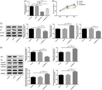
Next, SNHG14 expression was reduced by transfection with sh-SNHG14 into TPC-1 cells. Compared with the sh-NC group, expression of SNHG14 in the sh-SNHG14-1 group was lowest (Fig. 2A). Therefore, sh-SNHG14-1 was used in the follow-up experiments. CCK-8, western blot assays were performed to determine cell proliferation and apoptosis. As shown in Fig. 2B, knockdown of SNHG14 significantly inhibited cell viability compared with the sh-NC group. As shown in Fig. 2C, knockdown of SNHG14 significantly suppressed the expression of proliferation-related proteins CDK2, P21 and cyclin E1 compared with the sh-NC group. In addition, in the apoptosis experiment, knockdown of SNHG14 inhibited the expression of anti-apoptotic protein Bcl-2, whereas it promoted the expression of pro-apoptotic proteins Bax, cleaved-caspase3 and caspase9 (Fig. 2D).
Knockdown of SNHG14 inhibited proliferation of and promoted apoptosis of TPC-1 cells. (A) shRNA–SNHG14 knockdown efficiency was detected by qRT-PCR. (B) Effect of SNHG14 knockdown on the viability of TPC-1 cells was detected using CCK-8 assy. (C) Effect of SNHG14 knockdown on the expression of cell proliferation-associated proteins (CDK2, P21 and cyclin E1) in TPC-1 cells detected using western blotting. (D) Effect of SNHG14 knockdown on the expression of apoptosis-associated proteins (Bcl-2, Bax, cleaved-caspase3, caspase3 and caspase9) in TPC-1 cells detected using western blotting. **P < 0.01, ***P < 0.001.

Knockdown of SNHG14 inhibited the migration, invasion and EMT process of TPC-1 cells
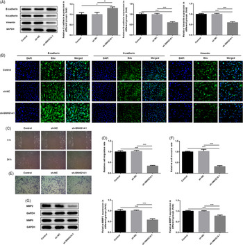
Thyroid tumour cells are very easily metastasized. The expression of epithelial–mesenchymal transition (EMT)-related proteins was measured using western blot and immunofluorescence assays. As shown in Fig. 3A, B, knockdown of SNHG14 significantly promoted the expression of E-cadherin, but decreased the expression of vimentin and N-cadherin compared with the sh-NC group. In addition, wound-healing and transwell assays were used to detect cell migration and invasion. As shown in Fig. 3C–F, knockdown of SNHG14 significantly reduced the migration and invasion of TPC-1 cells. Finally, western blot was used to detect the expression of invasion-related proteins MMP2 and MMP9. As shown in Fig. 3G, knockdown of SNHG14 significantly decreased the expression of MMP2 and MMP9 compared with the sh-NC group.
Knockdown of SNHG14 inhibited the migration, invasion and EMT process of TPC-1 cells. (A) Effect of SNHG14 knockdown on the expression of EMT-associated proteins (E-cadherin, N-cadherin and vimentin) in TPC-1 cells detected using western blotting. (B) Effect of SNHG14 knockdown on the expression of EMT-associated proteins (E-cadherin, N-cadherin and vimentin) in TPC-1 cells was measured using immunofluorescence analysis. (C, D) Effect of SNHG14 knockdown on migration of TPC-1 cells was detected using wound-healing assays. (E, F) Effect of SNHG14 knockdown on invasion of TPC-1 cells detected using transwell assays. (G) Effect of SNHG14 knockdown on the expression of MMP-related proteins (MMP-2 and MMP-9) in TPC-1 cells detected using western blotting. *P < 0.05, ***P < 0.001.

Knockdown of SNHG14 inhibited the growth of TPC-1 cells in mice
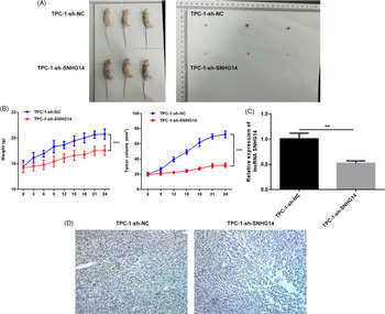
Next, the effect of SNHG14 on tumour growth in mice was detected using the xenograft tumour assay. As shown in Fig. 4A, 24 d after inoculation, anatomical observation of the tumour-bearing mice showed that knockdown of SNHG14 significantly reduced the xenograft tumour volumes. As shown in Fig. 4B, knockdown of SNHG14 significantly decreased the weight and tumour volume of mice compared with normal mice. Next, knockdown of SNHG14 significantly suppressed the expression of SNHG14 in tumour tissues (Fig. 4C). The results of immunohistochemistry showed that knockdown of SNHG14 significantly inhibited the expression of Ki67 compared with normal tissues (Fig. 4D).
Knockdown of SNHG14 inhibited the growth of TPC-1 cells in mice. (A) Gross observation of xenograft tumour size after transfection with sh-SNHG14. (B) Effect of SNHG14 knockdown on the body weight and tumour volume in BALB/c nude mice. (C) Effect of SNHG14 knockdown on the expression of SNHG14 in tumour tissue sections detected using qRT-PCR. (D) Effect of SNHG14 knockdown on the expression of Ki67 in tumour tissue sections analyzed by immunohistochemical staining. **P < 0.01, ***P < 0.001.

miR-93-5p was the target gene of SNHG14
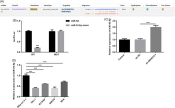
To further study the regulatory mechanism of SNHG14 in TPC-1 cells. miR-93-5p was selected as the target gene of SNHG14 using starBase v.2.0 software (http://starbase.sysu.edu.cn/browseNcRNA.php) (Fig. 5A). The results of the dual-luciferase reporter assay showed that the luciferase activity of WT group was significantly lower than that of miR-NC group, but there was no change in the Mut group, which indicated that SNHG14 could target miR-NC (Fig. 5B). In addition, knockdown of SNHG14 significantly increased the expression of miR-93-5p in TPC-1 cells (Fig. 5C) and the expression of miR-93-5p was downregulated in thyroid tumour cells (Fig. 5D).
miR-93-5p was the target gene of SNHG14. (A) Binding sequence between SNHG14 and miR-93-5p was detected using the starBase database. (B) Relationship between SNHG14 and miR-93-5p was analyzed using a luciferase reporter assay. (C) Effect of SNHG14 knockdown on the expression of miR-93-5p was detected using qRT-PCR. (D) Expression of miR-93-5p in thyroid normal follicular epithelial cells Nthy-ori3-1 and thyroid tumour cells (TPC-1, BMI CPAP, SW579 and 8505C) detected using qRT-PCR. ***P < 0.001.

miR-93-5p reversed the SNHG14-mediated promotion of proliferation and inhibition of apoptosis in TPC-1 cells
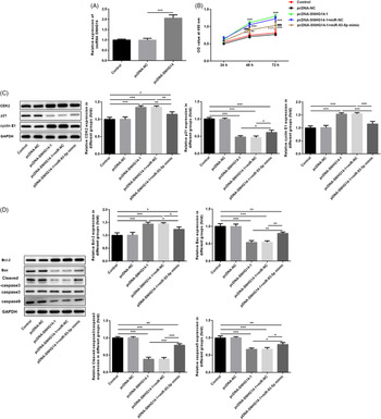
Next, the role of SNHG14 in TPC-1 cells by targeting miR-93-5p was determined. The expression of SNHG14 in TPC-1 cells was increased by SNHG14 overexpression vector construction and transfection (Fig. 6A). Results of CCK-8 and proliferation-related protein detection showed that miR-93-5p overexpression significantly inhibited the promoting effect of overexpression of SNHG14 on cell proliferation (Fig. 6B, C). Detection of apoptosis-related proteins showed that, compared with the PCDNA-SNHG14-1 + miR-NC group, miR-93-5p overexpression significantly promoted the expression of Bax, cleaved-caspase3 and caspase9, but decreased the expression of Bcl-2 (Fig. 6D), indicating that miR-93-5p overexpression reduced the inhibitory effect of SNHG14 overexpression on apoptosis of TPC-1 cells.
miR-93-5p reversed the SNHG14-mediated promotion of proliferation and inhibition of apoptosis in TPC-1 cells. (A) Efficiency of SNHG14 overexpression was determined using qRT-PCR. (B) Cell viability of TPC-1 was detected using CCK-8 assay. (C) Expression of cell proliferation-associated proteins (CDK2, P21 and cyclin E1) in TPC-1 cells detected using western blotting. (D) Expression of apoptosis-associated proteins (Bcl-2, Bax, cleaved-caspase3, caspase3 and caspase9) in TPC-1 cells detected using western blotting. *P < 0.05, **P < 0.01, ***P < 0.001.

miR-93-5p reversed the SNHG14-mediated promotion of migration, invasion and EMT process in TPC-1 cells
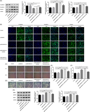
The results of EMT-related protein detection showed that, compared with PCDNA-SNHG14-1 + miR-NC group, miR-93-5p overexpression significantly inhibited the expression of N-cadherin and vimentin, but increased the expression of E-cadherin (Fig. 7A, B). The results of wound-healing and transwell assays showed that miR-93-5p overexpression partially reversed the promoting effects of SNHG14 overexpression on migration and invasion in TPC-1 cells (Fig. 7C–F). Finally, compared with the PCDNA-SNHG14-1 + miR-NC group, miR-93-5p overexpression significantly suppressed the promoting effect of overexpression of SNHG14 on the expression of MMP2 and MMP9 (Fig. 7E).
miR-93-5p reversed the SNHG14-mediated promotion of migration, invasion and EMT process in TPC-1 cells. (A) Expression of EMT-associated proteins (E-cadherin, N-cadherin and vimentin) in TPC-1 cells detected using western blotting. (B) Expression of EMT-associated proteins (E-cadherin, N-cadherin and vimentin) in TPC-1 cells was measured using immunofluorescence analysis. (C, D) Migration of TPC-1 cells was detected using wound-healing assays. (E, F) Invasion of TPC-1 cells was detected by transwell assay. (G) Expression of MMP-related proteins (MMP-2 and MMP-9) in TPC-1 cells was detected using western blotting. *P < 0.05, **P < 0.01, ***P < 0.001.

miR-93-5p reversed the SNHG14-mediated promotion of the growth of TPC-1 cells in mice
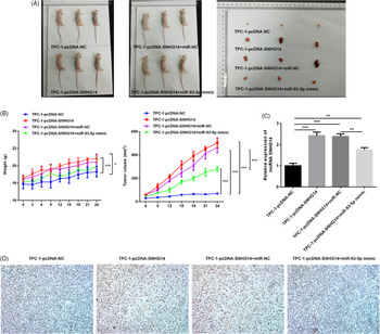
Next, a xenograft tumour assay was conducted to determine the effect of SNHG14 on tumour growth in mice by targeting miR-93-5p. As shown in Fig. 8A, 24 d after inoculation, anatomical observation of the tumour-bearing mice showed that overexpression of SNHG14 could significantly increase xenograft tumour volumes compared with normal mice. miR-93-5p overexpression significantly alleviated the promoting effect of overexpression of SNHG14 on xenograft tumour volumes. As shown in Fig. 8B, overexpression of SNHG14 significantly increased body weight and tumour volumes, while miR-93-5p overexpression reversed these above changes. In addition, results of tumour tissue detection showed that miR-93-5p overexpression significantly suppressed the expression of SNHG14 and Ki67 compared with normal tissue (Fig. 8C, D).
miR-93-5p reversed the SNHG14-mediated promotion of the growth of TPC-1 cells in mice. (A) Gross observation of xenograft tumour size after transfection with sh-SNHG14 and the miR-93-5p mimic. (B) Effect of SNHG14 overexpression together with miR-93-5p overexpression on the body weight and tumour volume in BALB/c nude mice. (C) Effect of SNHG14 overexpression together with miR-93-5p overexpression on the expression of SNHG14 in tumour tissue sections detected using qRT-PCR. (D) Effect of SNHG14 overexpression together with miR-93-5p overexpression on the expression of Ki67 in tumour tissue sections analyzed using immunohistochemical staining. *P < 0.05, **P < 0.01, ***P < 0.001.

Discussion
lncRNAs are a large class of non-coding transcriptions that interact with miRNA and play an important role in the occurrence and development of tumours. lncRNA can bind miRNA and therefore reduce the inhibition of miRNA on its target genes (Maldotti et al., Reference Maldotti, Incarnato, Neri, Krepelova, Rapelli, Anselmi, Parlato, Basile, Dettori, Calogero and Oliviero2016). Studies showed that abnormal lncRNA expression is closely related to the occurrence and metastasis of thyroid cancer (Yang J et al., 2014), but the mechanism of action of some lncRNAs in the process of thyroid occurrence and metastasis has not been fully elucidate.
SNHG14 plays a role as an oncogenic gene in various tumours. In clear cell renal cell carcinoma, overexpression of SNHG14 significantly promotes cell migration and invasion (Liu G et al., 2017). In non-small-cell lung carcinomas, SNHG14 may play a carcinogenic role by sponging miR-340 (Zhang Z et al., 2019b). In cervical cancer, SNHG14 could promote cervical cancer progression by regulating the miR-206/YWHAZ axis (Ji et al., Reference Ji, Wang, Bao, Yan and Ji2019). In ovarian cancer, SNHG14 expression is markedly enhanced and silenced SNHG14 hampers cell proliferation, migration and invasion in ovarian cancer (Li et al., Reference Li, Zhang and Li2020). SNHG14 may play a carcinogenic role in gastric cancer (Liu Z et al., 2018). In colorectal cancer, overexpression of SNHG14 restrains the proliferation, migration, and invasion of colorectal cancer cells (Zhang W et al., 2020c). Zhang P et al. (2020b) demonstrated that SNHG14 was underexpressed in endometrial cancer. In the present study, SNHG14 was upregulated in thyroid tumour cells, and knockdown of SNHG14 significantly inhibited the proliferation, migration, invasion, and EMT progression in TPC-1 cells, and promoted cell apoptosis.
Cyclin E1, P21 and CDK2 are proteins associated with cell proliferation. Bcl-2 and Bax are apoptosis-related proteins. Cyclin E1 is the key protein in cell cycle transformation from G1 phase to S phase, and its decreased expression activity could cause cell cycle stagnation. P21 could induce cell cycle arrest. CDK2 could promote DNA synthesis and induce cell cycle arrest from G1 phase to S phase. Bcl-2 and Bax proteins have anti-apoptotic and pro-apoptotic effects, respectively (Kaufmann and Earnshaw, Reference Kaufmann and Earnshaw2000; Zhang P et al., 2016). In addition, MMP-2 and MMP-9 belong to a class of matrix metalloproteinases that can degrade the extracellular matrix and increase tumour migration and invasion (Zhang X et al., 2020d). EMT is a process of cytoskeletal recombination and gradual transformation of epithelial cells into mesenchymal cells, and plays an important role in cell metastasis. During this process, the decrease in epithelial markers (E-cadherin) and the increase in mesenchymal markers (N-cadherin and vimentin) resulted in the stronger migration and motility of cancer cells (Zhang and Weinberg, Reference Zhang and Weinberg2018). Our results showed that knockdown of SNHG14 effectively inhibited the expression of cyclin E1, CDK2, Bcl-2, MMP2, MMP9, vimentin and N-cadherin, but enhanced the expression of P21, E-cadherin, Bax, cleaved-caspase3 and caspase9, thereby inhibiting cell activity and related biological functions. In vivo studies showed that SNHG14 knockdown could also effectively reduce tumour growth and its function as a tumour suppressor.
To further study the regulatory mechanism of SNHG14 in thyroid cancer, miR-93-5p was screened as the target gene of SNHG14 using the starBase database. mir-93-5p is abnormally expressed in many cancers except thyroid malignancies. For instance, in cervical cancer, miR-93-5p significantly promoted the development of cervical cancer by regulating THBS2 (Sun XY et al., Reference Sun, Han, Zhao, Cheng and Zhang2019). In lung cancer, miR-93-5p effectively enhanced the activity of non-small-cell lung cancer (Yang W et al., Reference Yang, Bai, Liu, Wang, Zhao, Che and Zhang2018). However, no studies have shown the specific role of miR-93-5p in thyroid cancer. In this study, miR-93-5p was downregulated in thyroid tumour cells. Luciferase assay results showed that SNHG14 could negatively target miR-93-5p. Overexpression of miR-93-5p effectively reduced the promotion effect of SNHG14 on thyroid cancer progression.
In conclusion, knockdown of SNHG14 could effectively inhibit the proliferation, migration, invasion and EMT process of thyroid tumour cells, and may be related to the negative targeting of miR-93-5p. Our results may provide a valuable reference for the clinic treatment of thyroid cancer. However, due to the limitation of time and funds, we only explored the effect of lncRNA SNHG14 and miR-93-5p on thyroid cancer cells. Further research is needed to focus on exploring the role of lncRNA SNHG14 and miR-93-5p in animal models, clinical samples and signalling pathways to further support the findings from our study.
Acknowledgements
Not applicable.
Funding
No funding was received.
Availability of data and materials
All data generated or analyzed during this study are included in this published article.
Competing interests
The authors declare that they have no competing interests.
Ethics approval and consent
This present study was performed based on the principles expressed in the Declaration of Helsinki. All animal experiments were approved by the Ethics Committee of the Zhejiang Chinese Medical University and conducted in accordance with the National Institutes of Health (NIH) Guide for the Care and Use of Laboratory Animals.