Introduction
Physical dormancy is determined by the impermeability of seed coats to water, which is caused by the presence of one or more layers of Malpighian cells that are tightly packed together and impregnated with water-repellant substances, such as lignin, callose and wax (Baskin, Reference Baskin2003; Smith et al., Reference Smith, Wang, Ben and Msanga2003). Seeds of species with physical dormancy are known in 17 families of angiosperms (Gama-Arachchige et al., Reference Gama-Arachchige, Baskin, Geneve and Baskin2010) and several types of specialized structures (‘water gaps’) have been found in 12 of the 17 families. The family Fabaceae has a large number of species with physically dormant seeds (Villers, Reference Villers1972; Morrison et al., Reference Morrison, McClay, Porter and Rish1998) and three types of water gaps are recognized: the lens, hilar slit (Baskin et al., Reference Baskin, Baskin and Li2000) and micropyle (Hu et al., Reference Hu, Wang, Wu, Nan and Baskin2008, Reference Hu, Wang, Wu and Baskin2009). Events in nature, such as high and alternating temperatures, are known to break physical dormancy effectively in some species, allowing water to enter through a gap (Fenner and Thompson, Reference Fenner and Thompson2005). Previous studies of species from several environments have shown that alternating temperatures between 15 and 35°C break physical dormancy of species that grow on dunes in Japan (Kondo and Takahashi, Reference Kondo and Takahashi2004), of the agricultural weed Ipomoea lacunosa (Jayasuriya et al., Reference Jayasuriya, Baskin, Geneve and Baskin2007) and in tree species that grow in gaps in a rain forest in Mexico (Vázquez-Yanes and Orozco-Segovia, Reference Vázquez-Yanes and Orozco-Segovia1982), in non-climax tree species from the evergreen Atlantic forest (Souza et al., Reference Souza, Voltolini, Santos and Paulilo2012) and a semi-deciduous forest in Brazil (Abdo and Paula, Reference Abdo and Paula2006). When exposed to alternating temperatures of 74/15°C and 60/15°C, the highest temperatures broke physical dormancy of seeds of species from an arid region of Australia (Quinlivan, Reference Quinlivan1966).
Among the Fabaceae, studies of physical dormancy of seeds have mainly focused on species within the subfamily Faboideae because this group contains important agricultural legumes (Baskin and Baskin, Reference Baskin and Baskin2001). There are fewer studies on the subfamily Caesalpinoideae and studies on effects of alternating temperatures to break physical dormancy are rare in seeds of tree species from Brazilian forest ecosystems, such as the Atlantic rain forest, one of the most threatened ecosystems in Latin America (Myers et al., Reference Myers, Mittermeier, Mittermeir, Fonseca and Kent2000).
Knowledge about the ecology of seed germination of species from the Atlantic rain forest is important for conservation programmes. Therefore, the goal of this work was to study how physical dormancy is broken in two tree species from the Atlantic rain forest that belong to the Caesalpinoideae. The taxa chosen, Cassia leptophylla and Senna macranthera, are non-climax tree species that were selected because of their commercial and ecological importance and because mature seeds of these taxa were available during the period of the experiment. These species are commercially valuable because of their wood, and C. leptophylla is also used in the production of honey and S. macranthera in the regeneration of degraded areas (Carvalho, Reference Carvalho2006). This work focused on the following: (1) the structure and chemical composition of the integument of the seeds; (2) the structure through which water enters the seed; and (3) whether the breaking of the physical dormancy in seeds is due to the high or the alternating temperature applied, similar to conditions required for other species of tropical rain forests.
Materials and methods
Seed collection
Fruits of Cassia leptophylla Vogel and Senna macranthera var. macranthera (DC. ex Collad.) H.S. Irwin & Barneby were collected from trees growing in Bosque do Alemão, in the city of Curitiba, Paraná, Brazil (25°24′22.89″S, 49°17′8.77″W) in July 2009. Seeds were removed from the fruits and stored in glass bottles, at room temperature, until they were used. The average mass of each seed was 0.17 g for C. leptophylla and 0.041 g for S. macranthera.
Structural analysis of the seed
For the morphological analysis, seeds were observed using a stereoscopic microscope (Leica EZ4D, Leica Microsystems, Wetzlar, Germany) and images were taken with a Sony digital camera. For analysis of the seed coats, seeds were adhered to a wooden block, with Super Bonder® (Henkel ltda., Itapevi, São Paulo, Brasil), and the hilar and extra-hilar regions were longitudinally and transversely sectioned (40 μm thick) with a sliding microtome (Micron HM400, Micron, Boise, Idaho, USA).
Some sections were prepared with only water (control) and others were exposed to the following histochemical reagents: Sudan IV for suberine, cutin, oils and waxes (Costa, Reference Costa1982); acid phloroglucinol or iron chloride for lignin (Costa, Reference Costa1982); toluidine blue for polychromatic reactions to lignin and cellulose (O'Brien et al., Reference O'Brien, Feder and McCully1965); and ruthenium red for pectic substances (Gerlach, Reference Gerlach1984). Samples were examined under a light microscope (Leica DMLS MPS 30) and images were taken with a Sony digital camera.
To verify the presence of callose in the seeds, sections were immersed in 0.05% aniline blue with a 0.1 M potassium phosphate buffer, at pH 8.3 (Ruzin, Reference Ruzin1951). As a control, some sections were immersed only in the potassium phosphate buffer. Sections were observed using an Olympus BX41 microscope (Olympus Corp., Tokyo, Japan), with a mercury vapour lamp (HBO 100) and a blue epifluorescence filter (UMWU2), at an excitation wavelength of 330–385 nm and a 420 nm emission wavelength. Images were taken with a Q-imaging digital camera (3.3 megapixel QColor 3C) and the software Q-captures Pro 5.1 (Q-Imaging, Surrey, British Columbia, Canada).
Some seeds were subjected to the process of cell dissociation (Franklin, Reference Franklin1945, modified by Kraus and Arduin, Reference Kraus and Arduin1997). Dissociated samples were stained with toluidine blue, mounted on slides with water and cover slips, and examined under a light microscope.
The surface of the hilar region was examined with a scanning electron microscope (SEM) to verify the effect of thermal scarification. Seeds were immersed in water for 2 min at 96°C and were then cut in half (the endosperm was removed, because it is rich in oleaginous substances). The sections of the treated and untreated seeds were then stored in a glass bottle, with silica gel, to dry for a month. The dried samples were adhered to aluminium supports with double-sided carbon tape, and sputter coated (using a Leica EM 500 SCD) with 20 nm of gold. The samples were analysed using a Jeol XL30 SEM. Five thermally scarified seeds and five untreated seeds, for each species, were analysed.
Localization of water entrance in the seeds after thermal scarification
To verify the path of water entry by treated seeds, a dye was used, as proposed by Jayasuriya et al. (Reference Jayasuriya, Baskin, Geneve and Baskin2007). For this, 20 seeds of each species were submitted to thermal scarification in water, for 2 min at 96°C. The 20 scarified seeds and 20 non-scarified seeds (control) were then immersed in an aqueous solution of 1% aniline blue. The scarified and non-scarified seeds were removed after intervals of 15 min, 30 min, 1 h, 2 h and 3 h of imbibition for analysis, four seeds were removed per interval for each treatment. For this analysis, the hilar and extra-hilar regions of the seeds were sectioned longitudinally. The sections were analysed using a light microscope (Leica EZ4D and Leica MPS 30 DMLS) and imaged with a Sony digital camera.
Seed germination and breaking of physical dormancy by alternating and high temperatures
Intact seeds were sterilized by immersing them in 5% sodium hypochlorite for 5 min, followed by washing them three times in distilled water. The seeds were then placed in transparent plastic boxes on two sheets of filter paper moistened with distilled water. The seeds were stored at 15, 20, 25, 30 or 35°C and at alternating temperatures of 35/25, 30/20, 25/15 and 30/15°C with a photoperiod of 12 h at high temperature and 12 h in the dark at low temperature. Germinated seeds were counted at 2-d intervals for 40 d, when the percentage of germination stabilized. Four boxes, each with 25 seeds, were utilized for each temperature level.
For high temperature treatments, seeds were exposed to 50°C for 4 h in an oven, for a single day or for seven consecutive days, in boxes with water (wet heat) or without water (dry heat). After the heat treatment, the seeds were incubated at 25°C in boxes with water for 3 or 7 d, according to the experiment. For the treatment that lasted 7 d a control was done with seeds not exposed to 50°C. The mass of seeds was measured after 24 and 72 h or daily, depending on the experiment. Four boxes, each with 25 seeds, were utilized for the heat treatments, except in the case of S. macranthera subjected to treatment at 50°C for 7 d, where 35 seeds were utilized for each box.
Results
Structural analysis of the seed
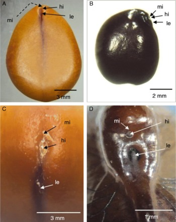
The hilar region of C. leptophylla and S. macranthera seeds consists of the micropyle, hilum and lens (Fig. 1). It is apical in C. leptophylla (Fig. 1A) and subapical in S. macranthera (Fig. 1B).
Hilar region of Cassia leptophylla (A, C) and Senna macranthera seeds (B, D) observed by stereoscopic microscopy. (A, B) External view of the seed; (C, D) detail of the hilar region of the seed; hi, hilum; le, lens; mi, micropyle. (A colour version of this figure can be found online at http://www.journals.cambridge.org/ssr)

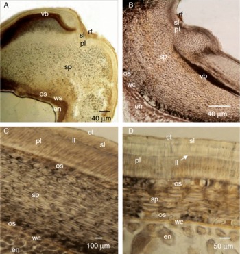
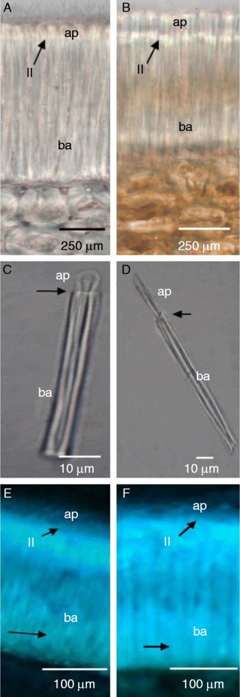
The testa of C. leptophylla and S. macranthera seeds consists of a palisade layer, osteosclereids and sclerified parenchyma (Fig. 2). The palisade layer consists of compactly disposed macrosclereids. In the hilar region this palisade layer is delimited externally by a subcuticular layer and remaining funicular tissue (Fig. 2A, B), and in the extra-hilar region, adjacent to the palisade layer, there is a subcuticular layer with transverse ribs and, more externally, a cuticular layer (Fig. 2). The macrosclereids are elongated and there is a refractive line (called a light line) that runs across the entire palisade layer (Fig. 3). This line distinguishes the apical and basal portions of the macrosclereids (Fig. 3C, D). In the macrosclereids, the apical and basal portions (not the region that corresponds to the light line) showed a positive reaction for lignin when stained with toluidine blue (Fig. 3E, F). The osteosclereids, which are cells with thickened walls, showed no positive reaction for lignin.
Photomicrography of longitudinal sections of the seed coat of Cassia leptophylla (A, C) and Senna macranthera (B, D) observed by light microscopy. (A, B) Hilar region; (C, D) extra-hilar region; ct, cuticle; en, endosperm; ll, lucid line; os, osteosclereids; pl, palisade layer; rf, remaining funicle; sl, subcuticular layer; sp, sclerified parenchyma; vb, vascular bundle; wc, white cells. (A colour version of this figure can be found online at http://www.journals.cambridge.org/ssr)

Photomicrography of the palisade layer longitudinal sections of the seed extrahilar region of Cassia leptophylla (A, E) and Senna macranthera (B, F). (A, B, E, F) Details of the palisade layer: light microscopy (A, B); fluorescence microscopy (E, F); arrows indicate the presence of callose. (C, D) Dissociated macrosclereids. ll, Lucid line; ap, apical portion; ba, basal portion. (A colour version of this figure can be found online at http://www.journals.cambridge.org/ssr)

The sclerified parenchyma was represented by a greater number of layers in the hilar region (Fig. 2A, B) than in extra-hilar regions (Fig. 2C, D). In extra-hilar regions, however, the number of layers was higher in C. leptophylla (Fig. 2C) than in S. macranthera (Fig. 2D). This tissue consists of cells with thickened walls, which showed no positive reaction for the presence of lignin.
Internally, the tissues that comprise the testa of the seeds have a thin layer of white cells (Fig. 2C, D), which constitute the tegmen. These cells have thin walls, and are very clear and almost imperceptible; they are horizontally elongated and there is a distinct cuticle between these cells and the testa.
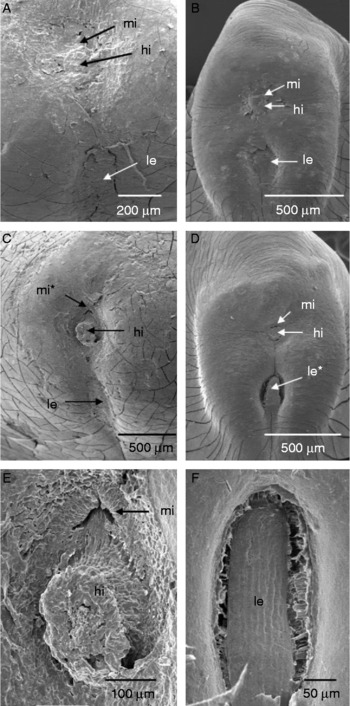
The ultrastructural analysis of the hilar region of C. leptophylla (Fig. 4A) and S. macranthera seeds (Fig. 4B) revealed that in all non-treated seeds the tissues of this region remained intact with no disruptions. However, when thermally scarified with hot water, the micropyle in most C. leptophylla seeds (Fig. 4C) and the lens in most S. macranthera seeds (Fig. 4D) changed. In C. leptophylla (Fig. 4E), the micropyle was opened more, and in S. macranthera (Fig. 4F) the lens region ruptured.
Electron micrography of the seeds of Cassia leptophylla (A, C, E) and Senna macranthera (B, D, F) showing the hilar region: (A, B) without thermal scarification; (C, D) with thermal hot-water scarification, structural changes are observed that enable water entry (*); (E) detail showing changes in the micropyle; (F) detail of the lens with the outline showing disruption. hi, Hilum; le, lens; mi, micropyle.

Localization of water entrance in the seeds after thermal scarification
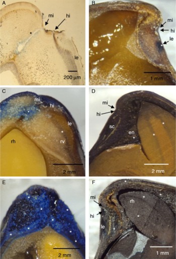
In dormant seeds of C. leptophylla and S. macranthera without thermal scarification, aniline blue did not enter the seeds after soaking them in the dye for 1–3 h; there was no penetration of seed coat, except for the subcuticular layer.
All of the thermally scarified seeds that were soaked in aniline blue for 15 min were bluish around the micropylar canal in C. leptophylla (Fig. 5A) and in the region of the lens in S. macranthera (Fig. 5B). In C. leptophylla, after 30 min the colour increased in the peripheral tissues of the micropylar canal and also in the hilar region and throughout the vascular bundle of raphe (Fig. 5C). In S. macranthera 30 min of soaking resulted in stained integuments and endosperm, and dye was absorbed by the radicle, where a bluish colour could be seen in the provascular tissue (Fig. 5D). After an hour of soaking the two species (Fig. 5E, F), the seed coats were completely stained. In S. macranthera, all of the internal structures of the seed were stained (Fig. 5F) and in C. leptophylla (Fig. 5E) there was no infiltration of the dye in the endosperm and embryo, even when subjected to a period of up to 3 h of immersion. In the extra-hilar region of C. leptophylla, the palisade layer was stained after 15 min of soaking and after 30 min the aniline blue advanced to the sclerified parenchyma, but did not stain the white cells; these, as well as the endosperm and cotyledons, did not stain after 1 h. In S. macranthera, initially only the palisade layer was stained, but after half an hour the stain had advanced through the other integuments, and after an hour of soaking the endosperm was stained. After 1 h, cracks were observed along the seed coat in both species.
Longitudinal sections of Cassia leptophylla (A, C, E) and Senna macranthera (B, D, F) seeds after thermal scarification, observed by light microscopy (A) and stereoscopic microscopy (B–F), showing the intake of aniline blue (*) in the hilar region. (A, B) After 15 min of soaking in dye; (C, D) after 30 min of soaking in dye; (E, F) after 1 h of soaking in dye; en, endosperm; hi, hilum; le, lens; mi, micropyle; rh, root–hypocotyl axis; rv, raphe vascular bundle; sc, seed coat. (A colour version of this figure can be found online at http://www.journals.cambridge.org/ssr)

Seed germination and breaking of physical dormancy by alternating and high temperatures
Regardless of the maximum and minimum limit of test temperatures and the interval between the alternating temperatures, there were no differences in germination percentages of C. leptophylla or S. macranthera seeds subjected to alternating temperatures between 15 and 35°C, but the results indicate a tendency for better germination in the range 20–25°C.
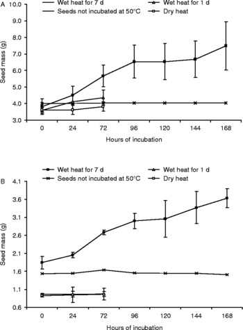
On the other hand, exposure of seeds on a moist substrate to 50°C for 4 h broke seed hardcoatedness, and after 7 d treated seeds had significantly greater mass than the controls (P ≤ 0.05). Seeds of C. leptophylla were more sensitive to wet heat at 50°C than to dry heat (P ≤ 0.05), while the opposite was true for seeds of S. macranthera (P ≤ 0.05) (Fig. 6A, B).
Imbibition curves for seeds of Cassia leptophylla (A) and Senna macranthera (B) after being subjected to 4 h in an oven at 50°C, in plastic boxes with (wet heat) and without (dry heat) water, and then transferred to 25°C with water, for 72 or 168 h. Bars indicate standard deviation. Each point represents 25 seeds, or 35 in the case of S. macranthera subjected to heat for 7 d.

Discussion
Structural analysis of the seed
Structural analysis of C. leptophylla and S. macranthera seeds confirmed features found in Caesalpinoideae. For example, the hilum was between the micropyle and the lens (Gunn, Reference Gunn1991) and the palisade layer was composed of elongated macrosclereids that were tightly packed together (Baskin, Reference Baskin2003; Smith et al., Reference Smith, Wang, Ben and Msanga2003). Callose in the light line has been reported in the subfamilies Faboideae (Bhalla and Slaterry, Reference Bhalla and Slaterry1984; Bevilacqua et al., Reference Bevilacqua, Fossati and Dondero1987; Serrato-Valenti et al., Reference Serrato-Valenti, Cornara, Ferrando and Modenesi1993; Ma et al., Reference Ma, Cholewa, Mohamed, Peterson and Jzen2004), Mimosoideae (Serrato-Valenti et al., Reference Serrato-Valenti, De Vries and Cornara1995) and Caesalpinioideae (Mosele et al., Reference Mosele, Hansen, Schulz and Martens2011). The present work also found callose in the light line of the two species of Caesalpinoideae, as well as the presence of lignin in the palisade layer. Lignin is present in several species of Fabaceae (Krzyzanowski et al., Reference Krzyzanowski, Neto, Mandarino and Kaster2008; Torres et al., Reference Torres, Santos, Schiavinato and Maldonado2009) but it is not an obligate feature and has been reported to be absent in species of Faboideae (Bevilacqua et al., Reference Bevilacqua, Fossati and Dondero1987; Serrato-Valenti et al., Reference Serrato-Valenti, Cornara, Ferrando and Modenesi1993), Mimosoideae (Serrato-Valenti et al., Reference Serrato-Valenti, De Vries and Cornara1995) and Caesalpinioideae (Souza, Reference Souza1982). Although the presence of lignin in the testa suggests impermeability to water, lignin is also ecologically important because it protects the seed against predation (Souza and Marcos Filho, Reference Souza and Marcos Filho2001).
Identification of the site where water enters the seeds
The lens is considered to be the initial site where water enters Fabaceae seeds after physical dormancy is broken (Baskin et al., Reference Baskin, Baskin and Li2000). This is true for several Fabaceae species, such as Schizolobium parahyba (Vell.) SF Blake, Caesalpinioideae (Souza et al., Reference Souza, Voltolini, Santos and Paulilo2012); Albizia lophanta (Willd.) Benth., Mimosoideae (Dell, Reference Dell1980), Sesbania punicea (Cav.) Benth., Faboideae (Manning et al., 1987) and for S. macranthera analysed in the present study. For C. leptophylla, however, water entered through the micropyle. Other works have reported the entry of water at sites other than the lens. For example, Rangaswany and Nandakumar (Reference Rangaswany and Nandakumar1985) reported the hilum and micropyle for Rhynchosia minima (L.) DC. (Faboideae) and Hu et al. (Reference Hu, Wang, Wu and Baskin2009) reported the hilum for Vigna oblongifolia A. Rich. (Faboideae), as the structures responsible for water absorption, and Bhattacharya and Saha (Reference Bhattacharya and Saha1990) demonstrated that water absorption in seeds of Cassia species was related to the opening of the micropyle (beside the lens).
Seed germination and breaking of physical dormancy by alternating and high temperatures
Studies with species from tropical forests (Vázques-Yanes and Orozco-Segovia, Reference Vázquez-Yanes and Orozco-Segovia1982; Souza et al., Reference Souza, Voltolini, Santos and Paulilo2012), agricultural areas growing cotton (Jayasuriya et al., Reference Jayasuriya, Baskin, Geneve and Baskin2007) and dunes (Kondo and Takahashi, Reference Kondo and Takahashi2004) have shown that alternating the temperature between approximately 15 and 20°C (minimum) and 30 and 35°C (maximum) effectively broke physical dormancy of seeds on a wet surface. However, in the present work these temperatures were not effective in breaking the physical dormancy of the two tropical tree species. Tropical forests have a large number of ecological niches that are occupied by different species, such as those that specialize in growing in big, medium or small gaps, or those that occupy the understorey (Deslow, Reference Deslow1980; Krischer, Reference Krischer2011). For this reason, the appropriate temperature required to break physical dormancy can be quite different and depends on the temperatures that occur in each ecological niche. In the case of C. leptophylla and S. macranthera, the effective temperature to break seed hardcoatedness was 50°C; presumably physical dormancy is indeed broken by a high temperature treatment, but since subsequent germination has not been followed, further experimentation on the matter is required to settle this issue. In a study still in process, the temperature measured in the centre of a large gap, in the Brazilian Atlantic rain forest, was around 50°C (data not published). C. leptophyla only occurs in south-eastern and southern Brazil in the secondary successional stages of the Atlantic rain forest and Atlantic semi-deciduous forest (Carvalho, Reference Carvalho2006), the two major vegetation types of the Atlantic forest (Morellato et al., Reference Morellato, Talora, Takahasi, Bencke, Romera and Zipparro2000). The Atlantic rain forest experiences a warm and wet climate without a dry season; and the Atlantic semi-deciduous forest experiences two seasons: a tropical season, with an intense rainy period in the summer preceded by accentuated droughts; and a subtropical season, which lacks a dry period but the plants experience a physiological drought provoked by a cold winter with temperatures that can be as low as 15°C (Velloso et al., Reference Velloso, Rangel Filho and Lima1991). In these two types of vegetation, there are forest gaps that reach the temperatures required to break the physical dormancy of C. leptophylla. On the other hand, S. macranthera has a wide distribution, occurring in humid forests to semi-arid areas at different elevations and successional phases (Carvalho, Reference Carvalho2006), and in these regions the high-temperature breaking of physical dormancy may occur.
Acknowledgements
The authors thank the Coordenação de Aperfeicoamento do Ensino Superior (CAPES) for their financial support and Tassiane T. Pinto for her aid in the experiments on breaking of physical dormancy of the seeds.