Introduction
Retiform haemangioendothelioma was first described only recently, in 1994.Reference Calonje, Fletcher, Wilson-Jones and Rosai1 It is a vascular neoplasm with a histopathological appearance intermediate between that of haemangioma and angiosarcoma, and thus represents a challenging, and potentially missed, diagnosis.
We present just such a misdiagnosed case of retiform haemangioendothelioma, previously managed as lymphangioma and treated with surgical excision. We describe the classical histological presentation of retiform haemangioendothelioma, as found in this case.
At presentation to our service, the patient had an extensive tumour, and subsequently required radical surgical excision and adjuvant radiotherapy, with associated cosmetic co-morbidity.
Case report
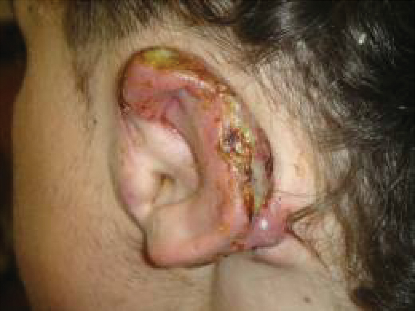
An 18-year-old young man presented with a one-year history of a discharging, ulcerated, thickened left pinna which had been repeatedly aggravated by rugby injuries (Figure 1). At the age of 11 years, he had been diagnosed with a congenital lymphangioma of the left side of the head and neck, which had been surgically removed but required further excision at the age of 13 years.
The patient's left ear on presentation, showing the retiform haemangioendothelioma tumour.
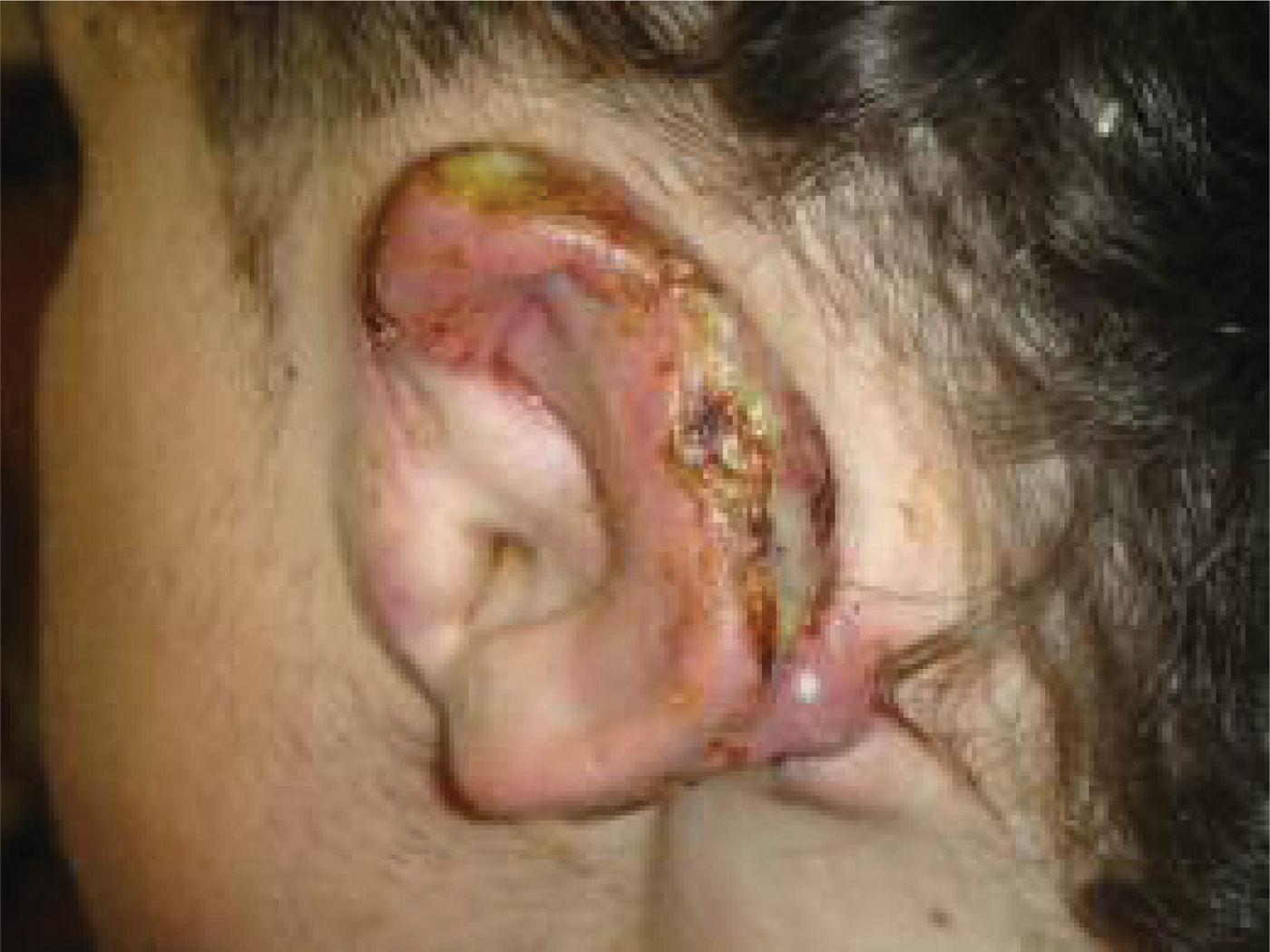
On examination, the patient had several areas of ulceration along the left pinna rim and post-auricular region.
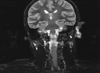
Radiological examination of the head and neck identified a metabolically active mass involving the left pinna, suggestive of a vascular tumour, with signs of left mastoid osseous involvement (Figure 2). Enlarged level two and facial lymph nodes were also present.
Coronal computed tomography brain scan, showing a metabolically active mass involving the left pinna, suggestive of a vascular tumour, with signs of left mastoid osseous involvement.

Histological examination of a biopsy specimen indicated a vascular neoplasm.
Therefore, the patient underwent wide local excision, including total pinnectomy, with lateral temporal bone resection and neck dissection.
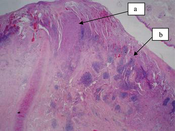
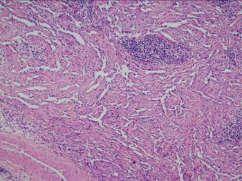
Macroscopic examination of the surgical specimen revealed a multifocal, tumourous mass 11 cm in maximum dimension. Histological examination showed numerous arborising, thin-walled, vascular channels in a retiform pattern, lined by plump endothelial cells, with a prominent lymphocytic infiltrate (Figures 3 and 4). Some areas showed a more solid growth pattern, with moderate but not severe atypia. A histological diagnosis of retiform haemangioendothelioma was made. Residual benign lymphangioma was also identified. A single, 2 mm metastatic deposit was found in a lymph node within the parotid tail.
Photomicrographs of the surgical specimen, showing (a) a solid area, and (b) a typical retiform area.

Photomicrograph of the surgical specimen, showing retiform growth with lymphoid infiltrate.

The patient also underwent adjuvant radiotherapy. This, plus the radical nature of his tumour excision, resulted in moderate cosmetic co-morbidity.
Discussion
Retiform haemangioendothelioma was first described in 1994 as an extremely rare variant of low-grade angiosarcoma characterised by a high local recurrence rate and very low metastasis rate.Reference Calonje, Fletcher, Wilson-Jones and Rosai1 The majority of cases present on the trunk and limbs, with some isolated cases involving the head and penis,Reference Tan, Kraybill, Cheney and Khoury2 although there is a female sex predilection.Reference Calonje, Fletcher, Wilson-Jones and Rosai1–Reference Bhutoria, Konar, Chakrabarti and Das3
The tumour presents clinically as single, slowly enlarging, exophytic mass or a plaque-like tumour of the dermis and subcutaneous tissues.
There have been 31 previously reported cases of retiform haemangioendothelioma, and three cases (including the present case) of lymph node metastasis.Reference Calonje, Fletcher, Wilson-Jones and Rosai1, Reference Bhutoria, Konar, Chakrabarti and Das3 The existence of soft tissue metastasis remains controversial.Reference Duke, Dvorak, Harris and Cohen4 Our finding of a third case with lymph node metastasis highlights the need to continue to screen for metastasis, as the true frequency of metastatic spread cannot be definitively predicted from the current literature.
The diagnosis of retiform haemangioendothelioma is based on the presence of characteristic histological features. The differential diagnosis prior to biopsy includes lymphoma, haemangioma, dermatofibrosarcoma protuberans, cutaneous angiosarcoma, bacillary angiomatosis and Kaposi's sarcoma.Reference Duke, Dvorak, Harris and Cohen4, Reference Darouti, Marzouk and Sobhi5 An association with previous lymphangioma or lymphoedema has been described. Retiform haemangioendothelioma shares some clinical characteristics with Kaposi's sarcoma, a tumour with known human herpes virus (HHV) 8 association. There has been a single reported case of retiform haemangioendothelioma in which HHV8 DNA sequences were detected, suggesting a potential viral aetiology.Reference Schommer, Herbst, Brodersen, Kiehl, Katenkamp and Kapp6
The term haemangioendothelioma refers to a group of vascular neoplasms that have a histopathological appearance intermediate between that of haemangioma and angiosarcoma.
In the present case, the histological appearance consisted of numerous elongated, arborising, thin-walled, vascular channels arranged in a retiform pattern, i.e. reminiscent of normal rete testis.Reference Calonje, Fletcher, Wilson-Jones and Rosai1 Some small areas of this tumour showed a more solid growth pattern (Figure 3), as first described in 1994 by Calonje et al. Other commonly seen histological features present in this case included a prominent lymphocytic infiltrate and papillae with hyaline collagenous cores (Figure 4), similar to a Dabska tumour. Cytological atypia is minimal and mitotic figures virtually absent.Reference Caljonje, Fletcher and Fletcher7 Histological features associated with improved prognosis include the presence of differentiation toward the high endothelial cells of post capillary venules in retiform haemangioendothelioma.Reference Calonje, Fletcher, Wilson-Jones and Rosai1
There have been no reported deaths associated with retiform haemangioendothelioma, although few cases have had long term follow up. The initial case series reported a median follow up of 7.35 years, with no mortality.Reference Calonje, Fletcher, Wilson-Jones and Rosai1–Reference Bhutoria, Konar, Chakrabarti and Das3, Reference Zhang, Lu, Yin, Wen, Su and Li8
The treatment of choice is surgical excision with histopathologically proven tumour-free margins. Since our patient's tumour was large and the margins close, and there was a single lymph node metastasis, he underwent adjuvant radiation therapy. This has previously been effective in cases with lymph node metastasis, large tumour size and local recurrence.Reference Calonje, Fletcher, Wilson-Jones and Rosai1, Reference Schommer, Herbst, Brodersen, Kiehl, Katenkamp and Kapp6
Sixteen months post-operatively, our patient remained well. However, given his tumour's persistent recurrence, and the presence of a metastatic lymph node, the prognosis remained somewhat guarded.
Acknowledgements
We thank Prof Chris Fletcher, Harvard University, Boston, Massachusetts, USA, and Dr Mairin McMenamin, St James's Hospital, Dublin, Ireland, for their expert opinion on the pathology of this case.