Introduction
Cancer is considered to rank as the leading cause of death and the major barrier to improve lifetime worldwide in the twenty-first century (Ref. Reference Bray1). Up to now, tumour metastasis remains the leading cause of cancer-related death (Ref. Reference Hanahan and Weinberg2). Tumour metastasis is a complex process that tumour cells need to adopt invasive properties to escape from the primary tumour mass, invade through surrounding tissue, intravasate and survive in the circulation and spread to distant sites to form secondary tumours, and is the leading cause of mortality among patients with cancer (Refs Reference Valastyan and Weinberg3, Reference Obenauf and Massague4). Epithelial–mesenchymal transition (EMT), as a cellular process in which epithelial tumour cells acquire an invasive phenotype and enhanced stemness and increased resistance to immune clearance and various therapeutic assaults, has been shown to play an important role in tumour metastasis (Ref. Reference Lu and Kang5). Therefore, exploring the underlying mechanisms of EMT has great importance for further understanding the tumour metastatic process.
The occurrence and maintenance of the EMT programme is a complex process involving multiple factors and molecular mechanisms. Tumour microenvironment (TME), referring to the local environment in which tumour cells produce and live, has been proved to play a key role in regulating tumour cells' EMT programme. TME consists of not only tumour cells themselves, but also fibroblasts, immune and inflammatory cells, glial cells and other cells around them, as well as intercellular stroma, micro vessels and infiltrated biomolecules in the nearby areas (Ref. Reference Hui and Chen6). Emerging and accumulating studies indicate that, on one hand, mesenchymal cells in TME can establish ‘crosstalk’ with tumour cells to regulate their EMT programme; on the other, EMT-tumour cells can create a favourable environment for their own growth via educating stromal cells. Over the past few years, our group has conducted a series of studies on the interaction between tumour-associated macrophages (TAMs) and colorectal cancer (CRC) cells in TME (Refs Reference Wei7, Reference Wei8, Reference Yang9). TAMs, as the most abundant immune cells in TME, can secrete various mediators such as cytokines or chemokines or exosomes to suppress the anti-tumour immune response, stimulate angiogenesis and finally enhance cancer cells proliferation, invasion, intravasation and dissemination for metastasis (Ref. Reference Noy and Pollard10). Our previous studies demonstrated that TAMs could induce EMT of CRC cells via secreting interleukin-6 (IL-6) to activate the JAK2/STAT3/miR-506-3p/FoxQ1 signalling cascade axis (Ref. Reference Wei7); meanwhile, we also found that EMT-programmed CRC cells could remodel the TME and polarise TAMs to M2 phenotype via release cytokines (such as interleukin-4(IL-4) and interleukin-10(IL-10)) and exosomes, thereby facilitating tumour progression and metastasis (Refs Reference Lin11, Reference Yang12). However, in addition to TAMs, there are fibroblasts, T cells, NK cells and neutrophils in TME, which also play an important role in the EMT process of CRC cells.
In the present review, we aim to describe the current knowledge about EMT in cancer, summarise the current findings about the role of TME in EMT, and provide an update on the mechanisms of TME-induced EMT in CRC, hoping to provide new ideas for understanding and resisting tumour metastasis.
EMT and cancer
EMT is a cellular programme from epithelial to mesenchymal phenotypes, during which epithelial cells undergo multiple biochemical changes to enable them to acquire mesenchymal characteristics (Ref. Reference Kalluri and Weinberg13). These changes result in cellular changes that include the loss of epithelial cell polarity, disruption of junctional complexes, degradation of the underlying basement membrane and reorganisation of the extracellular matrix (ECM) (Ref. Reference Lamouille, Xu and Derynck14). EMT plays essential functions in a diverse range of physiological and pathological conditions including embryogenesis, wound healing and malignant progression and is executed by EMT-inducing transcriptions factors (EMT-TFs), non-coding RNAs together with epigenetic and post-translational regulators (Refs Reference Yang15, Reference Nieto16, Reference De Craene and Berx17, Reference Dongre and Weinberg18). The reverse of this mesenchymal–epithelial transition (MET) process enables mesenchymal cells to progressively establish apicobasal polarity through an evolutionarily conserved group of proteins and distinct but well-defined mechanisms (Ref. Reference Pei19).
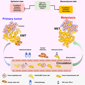
The EMT process plays diverse functions in cancer progression and is mainly associated with tumorigenesis, invasion, tumour stemness, metastasis and resistance to therapy (Refs Reference Pastushenko20, Reference Gupta21). We described the EMT programme in cancer progression in Figure 1. Many research studies indicate that EMT-TFs might play oncogenic functions and can promote malignant transformation and the development of primary tumour by cooperating with mitogenic oncoproteins (Refs Reference Ansieau22, Reference Maestro23, Reference Valsesia-Wittmann24, Reference Puisieux, Brabletz and Caramel25, Reference Liu26, Reference Phillips27, Reference Krebs28, Reference Morel29, Reference Ni30). Several studies have demonstrated an association between EMT and acquisition of stem cell-like properties in several different types of cancer, such as breast, lung, pancreatic and colon cancers (Refs Reference Wilson31, Reference Shibue and Weinberg32, Reference Pradella33, Reference Lambert and Weinberg34, Reference Scheel and Weinberg35). EMT is associated with cancer stemness, as evidenced by an increase in the number of cancer stem cells (CSCs) (Ref. Reference Zheng36). CSCs facilitates tumour metastasis by regulating the plasticity among epithelial or mesenchymal states (Ref. Reference Jayachandran, Dhungel and Steel37). Moreover, CSCs are highly related to chemoresistance, in particular, the cells exhibiting EMT exist in CSCs population (Ref. Reference Tanabe38). Emerging evidence suggests that tumour cells undergoing EMT acquire greater chemoresistance and resistance to immunotherapy in multiple cancer types, such as resistance to oxaliplatin and cisplatin chemotherapies in breast, ovarian, colon and pancreatic cancers (Refs Reference Lu and Kang5, Reference Wang39, Reference Ramesh, Brabletz and Ceppi40). Multidrug Resistance Gene1 (MDR1), as a central resistance-associated gene chemo-drug efflux, is found a higher-level expression in mesenchymal phenotype cells (Ref. Reference Cao41). In addition to CSCs, EMT contributes to enhance the resistance of cancer cells to anticancer therapy by the regulation of multiple apoptotic signalling pathways, the increase of drug efflux and the slowing of cell proliferation etc (Ref. Reference Shibue and Weinberg32). The release of immunoregulatory cytokines and chemokines induced by EMT programme results in the immunosuppressive effects (Refs Reference Wrzesinski, Wan and Flavell42, Reference Kudo-Saito43). Besides, EMT can reduce the expression of cancer cell surface major histocompatibility complex (MHC) class I to evade the cytolytic effects of T cells and mediate the expression of programmed cell death 1 ligand 1 (PD-L1) in cancer cells (Ref. Reference Dongre44). Metastasis is the main cause of cancer death. It is now widely accepted that EMT-associated features support tumour invasion, dissemination and metastasis (Refs Reference Lambert and Weinberg34, Reference Aiello45, Reference Ye46). Metastasis-related genes may activate the transcription of EMT-associated molecular, such as c-Myc binds to the promoters of ZEB1, ZEB2 and SNAIL genes (Ref. Reference Zhong47). By changing key EMT-TFs, many studies have reported the activation of EMT is the major drivers of tumour metastasis (Refs Reference Krebs28, Reference Spaderna48, Reference Yang49, Reference Tran50). It is already clear that the neoplastic cells in early cancer are in an epithelial-like state, and as the tumour progresses, they gradually acquire more mesenchymal characteristics (Ref. Reference Dongre and Weinberg18). Tumour cells undergo EMT to acquire increased motility and facilitate its dissociation from primary tumour and dissemination into surrounding, blood circulation or distant tissues (Ref. Reference Nieto16). Although EMT is essential for spreading from primary tumours, the process fails to explain the last step of metastasis that how to colonise distant organs. Therefore, some evidences point that tumour cells need to undergo the reversed process of EMT to recover epithelial traits for metastatic colonisation (Refs Reference Lu and Kang5, Reference Ocana51). It's not an easy task to acquire the EMT for tumour cells, because the process of EMT requires the cooperation of a complex network, consisting of various molecular signalling pathways and regulators. For these factors, we can categorise them into three groups: the first is EMT effectors which execute the EMT programme, such as epithelial cell junction proteins (E-cadherin, claudins and occludins) and mesenchymal adhesion proteins (vimentin, fibronectin and N-cadherin). The second is EMT-TFs which orchestrate the EMT programme, including the Snail family of zinc-finger transcription factors Snail/Slug, the zinc finger E-box binding homeobox (ZEB) family of transcription factors ZEB1/ZEB2, and the TWIST family of basic helix−loop−helix (bHLH) transcription factors TWIST1/TWIST2. And the third is EMT inducers which activate the EMT programme, such as transforming growth factor-beta (TGF-β), IL-6, tumour necrosis factor-alpha (TNF-α) and exosomes etc from TME and several intracellular signalling pathways. The canonical signalling pathways including TGFβ/SMAD, WNT,NOTCH and PI3 K/AKT and Janus kinases (JAKs)/ signal transducer and activator of transcription proteins(STATs) etc activate EMT through inducing the expression of EMT-TFs when cancer cells are exposed to specific signals released from different cells in the TME (Ref. Reference Dongre and Weinberg18). In addition, there are a number of novel signalling pathways found to activate EMT. For instance, our group found the activation of IL6/STAT3/miR-506-3p/FoxQ1 axis promoted EMT in CRC cells. Of note, the IL6 is released from TAMs. Therefore, the interplay between the TME and EMT promotes tumour progression.
Epithelial–mesenchymal transition (EMT) programme in cancer progression is displayed in this figure. Tumour epithelial cells express EMT -inducing transcription factors (EMT-TFs) ZEB family, Snail/Slug and TWIST, which repress the expression of genes associated with the epithelial state (E-cadherin, Claudins, Occludins and Cytokeratin) and activate the expression of genes associated with the mesenchymal state(vimentin, fibronectin and N-cadherin) in the primary tumour. Cancer epithelial cells undergoing EMT may become motile and acquire invasive capacities. The reverse of this process MET enables mesenchymal cells to progressively revert to the epithelial state. These changes contribute to the invasion through surrounding tissue, intravasation, extravasation and survival in the circulation and colonisation in metastatic sites. In addition to promoting tumour metastasis, EMT also induces other malignant features of cancer cells, such as chemoresistance, cancer stemness and immune evasion etc.

TME, EMT and cancer
The tumour is an organ-like structure that is a combination of malignant cells and their microenvironment, named TME (Ref. Reference Hanahan and Weinberg2). TME consists of a variety of cell types (endothelial cells, fibroblasts, stromal cells and immune cells etc) and extracellular components (cytokines, growth factors, hormones, ECM etc) that are surrounding tumour cells and nourished by a vascular network. Bidirectional interactions between cancer cells and TME promote tumour progression, resistance to therapy, invasion and metastasis (Ref. Reference Hanahan and Coussens52). Many studies indicate that the tumour cells associated-EMT is triggered in response to signals that cells receive from their microenvironment (Refs Reference Fan53, Reference Yu54, Reference Pistore55, Reference Qian and Pollard56, Reference Su57, Reference Che58). A large number of stromal cells in TME secrete various cytokines and chemokines, which act on nearby cancer cells in a paracrine manner to induce EMT in cancer cells, thus promoting tumour progression and metastasis (Ref. Reference Dongre and Weinberg18). In particular, the role of TGF-β, epidermal growth factor (EGF), fibroblast growth factor (FGF), insulin-like growth factors (IGF) and WNT ligands have been extensively studied in multiple model systems (Refs Reference Sakuma, Aoki and Kannagi59, Reference Strutz60, Reference Steinway61). Cytokines such as TGF-β, TNF-α and IL6 and hypoxia are capable of inducing EMT in various tumours. Several ECM proteins, including collagen-I, fibronectin and hyaluronan, and ECM remodelling via extracellular lysyl oxidase are also implicated in regulating EMT (Ref. Reference Jung, Fattet and Yang62). Meanwhile, tumour cells undergoing EMT promote tumour self-progression by secreting large amounts of cytokines and chemokines to regulate other cells in the TME in an autocrine or paracrine manner or by promoting autophagy of target cells (Refs Reference Akalay63, Reference Hsu64). The potential link between tumour inflammation milieu in the TME and EMT at the site of the primary tumour is reported, involving that inflammation triggers and sustains tumour EMT and EMT induces the secretion of inflammatory soluble factors, the main of which are interleukin-8(IL-8) and IL-6 (Refs Reference Dominguez, David and Palena65, Reference Fernando66). Although multiple studies have concentrated on understanding the signals that trigger EMT in the context of a tumour, recent reports have begun to investigate how the acquisition of mesenchymal features by carcinoma cells could also contribute to the development of TME. These ‘mesenchymalised’ tumour cells are also prone to secrete high levels of IL-6, IL-8 and other factors that could further contribute to the TME (Ref. Reference Fernando66). EMT has also been reported to be associated with immunosuppression in the TME. Immune cells in the TME secrete cytokines and chemokines to regulate EMT. Conversely, tumour cells undergoing EMT induce an immunosuppressive environment around the tumour by producing immunosuppressive cytokines and chemokines to recruit or expand the immunosuppressive cells (Ref. Reference Erin67). Thus, it will be seen that EMT is closely associated with the TME in cancer.
TME, EMT and CRC
In this section, we will focus on the interaction between EMT and TME in CRC. Understanding the relationship between the EMT programme and TME in CRC provides a better basis for the development of innovative therapeutic strategies.
TME as a driver for EMT programme of CRC cells
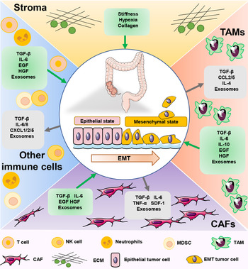
In TME, there are many non-tumour cells components that regulate phenotypes and functions of tumour cells though communicating with them. Among these, TME induces the EMT programme of CRC cells that attract our attention. Next, we will discuss the role of the various components of TME on the EMT programme in tumour cells and summary the relationship of them in Figure 2.
Molecular mechanisms of EMT programme induction in CRC cells through components of the TME. TME consists of a variety of cell types, such as endothelial cells, fibroblasts, stromal cells and immune cells etc and extracellular components. These non-tumour cells components communicate with tumour cells to induce EMT programme. We display the molecular mechanisms involved in CRC. these cells components like TAMs, CAFs and various immune cells contribute to the EMT programme of CRC cells by producing all kinds of cytokines and exosomes etc. Besides, extracellular components like ECM regulate this process through increase the stiffness of stroma or promote the formation of an anoxic and acidic environment. Likewise, CRC cells undergoing EMT also educate these non-tumour cells in TME by the secretion of cytokines, chemokines and exosomes.

TAMs Macrophages that recruited from circulating monocytes to tumours and affected the progression of cancer, named TAMs (Ref. Reference Cassetta and Pollard68), are major components of the innate immune system in TME, account for a dominant proportion of tumour-infiltrating leukocytes and present at all stages of tumour progression (Refs Reference Solinas69, Reference Biswas and Mantovani70). In primary tumours, TAMs regulate tumour growth by stimulating angiogenesis and enhancing the aggressiveness, motility and intravascular invasion of tumour cells. During metastasis, TAMs can trigger EMT and promote tumour cell extravasation, survival, immune escape and sustained growth of cancer cells in distant organs. TAMs not only promote tumour progression in the primary tumour, but also at metastatic sites, secrete interleukin-35(IL-35) to promote metastatic colonisation through activation of JAK2-STAT6-GATA3 signalling to reverse EMT programme in breast cancer (Ref. Reference Lee71). TAMs also have immunosuppressive effects, preventing natural killer cells and T cells from attacking tumour cells during tumour progression and after recovery from chemotherapy or immunotherapy (Ref. Reference Noy and Pollard10). Many studies have confirmed TAMs as prognostic indicators of tumour progression and a key determinant of anticancer therapy (Refs Reference Mantovani72, Reference Komohara73, Reference Steidl74, Reference Abdou75, Reference DeNardo and Ruffell76, Reference De Palma, Nassiri and Cianciaruso77, Reference Mantovani and Allavena78). TAMs can be divided into M1-like (pro-inflammatory, anti-tumour) and M2-like (anti-inflammatory and pro-tumour) (Refs Reference Biswas and Mantovani70, Reference Vitale79), a dual role in CRC progression (Ref. Reference Yang80). The role of TAMs in inducing EMT by secreting different types of cytokines and chemokines has been extensively studied in CRC. In vitro experiment, E-cadherin is reduced and vimentin is increased in CRC cells with M2 macrophages medium, indicating that M2 macrophages can induce EMT programme of CRC cells (Ref. Reference Vinnakota81). Previously, our group reported the interactions between TAMs and tumour cells for promoting metastasis in CRC in several articles (Refs Reference Wei7, Reference Wei8, Reference Yang9, Reference Lin11, Reference Yang12). In clinical specimen, TAMs, identified by CD163+ (a M2-like macrophages markers), are mainly detected at the tumour invasive front and stroma and the presence of CD163+ TAMs at tumour invasive front is associated with the EMT programme in CRC patients (Ref. Reference Wei7). We also found that a higher CD163+/CD68+ ratio was significantly correlated with the EMT programme in 81 CRC patients (Ref. Reference Yang9). Presence of CD68+CD163− macrophages is associated with lower Snail expression and predict better long term overall survival in CRC patients (Ref. Reference Li82). Inagaki et al. also demonstrated higher M2 macrophages count at the invasive front was related to lymph vascular invasion, low differentiation of histology and lymph node metastasis by double immunofluorescence (Ref. Reference Inagaki83). According to our research, IL6 is required for TAMs to induce EMT of CRC cells. IL6 derived from TAMs elicits EMT through IL6/STAT3/miR-506-3p/ forkhead box Q1(FoxQ1) axis in CRC cells (Ref. Reference Wei7). Zhong et al. found that cytoplasmic polyadenylation element-binding protein 3 (CPEB3) inhibits EMT in CRC cells through directly binging to the IL-6R mRNA, contributing to the reduction of phosphorylated-STAT3 expression in CRC cells, and CPEB3 also inhibits M2-like TAM polarisation in vivo (Ref. Reference Zhong84). Protein phosphatase of regenerating liver-3 (PRL-3) expression in TAMs promotes CRC cells EMT by activating mitogen-activated protein kinase (MAPK) pathway in TAMs and upregulating the expression of IL6 and IL8 (Ref. Reference Zhang85). Santiago et al. showed that extracellular GzmA promotes NF-κB-dependent IL-6 production in macrophages, which in turn induces STAT3 activation in cultured CRC cells (Ref. Reference Santiago86). Wan et al. suggested that dysbiosis of the intestinal flora stimulates macrophages activation and subsequently induces the EMT process through the secreted pro-inflammatory cytokines IL-6 and TNF-α (Ref. Reference Wan87). In addition to IL6, TGF-β and TNF-α can also serve as important signals to activate EMT in CRC cells. We also indicated that M2-polarised TAMs secreted TGF-β to facilitate the EMT, growth, proliferation and invasion of CRC cells by in vivo and in vitro experiments (Ref. Reference Yang9). TGF-β derived from TAMs could facilitate EMT progression of CRC cells through the TGF-β/Smad2,3-4/Snail signalling pathway (Ref. Reference Cai88). M2 polarised macrophages promote cancer metastasis by enhancing EMT of CRC cells and secreting vascular endothelial growth factor (VEGF) (Ref. Reference Wang89). C-C motif chemokine ligand 22 (CCL22) released from M2 macrophages activate EMT programme via CCL22/ PI3 K/AKT signal pathway, which in turn regulates 5-FU-mediated CRC chemoresistance (Ref. Reference Wei8). CRC cells often secrete cytokines to mediate macrophage polarisation and recruitment of macrophages, thereby promoting their own EMT programme, such as C-C motif chemokine ligand 2(CCL2) (Ref. Reference Wei7), IL4 (Ref. Reference Lin11) and exosomes (Ref. Reference Wang89). Expect for these secreted factors and cytokines, exosomes play an important role in the interaction between macrophages and tumour cells as a tool for intercellular communication (Ref. Reference Wu90). Our group previously indicated that exosomes derived from CRC cells have a significant role in intercellular communication for inducing M2 macrophage polarisation (Ref. Reference Yang12). Zhang et al. found that macrophages conditioned with profilin 1(PFN1) in CRC-derived EV spike CRC cell proliferation and migration in vitro and in vivo (Ref. Reference Zhang91). Several miRNAs (miR-25-3p, miR-130b-3p, miR-425-5p) upregulated in CRC cells by activating the CXCL12/CXCR4 axis can be transferred to macrophages via exosomes, which regulate phosphatase and tensin homolog (PTEN) through activation of PI3 K/Akt signalling pathway to mediate M2 polarisation in macrophages (Ref. Reference Wang89). Exosomes derived from CRC cells transport lncRNA RPPH1 into macrophages which induce macrophage M2 polarisation, thereby, in turn, promoting EMT of CRC cells (Ref. Reference Liang92). Besides, M1-macrophages, which are pro-inflammatory and anti-tumour, have also been reported to play a role in tumour progression (Ref. Reference Cho93). Very recently, it has been revealed that M1 macrophages contributed to the initiation of CSC in breast cancer cells through EMT process in a paracrine manner (Ref. Reference Guo94). Studies of M1-macrophages on tumour cell EMT programme are scarce, probably because as tumours develop, macrophages tend to be cultured by tumour cells into an immunosuppressive phenotype (M2) to aid tumour progression. In conclusion, M2-like macrophages induce EMT programme of CRC cells through secreting various cytokines and chemokines, meanwhile, CRC cells also produce secreted factors and exosomes to promote M2 polarisation in macrophages, thereby, in turn, enhancing this feedback progression.
Cancer-associated fibroblasts (CAFs) are the largest component of the stroma, that known as the cells negative for epithelial, endothelial and leukocyte markers with an elongated morphology and lacking the mutations found within cancer cells (Ref. Reference Sahai95). CAFs can be derived from various cell types, such as normal-associated fibroblasts, epithelial cells following EMT, endothelial cells via endothelial-to-mesenchymal transition (EndMT), bone marrow-derived cells (BMDCs), adipocytes and stellate cells etc (Ref. Reference Prakash96), the majority of CAFs are generated by differentiation of resident fibroblasts due to the Stimulation from TME. CAFs are a key determinant in the malignant progression of cancer and represent an important target for cancer therapies. Numerous evidences demonstrate that CAFs play prominent roles in cancer progression and have major clinical significance, through which remodel the ECM structure and interact with cancer cells or other stromal cells (Ref. Reference Chen and Song97). In CRC, CAFs are highly abundant and associated with 10 cancer hallmarks described by Hanahan and Weinberg (Ref. Reference Tommelein98). Clinically, the markers of CAFs such as α-SMA, FSP1 and fibroblast activation protein alpha (FAP), can predict outcome in CRC patients (Ref. Reference Herrera99). We will focus on how CAF regulates the EMT programme in CRC cells. Indeed, a large body of evidence points to the ability of CAFs to induce EMT in carcinoma cells through various paracrine signalling channels. CAFs secrete an array of cytokines and growth factors, such as TGFβ, IL-6, EGF and HGF, that can co-ordinately induce EMT in carcinoma cells (Ref. Reference Dongre and Weinberg18). In vitro studies indicate SW480 CRC cells treated with CAF-CM significantly upregulate the expression of EMT-TFs and mesenchymal markers, which reveals that CAFs secretions induce EMT of CRC cells (Ref. Reference Jahangiri100). Previous studies have demonstrated that CAFs drive the metastasis of cancer through mechanical force on cancer tissue and paracrine action on tumour cells (Ref. Reference Karagiannis101). Activin A released from CAFs increase matrix stiffness, which in turn may increase Snail expression and induce CRC cells EMT (Ref. Reference Bauer102). In the same way that macrophages promote EMT in CRC cells, CAFs connect with CRC cells through the secretion of cytokines, chemokines and exosomes. CCL2 derived from CAFs lead to the EMT programme in CRC cells through upregulating FGFR4 in bystander CRC cells in an Ets-1-dependent manner (Ref. Reference Liu103). Exosomal miR-92a-3p from CAFs is transferred to CRC cells, leading to the EMT programme and 5-FU/L-OHP chemotherapy resistance by activating Wnt/β-catenin pathway and inhibiting F-box and WD repeat domain containing 7 (FBXW7) and modulator of apoptosis 1 (MOAP1) (Ref. Reference Hu104). Exosomal LINC00659 is transferred from CAFs to CRC cells to promote EMT via interacting directly with miR-342-3p to increase annexin A2 (ANXA2) expression (Ref. Reference Zhou105). Same as macrophages, CAFs are divided into inflammatory-like CAFs(iCAFs) and myofibroblasts(myCAFs) via their phenotypically heterogenous, with pro-tumorigenic and anti-tumorigenic functions, respectively (Refs Reference Ganguly106, Reference Ohlund, Elyada and Tuveson107). Different subtypes of CAFs play different functions in the CRC EMT programme. The coculture of CRC cells and iCAFs leads to the significant upregulation of EMT markers, while the myCAFs are opposite of this phenotype (Ref. Reference Mosa108). However, Cao et al. studies showed a converse result that the EMT induction potential of CM derived from i-state CAF is reduced compared to that of m-state CAF CM in a 3D physio-mimetic interpenetrating network-based platform (Ref. Reference Cao109), which suggest the additional heterogeneity within the iCAF subsets may exist different functions. Collectively, there is no doubt that CAF induces EMT in CRC cells, mainly through the secretion of EMT inducers. Interestingly, Single-cell sequencing analysis of CRC tissue has shown that upregulation of EMT-related genes occurs in CAF in tumour tissue but not in tumour epithelial cells (Ref. Reference Li110). CAFs could easily mask the expression of EMT-related genes in unsegregated tumour material (Ref. Reference Vellinga111). This leads us to think about who exactly expresses EMT-related genes when we analyse EMT in tumour tissue, which will probably be the next step in CRC research. But in general, CAFs exert an important role in the progression of CRC and may be a prominent therapeutic target of stroma-based therapy in CRC treatment.
Other immune cells In TME, in addition to macrophages, the innate immune cell types also include T lymphocytes, dendritic cells, neutrophils, myeloid-derived suppressor cells, natural killer cells and innate lymphoid cells. Various immune cells that accumulate in the tumour stroma can crosstalk with adjacent cancer cells to activate previously latent EMT processes. CD8+ cytotoxic T cells (CTLs) act as the focus of anti-tumour immunity, which make other cells in TME produce immunosuppressive factors to avoid the attack of CTLs for survival, which in turn contribute to EMT programmes passively (Ref. Reference Erin67). ITGAE+ defined CD8+ T lymphocytes are associated with the EMT with decreased Snail and increased E-cadherin expression accompanied in CRC patients (Ref. Reference Hu112). Tumour-associated dendritic cells (TADCs)-derived CCL5 contribute to enhance EMT of CRC cells via metastasis-associated lung adenocarcinoma transcript 1 (MALAT-1)/Snail pathway (Ref. Reference Kan113). CXCL1 released from dendritic cells increases the metastatic ability of colon cancer by enhancing EMT (Ref. Reference Hsu114). As the most abundant leucocytes in circulation, neutrophils are known as the ‘first responder’ cells of the innate immune system. Recent studies have also shown that neutrophil infiltration can be substantial at the site of a primary tumour, where the degree of infiltration has been positively correlated with poor prognosis in CRC patients (Ref. Reference Templeton115). In response to the presence of soluble immune-mediators at the tumour site, these tumour-associated neutrophils (termed TANs) acquire either anti-tumoral or pro-tumoral functions, depending on their polarisation towards a TAN1 versus TAN2 phenotype, respectively (Ref. Reference Fridlender116). Some studies have demonstrated that TANs promote the spread of cancer cells through the release of matrix metallopeptidase (MMP)9, VEGF and HGF in the primary and metastatic sites (Ref. Reference Mizuno117). So far, there are few studies on the EMT programme of tumour cells that are activated by immune cells other than macrophages in CRC. However, there is no doubt that immune cells can influence the EMT programme of CRC cells.
ECM is the non-cellular component of TME, composed of water, proteins and polysaccharides (Ref. Reference Mohan, Das and Sagi118). ECM contributes to the EMT programme of tumour cells (Ref. Reference Bulle and Lim119). Among the ECM, collagen is the most abundant component. By generating a stroma gene filter, Vellinga et al. suggested that neoplastic cells in a high stroma gene tumour express EMT drivers, in the particular collagen-rich stroma (Ref. Reference Kirkland120). Decorin is one of the main components of the tumour stroma, lack of this proteoglycan increases the protein levels of EMT-TFs including Snail, Slug, Twist and MMP2 in CRC (Ref. Reference Mao121). The vascular system as a major component in tumour tissue, where platelets extravasate into the microenvironment of CRC and interact with CRC cells that induces EMT and increase the adhesion of CRC cells and endothelium in the context of circulation and metastasis (Ref. Reference Plantureux122). Besides, the acidic, hypoxic environment exposed to CRC cells also induces EMT programmes. Acidosis, as a common characteristic of TME, induces partial EMT of CRC cells via promoting autocrine TGF-β2 signalling (Ref. Reference Corbet123). Hypoxia induces EMT of CRC cells through ubiquitin-specific protease 47-mediated stabilisation of Snail (Ref. Reference Choi124). Collectively, TME, as a complexly composed environment, various components of which have the potential to contribute to the EMT programme of tumour cells and promote tumour progression.
EMT-programmed CRC cells as an inducer for TME education
TME can induce EMT in tumour cells, in turn, how do tumour cells affect the TME after the activation of the EMT programme? Now we will discuss the impacts of CRC cells after undergoing EMT (EMT-CRC) on the TME. As discussed above, tumour cells can induce M2 polarisation in macrophages to promote metastasis. Our data indicated that NOTCH2 activation enhanced CRC cells EMT and NOTCH2/GATA3-mediated IL-4 secretion, ultimately promoting M2-like TAM polarisation (Ref. Reference Lin11). We also found that exosomal miR-106b derived from EMT-CRC cells promoted the M2-like polarisation of macrophages through increasing the level of miR-106b which activating the PI3Kγ/AKT/mTOR signalling pathway by directly suppressing programmed cell death 4 (PDCD4) in a post-transcription level (Ref. Reference Yang12). Izabela et al. suggested that extracellular vesicles (EVs) released from CRC cells undergoing EMT might result in increasing lung inflammation that forms the lung pre-metastatic niche and facilitates cancer progression. EVs from HT29-cells in the early EMT stage activated by Snail transcription factor (HT29-Snail), as compared to those from epithelial-like HT29, tend to secrete less IL8 from macrophages (Ref. Reference Papiewska-Pajak125). The above studies indicate that CRC cells undergoing EMT not only affect the TME in primary tumour, but also create the niches for metastasis. For CAFs, the epithelial (E) or mesenchymal (M) states of hybrid E/M CRC cells contribute differential ability for the activation of fibroblasts. The more epithelial cells activate the fibroblasts more strongly due to the contribution more to collagen remodelling, while the less differentiated more-mesenchymal-alike cells do them less degree in vitro (Ref. Reference Druzhkova126). EMT-programme cancer cells governed CAFs heterogeneity through a ZEB1-dependent secretory programme in lung adenocarcinoma (Ref. Reference Bota-Rabassedas127), and it's reported that CSCs promoted the resident fibroblast to CAFs and hijacked them for their own benefit (Ref. Reference Fiori128). Thus, it can be seen that EMT-CRC cells promote their progression by secreting various factors and exosomes that regulate other non-tumour cells in TME. However, there are few related reports in CRC, which may become a future research direction.
Conclusion and perspective
Herein, we review the EMT programme and TME in cancer, and the focused discussion of EMT driven by TME in CRC. The current research about the occurrence of the EMT programme is that the alteration of mutated genes or oncogenes in tumour cells activates some intracellular signalling pathways to promote EMT. But EMT does not merely occur in tumour cells at the beginning of tumorigenesis. In the process of tumour development, the TME will be formed, and the EMT process in tumour cells can also be activated by various factors secreted in the TME. Currently, most of the research in this field is based on basic studies, and it will be a great challenge for how to use TME and EMT in the clinic.
For a therapeutic perspective, targeting the EMT programme directly is a promising treatment strategy. Therapies for EMT are still under investigation, including inhibitors of the TGF signalling pathway and some small-molecule inhibitors of the enzymatic activity of the HGFR tyrosine-kinase domain that are undergoing preclinical and clinical trials (Refs Reference Scagliotti, Novello and von Pawel129, Reference Neuzillet130). CBUD-1001, a novel fluorinated aminophenyl-benzamide-based compound, may prove to be a promising new drug candidate for CRC therapy by deregulating the EMT signalling pathway (Ref. Reference Kim131). In addition to these signalling pathways and drugs that target EMT, we can highlight the changes in TME to block the EMT programme of tumour cells. As previously described, TAMs, TANs and CAFs in TME with different phenotypes exert opposite effects on the tumour. Based on their different phenotypes, we can determine the prognosis and therapeutic target of different tumours, which requires some new technologies, such as single-cell sequencing, which can lead to a clearer analysis of TME. Besides, we are able to target therapeutic and prognostic analysis of secreted factors in TME that contribute not only to the EMT programme, but also to other activities of tumour cells. Exosomes are important mediators of intercellular communication and also have a significant role in the EMT of tumour cells in TME that we described. Exosomes are not only present in tumour tissues but also in circulation as liquid biopsies that play a role in the diagnosis, treatment and prognosis of tumours. However, there are still few studies on exosomes in the TME, which may be the priority of our future research. EMT-induced immune escape promotes cancer progression and may also provide a platform for the discovery of novel therapeutic approaches, but there are few studies about EMT cancer cells that induce the changes of TME. Collectively, TME, as a non-negligible driver for EMT, may provide important clues for tumour therapy.
Financial support
This work was supported by grants from the Fundamental Research Funds for the Central Universities (2042021kf0143), Zhongnan Hospital of Wuhan University, Excellent Doctor Fund Project (ZNYB2020002), Zhongnan Hospital of Wuhan University, Technology and Innovation Seed Found (CXPY2020025), and The Clinical Medical Research Centre of Peritoneal Cancer of Wuhan (2015060911020462).
Conflicts of interest
The authors declare that they have no competing interests.