The lysosomal storage diseases are a privileged group of genetic disorders by virtue of the speed by which these disorders have progressed from biochemical and genetic characterisation to the establishment of therapeutic regimens. Although many metabolic diseases have been found to be amenable to treatment by manipulation of the respective metabolic pathway, lysosomal disorders are notable for their responsiveness to direct replacement of the defective enzyme. The mucopolysaccharidoses (MPSs) have recently entered the category of lysosomal disorders in which enzyme-replacement strategies are available, with recombinant enzyme-replacement products approved for MPS type I (MPS I) (laronidase), MPS II (idursulfase) and MPS VI (galsulfase). Each of these enzyme-replacement approaches has shown significant impact on disease parameters and holds much hope for improving the quality of life of affected individuals.
With the introduction of treatment regimens for the MPSs, there has been increasing interest in the identification of biomarkers of disease, methods for rapid and early diagnosis (including newborn screening), and alternative and/or additional therapeutic approaches. Inherent in exploring these areas is a need for a thorough understanding of the pathogenesis of disease. The exact mechanisms by which deficiencies of lysosomal hydrolases ultimately lead to disease manifestations are not clear. In this 110th anniversary year of Archibald Garrod's Croonian lecture highlighting the importance of inherited chemical individuality – that is, inborn errors of metabolism – as a central component of disease, it is interesting that although many lysosomal disorders have progressed to enzyme-replacement therapy (ERT) there is a poor understanding of the underlying metabolic perturbations. This review explores recent observations in the lysosomal storage diseases and highlights mechanisms that likely underlie the pathogenesis of the MPSs. The emerging picture from the study of individuals and animal models is one of a complex combination of primary substrate storage and secondary effects.
Fundamentals of the diseases
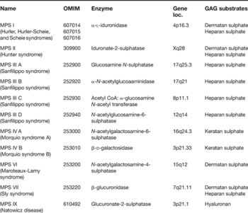
The MPSs are a group of lysosomal storage diseases that are characterised by defects in the degradation of macromolecules termed mucopolysaccharides, or more accurately named glycosaminoglycans (GAGs) (Ref. Reference Neufeld, Muenzer and Scriver1). The rich history of these disorders is well reviewed by Victor McKusick and colleagues (Refs Reference McKusick2, Reference McKusick3). The alteration of GAG degradation is caused by mutations in genes coding for key lysosomal hydrolases (Table 1).
Classification of the MPSs

Abbreviations: GAG, glycosaminoglycan; loc., localisation; MPS I, mucopolysaccharidosis type I.
Each of the disorders is characterised by progressive multisystem disease with key morbid manifestations involving the skeleton, joints, somatic tissues, heart and, in some disorders, the central nervous system (CNS). Interestingly, each of the disorders is characterised by a spectrum of disease manifestations and clinical course, which ranges from early-onset rapid progression (leading to early death) to attenuated forms characterised by later onset and slower progression (often resulting in a normal lifespan). Patients are thus often classified as severe or attenutated. This disease spectrum is most evident in MPS I and MPS II: severely affected individuals have progressive CNS disease and a shortened lifespan, in addition to somatic features, whereas patients with attenuated disease can have no significant CNS disease and a normal lifespan albeit suffer from significant disease morbidity related to skeletal, joint and cardiopulmonary manifestations (Refs Reference Neufeld, Muenzer and Scriver1, Reference Roubicek, Gehler and Spranger4, Reference Young5).
The disease spectrum seen in the MPSs is attributable to allelic heterogeneity and thus differences in residual enzyme activity. This is best illustrated by detailed biochemical analysis in MPS I and MPS II, utilising techniques to measure iduronidase and iduronate-2-sulphatase activity and protein mass, where the amount of residual activity and protein was found to correlate with disease severity (Refs Reference Ashton6, Reference Parkinson-Lawrence7). Recently, electrospray ionisation tandem mass spectrometry to measure oligosaccharides derived from the GAGs dermatan sulphate and heparan sulphate in cultured MSP I fibroblasts, combined with an accurate residual iduronidase activity measurement, was shown to distinguish patients with and without CNS involvement – that is, severe versus attenuated disease (Ref. Reference Fuller8). How practical these measures will be in the clinical setting remains to be determined. These studies clearly indicate that the severity of the disease phenotype in MPS I is directly related to ‘residual’ flux through this degradation pathway. In addition, studies correlating genotype to phenotype in the MPSs, particularly MPS I (Refs Reference Beesley9, Reference Bunge10, Reference Terlato and Cox11, Reference Brooks12), MPS II (Refs Reference Timms13, Reference Vafiadaki14, Reference Froissart15) and MPS VI (Refs Reference Karageorgos16, Reference Litjens17), and the prediction of the mutation effect based on the position of missense mutations on the proposed crystal structure of iduronidase in MPS I (Ref. Reference Rempel, Clarke and Withers18), provide evidence against other loci significantly influencing disease severity.
Fundamentals of the pathway
Although classical in its use, the nosology in Table 1 does not reflect the true nature of the complexity of these disorders. For example, this classification scheme would suggest that MPS I and MPS II should have identical phenotypes by virtue of the fact that both conditions lead to the storage of heparan and dermatan sulphate. Although the phenotypes of severely affected MPS I and MPS II patients are similar, they differ considerably in key aspects of disease – notably the degree of corneal involvement, the age of onset and rate of progression of CNS effects, as well as the phenotype of the CNS dysfunction particularly at the end stages of disease. To make sense of these observations, a more-detailed understanding of the metabolic effects of altered GAG metabolism is required.
The study of inborn errors of metabolism has highlighted the importance of understanding the alteration of homeostasis that occurs as a result of a metabolic block in a pathway (Ref. Reference Treacy, Childs and Scriver19). Historically, a relatively straightforward metabolic model for the MPSs has been considered: specific substrate (GAG) accumulation within the lysosome directly leading to disease symptoms. The study of animal models of the MPSs and limited studies in humans indicate that the alteration of cellular homeostasis resultant from altered GAG degradation is profound and even involves pathways unrelated to GAG metabolism (Refs Reference Walkley and March20, Reference Walkley21, Reference Walkley, Zervas and Wiseman22, Reference Walkley23, Reference Jolly and Walkley24). Recent observations indicate that defects in the lysosomal degradation of GAGs may influence diverse biochemical and physiological processes, including: trafficking of molecules through the endosomal network; the activity of other lysosomal enzymes; the steady state of intracellular and extracellular GAGs, oligosaccharides and proteoglycans; metabolic pathways involved in ganglioside and cholesterol metabolism; proteinases that are regulated by GAG-dependent inhibitors (serpins, serine proteinase inhibitors and cathepsins); and inflammation.
Homeostasis and GAGs: biological function of GAGs
A key feature often overlooked in discussion of the MPSs is the fact that the main storage products – GAGs – are enormously diverse molecules. In fact, the size of the GAG chains and the possible modifications of sugar residues suggest GAGs may represent the most information-dense biological molecule (Refs Reference Turnbull, Powell and Guimond25, Reference Bülow and Hobert26, Reference Taylor and Gallo27, Reference Jackson, Busch and Cardin28). As such, it is not surprising that defects in the ability to degrade GAGs result in complex multisystem disease.
GAGs are linear heteropolysaccharide molecules composed of characteristic repeating disaccharide units. The typical unit length of GAG molecules is of the order of 50−150 disaccharide units. The specific individual monosaccharide units define the GAG subtype: heparan sulphate is composed of glucosamine and the hexuronic acids iduronate or glucuronate; dermatan sulphate comprises galactosamine and the hexuronic acid iduronate; chondrotin sulphate comprises galactosamine and the hexuronic acid glucuronate; keratan sulphate comprises galactose and glucosamine; and hyaluronan comprises glucuronate acid and glucosamine. In addition to the core sugar units, all GAG chains, with the exception of hyaluronan, are extensively modified by N-acetylation, N-sulphation, O-sulphation and epimerisation. These modifications occur in various sections of the GAG chain, thus creating enormous molecular diversity. With the exception of hyaluronic acid, GAGs are attached to a core protein, through serine or asparagine residues, the complex of which is termed a proteoglycan. The function of each specific proteoglycan is largely determined by the type of GAG molecules complexed to it. In addition, the GAG components of proteoglycans provide functions that are independent of the core protein.
GAGs are synthesised from nucleotide sugars and assembled onto core proteins within the Golgi complex and undergo simultaneous modification. The process of GAG polymerisation, modification and attachment to core proteins is discussed extensively in Ref. Reference Prydz and Dalen29. Factors involved in the regulation of GAG synthesis and the determination of the specific patterns of modification remain largely unknown. Once produced, proteoglycans are targeted either to the cell surface or to the extracellular matrix, where further sugar modifications may take place. At the cell surface, particularly in the case of heparan sulphate proteoglycans, the heparan sulphate side chains may be released into the extracellular matrix by heparanase activity. Thus, GAGs reside primarily at the cell surface and within the extracellular matrix and can be free, particularly within the extracellular matrix, or covalently bound to proteins forming proteoglycans.
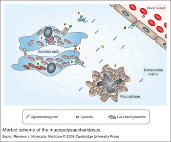
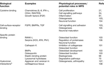
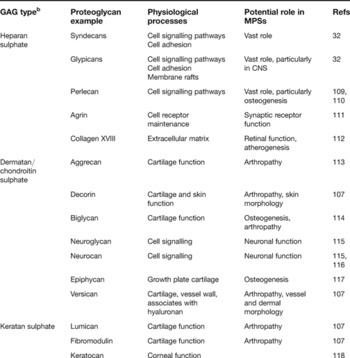
By virtue of their negative charge, GAGs influence ligand–receptor interaction by stabilising receptors at the cell surface and by direct binding and presentation of ligands to their respective receptor. Free GAGs within the extracellular matrix can also sequester humoral factors, thus modulating their biological effects (Fig. 1). Thus, GAGs ultimately affect the continued crosstalk that occurs between cells. The critical importance of proteoglycans in the regulation of developmental processes and metabolism has been extensively reviewed (Refs Reference Prydz and Dalen29, Reference Bishop, Schuksz and Esko30, Reference Perrimon and Bernfield31, Reference Bernfield32, Reference Yamaguchi33, Reference Handel34). When this paradigm is expanded to include different tissues, the complexity and profound nature of GAGs and proteoglycans in metabolism of an intact organism is evident (Tables 2 and 3). Tables 2 and 3 list the known functions of GAGs and key proteoglycans that are likely to underlie pathological features of the MPSs. As discussed below, very few of these have been clearly demonstrated to be involved in the pathogenesis of MPS; the remainder have not been investigated.
Morbid scheme of the mucopolysaccharidoses. The figure illustrates the various physiological processes that may be affected by defective glycosaminoglycan (GAG) degradation, which may lead to increased storage of GAGs in lysosomes and increased levels in the extracellular matrix. (a) Macrophage dysfunction related to GAG-filled lysosomes; (b) altered assembly of plasma membrane receptors; (c) altered sequestration of cytokines and growth factors; (d) altered recruitment of circulating cytokines; (e) altered presentation of cytokines to signalling receptors; (f) interference with cellular trafficking; (g) additional lysosomal storage by interference of lysosomal hydrolases; (h) abnormal extracellular matrix crosslinks; (i) altered cell attachment.

Biological roles of GAGs (independent of core protein) relevant to the MPSsa

aThe table is based on information reviewed in Refs 26 and 30.
bAlways independent of core protein.
Abbreviations: ATIII, antithrombin III; BMP, bone morphogenic proteins; BMPR, BMP receptor; FGF, fibroblast growth factor; FGFR, FGF receptor; HCII, heparin cofactor II; IFN, interferon; IL, interleukin; MPS, mucopolysaccharidosis; RANK-L, ligand of receptor activator of NF-κB; RANTES, ‘regulated upon activation, normal T-cell expressed and secreted’; TGF, transforming growth factor.
Biological roles of GAGs (as proteoglycans) relevant to the MPSsa

aThe table is based on information reviewed in Refs 26 and 30.
bProteoglycans may be decorated with more than one GAG type; the preferential GAG type is listed here.
Abbreviations: CNS, central nervous system; MPSs, mucopolysaccharidoses.
The degradation of GAGs occurs primarily in the lysosomal compartment through exoglycosidic cleavage (restricted to the nonreducing terminal portion of the chain) of specific terminal sugars. Molecules destined for lysosomal degradation travel through the endosomal network after endocytosis from the cell surface via clathrin-coated pits, or through the process of autophagy of organelles, membranes and cytoplasmic material. However, cleavage of proteoglycans by proteases and endoglycosidic cleavage of GAGs can occur within the extracellular matrix as well as in the endosomal compartment. Ultimately, the monosaccharides and inorganic sulphate generated from the complete degradation of GAGs are actively transported out of the lysosome. Each of the MPSs is caused by deficiency of one of the enzymes responsible for exoglycosidic cleavage (Table 1), thus leading to the accumulation of oligosaccharides and high molecular weight GAGs with specific terminal saccharide units. Although this review is centred on the MPSs, it is critical to note that the lysosome is central to the metabolism of many other compounds, including sphingolipids, glycogen, cholesterol, cystine and cobalamine.
Treatment of the MPSs
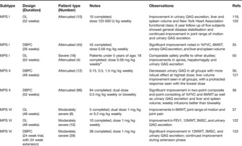
Historically, symptom management represented the mainstay of treatment for the MPSs. Recent progress has led to the development and introduction of direct ERT for MPS I (laronidase) (Ref. Reference Wraith35), MPS II (idursulfase) (Ref. Reference Muenzer36) and MPS VI (galsulfase) (Ref. Reference Harmatz37). This progress clearly resulted from advances in the the identification of lysosomal corrective factors and the understanding of lysosomal targeting of enzymes – specifically that terminal mannose and mannose-6-phosphate residues of the oligosaccharide chains could be used to target recombinant enzyme back to the lysosome from the cell surface. The early comprehensive studies of ERT, particularly in the MPS VII murine model as well as other animal studies, were cardinal in setting the stage for human trials of ERT (Ref. Reference Sands38). The respective double-blind placebo-control and open-label clinical trials of ERT for each of these disorders have demonstrated improvement in disease manifestations in patients with significant degrees of pre-existing disease (Table 4).
Summary of enzyme-replacement therapy trials for the MPSs

aDose for four patients increased to 1.16 mg/kg at 26 weeks.
Abbreviations: DBPC, double-blind placebo-control trial; FEV1, forced expiratory volume at 1 s; %FVC, percent predicted forced vital capacity; GAG, glycosaminoglycan; MPSs, mucopolysaccharidoses; 3MSC, 3 min stair climb; 6MWT, 6 min walk test; 12MWT, 12 min walk test; OL, open-label trial.
The intravenous use of recombinant enzyme is not expected to alter the CNS manifestations of disease, as recombinant enzyme cannot readily cross the blood–brain barrier. This is particularly important for MPS I and MPS II where a significant portion of patients are severely affected and thus have progressive or are predicted to have progressive CNS disease. In the case of patients predicted to have severe MPS I, haematopoietic stem cell transplantation (HSCT) performed before the onset of significant CNS disease (i.e. before the age of 18 months) is associated with stabilisation of CNS findings. Long-term follow-up of these patients revealed that although many aspects of the somatic and CNS disease is altered by early transplantation, there is only a minimal effect on skeletal disease and cardiac involvement (Refs Reference Guffon39, Reference Peters40, Reference Peters41, Reference Souillet42, Reference Staba43, Reference Vellodi44, Reference Whitley45). Interestingly, HSCT does not appear to have the same clinical effect on the CNS disease in MPS II (Refs Reference Guffon46, Reference Vellodi47) or MPS III (Ref. Reference Sivakumur and Wraith48), although the number of patients with these disorders who have been transplanted and reported in the literature is considerably smaller. It is not clear whether very early transplantation in the latter group of patients would result in improved outcome. Whether ERT in combination with HSCT used either in the peritransplant period or on a long-term basis improves the outcome for severely affected MPS I patients remains unknown. Studies of severely affected MPS I patients who received ERT before bone marrow transplantation (Ref. Reference Grewal49), and a 52-week open-label trial of ERT in severely affected MPS I patients who were less than 5 years of age (Ref. Reference Wraith50), have shown that that ERT is well tolerated in this group and did result in alterations in disease measures.
Altered homeostasis in MPS diseases
The recent clinical trials of ERT in MPS I, MPS II and MPS VI have demonstrated significant improvements in disease manifestations in patients with pre-existing disease (Ref. Reference Beck51). In these trials, ERT has not been sufficient to correct the disease state completely within the reported observation period. This is of course the conclusion also noted for bone marrow transplantation outcomes for the MPSs. Although insufficient targeting of enzyme to certain tissues – including brain, connective tissue and bone – may underlie some of these observations, a key factor is likely to be secondary biochemical changes (i.e. altered homeostasis) that occur as part of the disease progression. Animal studies of ERT or gene-based therapy in the MPSs have shown a more robust clinical response, albeit still not a complete cure, when started very early in the disease course, thus confirming that both factors are likely at play (Refs Reference Sands and Davidson52, Reference Vogler53). The storage of GAGs themselves is thought to be predominantly responsible for the organ enlargement seen in these disorders – that is, hepatomegaly, splenomegaly and coarse facial features noted in the MPSs. It is thus not surprising that these features of disease respond very effectively to ERT. However, evidence would suggest that the other disease manifestations – progressive skeletal disease, arthropathy, CNS dysfunction and cardiac involvement – likely result from additional complex metabolic perturbations. These are unlikely to respond to ERT strategies and may require the development of additional therapeutic approaches. Studies of both animal models and humans with a variety of lysosomal storage diseases have begun to highlight the complex alteration of homeostasis that results from primary lysosomal substrate storage. The following key areas have been uncovered.
Tissue storage of GAGs
Extensive studies of tissue levels of GAGs in animal models of the various MPSs performed by either analysis of soluble Alcian-Blue precipitable GAGs or total uronic acid content reveal that there is massive storage of GAGs in liver, kidney, lung, spleen and heart but relatively mild to modest storage of GAGs within the CNS (Refs Reference Li54, Reference Vogler55, Reference Bhaumik56). Microscopic analysis of bone has shown retained GAG within the trabecular matrix and bone cortex, but no quantitative studies of GAG content in bone or growth plate tissue have been reported (Refs Reference Russell57, Reference Nuttall58). It has been suggested that the stored tissue GAGs are contained intracellularly within lysosomes, although no comprehensive studies examining the extracellular levels or specific content of GAGs within the extracellular matrix have been published. McGlynn et al. (Ref. Reference McGlynn, Dobrenis and Walkley59) note that immunohistochemical staining with monoclonal antibody against heparan sulphate in the MPS I, MPS IIIB and MPS VII murine models showed uniformly increased immunoreactivity in all brain regions with clear labelling of storage bodies within perkarya as well as increased nuclear and membrane staining in virtually all neurons. Observations in the MPS IIIA murine model showed greater variation in these neuronal findings. The increased urinary levels of GAGs seen in all of the MPS disorders as well as the evidence of increased plasma GAGs as determined by GAG ELISA analysis in MPS I, MPS II and MPS VI suggest that extracellular GAG levels are indeed altered in these disorders (Ref. Reference Tomatsu60). What role increased extracellular GAG may play in the MPS diseases is discussed below.
Analysis of various MPS animal models has provided interesting insights into the time- course of GAG storage and the natural history of pathological changes in these diseases. Interestingly, storage is seen in many tissues in utero, as documented in humans (Refs Reference Ramsay61, Reference Greenwood62, Reference Wiesmann63) and animal models (Refs Reference Vogler55, Reference Casal and Wolfe64, Reference Jones65). Lysosomal vacuolation appears to be evident first within the reticuloendothelial cells and then proceeds to other cell types (Refs Reference Li54, Reference Vogler55, Reference Clarke66). These observations indicate a central role for tissue GAG degradation and clearance by cells of the macrophage/monocyte lineage. Perhaps the therapeutic effect of HSCT in the MPSs may relate primarily to the provision of enzyme-competent macrophage/monocyte lineages, with the additional role of secretion and reuptake of enzyme from donor macrophages by neighbouring somatic cells being less important. These observations would also suggest that ERT regimens for the MPSs may benefit from additional enzyme specifically targeted to macrophage/monocyte cells. This has been accomplished in Gaucher disease by the processing of the oligosaccharide side chains of glucocerebrosidase to high mannose (cerezyme) rather than mannose-6-phosphate (Ref. Reference Beutler67). In addition, perhaps the outcome of long-term combined ERT and bone marrow transplantation in some of the MPSs may result in improved outcome.
Alteration of lysosomal enzyme activities
Deficiency of one lysosomal hydrolase appears to lead to alterations in the enzyme activities of other lysosomal hydrolases. This has been well documented in MPS I, MPS II, MPS III and MPS VII (Refs Reference Li54, Reference Van Hoof and Hers68, Reference Sands69). A complex pattern that can be tissue and cell-type specific has been noted: consistent observations within the MPSs include increased activity of β-hexosaminidase, α-glucosidase, β-galactosidase, and β-gluronidase within the liver and brain with decreased activities of sialidase (also known as neuraminidase) and N-acetylglucosaminyltransferase (GalNAc-T). The reason for the increases in other lysosomal enzymes is largely unknown but has been postulated to reflect the increased cellular mass of lysosomes or the stabilisation of enzymes by the storage material. Interestingly, the increased enzyme activities of many lysosomal enzymes in tissue homogenates of the MPSs is sharply contrasted by in vitro studies that show strong inhibition of leukocyte lysosomal enzymes by heparin, chondroitin sulphate and hyaluronic acid, particularly at conditions that mimic the lysosomal environment (Ref. Reference Avila and Convit70). Although the alteration of sialidase activity may play a role in the increases in brain glycosphingolipids noted in the MPSs, what role, if any, these alterations of unrelated enzymes have in the pathophysiology of the MPSs is largely unknown.
Alteration of ganglioside metabolism
Significant accumulation of glycosphingolipids, most notably GM2 and GM3 gangliosides, has been noted in human MPS I, MPS II and MPS IIIA (Refs Reference Walkley21, Reference Canstantopoulous and Dekaban71, Reference Jones72) and has been extensively studied in various animal models of MPS I, MPS II, MPS IIIA, MPS IIIB, MPS IIID, MPS VI and MPS VII (Refs Reference Walkley and March20, Reference Walkley21, Reference Walkley, Zervas and Wiseman22, Reference Walkley23, Reference Li54, Reference Russell57, Reference Jones73, Reference Liour74). This finding is surprising as the degradation of glycosphingolipids does not require the involvement of any of the hydrolases that are deficient in the MPSs. In addition to these gangliosides, extensive studies of the caprine model of MPS IIID reveal significant elevations of the glycoshingolipid GD3 and lactosyl ceramide in the prenatal, neonatal and adult caprine brain (Ref. Reference Jones65).
These studies suggest that the storage of gangliosides occurs very early in the disease course and could represent a main causative factor ultimately responsible for the progressive CNS dysfunction that occurs in the MPSs. The consequences of storage of gangliosides, particularly GM2, are thought to include alterations of dendritic and axonal morphology, thus leading to reinitiation of dendritic growth and altered synapses. One such abnormality, termed ectopic dendritogenesis, appears to be formation of meganeurites consisting of new synapse-covered dendritic neurites at the axonal hillocks and axonal hillock enlargements (Ref. Reference Walkley21). These same features are seen in GM2 gangliosidosis (Tay–Sachs disease), GM1 gangliosidosis, Niemann–Pick A and C, and α-mannosidosis (Refs Reference Walkley21, Reference Walkley, Platt and Walkley75).
The primary reason for the increased glycosphingolipids in the MPSs is not entirely clear. Comprehensive studies performed in the murine models of MPS I, MPS IIIA, MPS IIIB and MPS VII (Ref. Reference McGlynn, Dobrenis and Walkley59) indicate that the subcellular localisations of heparan sulphate, GM2 and GM3 show only partial colocalisation. Particularly striking differences were noted in the localisation of GM2, which more commonly localised independently of the other accumulated molecules. In addition to storage of glycosphingolipids, significant storage of unesterified cholesterol was also noted (see below). Extensive studies of caprine MPS IIID brain have shown significant accumulation of simple glycosphingolipids such as GM2, GM3, GD3 and lactosyl ceramide with little evidence of increases in more complex glycosphingolipids (Refs Reference Jones73, Reference Liour74). Importantly, the increases in glycosphingolipids among affected animals were apparent in the fetal brain. The increases in sphingolipids are thought to be partially related to the ability of GAGs to inhibit the activity of other lysosomal hydrolases, particularly sialidase. In fact, sialidase is noted to be reduced to as much as 25% of normal in brain tissue homogenates from various MPS animal and human studies (Refs Reference Jones72, Reference Jones73, Reference Liour74). The pattern of glycosphingolipid storage noted in the caprine MPS IIID model would also suggest that, in addition to a block in the degradation of glycosphingolipids, a defect in the synthesis of complex lipids must also be present. Indeed, GalNAc-T, a key enzyme in the biosynthesis of complex glycosphingolipids, is significantly impaired as well, likely secondary to its inhibition by GAG storage (Ref. Reference Liour74). It has been suggested that GAG interference with enzymes involved in glycosphingolipid degradation and synthesis is probably only part of the story, and that additional components of altered feedback of glycosphingolipid synthesis and altered intracellular trafficking must be at play to account for the results of localisation studies (Ref. Reference McGlynn, Dobrenis and Walkley59).
Alteration of intracellular cholesterol
Recent studies in murine models of the MPSs have shown accumulation of free unesterified cholesterol in neurons in MPS I, MPS IIIA and MPS IIIB (Ref. Reference McGlynn, Dobrenis and Walkley59). Interestingly, in these murine models cholesterol appeared to be stored only in neurons that also stored gangliosides, while some neurons stored gangliosides without cholesterol: thus, cholesterol storage appears to be secondary to ganglioside storage, which in turn is secondary to GAG storage. Neuronal cholesterol accumulation occurs as the primary storage material in the neurodegenerative lysosomal disease Niemann–Pick C, which is caused by alterations of one of two genes (NPC1 and NPC2) involved in the intracellular trafficking of cholesterol (Ref. Reference Walkley and Suzuki76). Secondary intraneuronal accumulation of cholesterol has been noted in GM2 gangliosidosis and other primary glycosphingolipid disorders (Ref. Reference Pagano77). It has been postulated that the altered intracellular distribution of cholesterol noted in the glycosphingolipidoses may in fact lead to defective transport of glycosphingolipids themselves, and that the accumulation of these two molecules within late endosomes and lysosomes reflects the sequestration of membrane rafts, an important component of the pathophysiology of disease. The secondary accumulations, particularly within the brain, in the MPSs may represent relevant and important contributors to the pathophysiology of disease (Ref. Reference McGlynn, Dobrenis and Walkley59).
Inflammation
Activation of inflammatory responses has been implicated in several lysosomal storage diseases and most recently the MPSs. Microarray studies provided a basis for the initial observation of microglial activation in a murine model of GM2 gangliosidosis (Ref. Reference Jeyakumar78). Subsequently, studies of Sandhoff human brain tissue (Ref. Reference Myerowitz79) as well as murine models of GM1 gangliosidosis, Niemann–Pick C (Ref. Reference Langmade80), multiple sulphatase deficiency (Ref. Reference Settembre81), MPS I and MPS IIIB (Ref. Reference Ohmi82) indicate that activation of an inflammatory cascade within the CNS is a component of disease. Studies in the Sandhoff murine model indicate that the activation of microglia precedes the more rapid phase of neurodegeneration noted in that model (Ref. Reference Jeyakumar78). Furthermore, studies of murine models of MPS I and MPS IIIB revealed perineuronal microglial activation within the CNS and increased transcripts for CD38, lysozyme M, cathepsins S and Z, cytochrome b558, DAP12 and complements C1q and C4, all of which relate to macrophage/monocyte activation (Ref. Reference Ohmi82). As is noted in the Sandhoff murine model, the increases in the transcripts in the murine MPS IIIB model occurred before the detection of behavioural abnormalities.
The studies to date cannot distinguish between activation of microglia resident within the brain and/or recruitment of activated monocytes from the circulation. The observation that activation of macrophages within the CNS occurs before evidence of significant CNS pathology would indicate that this phenomenon is not primarily related to neuronal cell loss. What direct role the activation of microglia has in the pathogenesis and progression of CNS disease in the MPSs remains unknown, but the observation that treatment of the murine Sandhoff model with the anti-inflammatory indomethacin slowed the rate of neurological deterioration (Ref. Reference Jeyakumar83) would indicate that inflammatory responses within the CNS are likely to be one of many factors involved in the progression of disease. Interestingly, studies performed in chimaeric Niemann–Pick C mice that have both wild-type (Npc +/+) and mutant (Npc −/−) Purkinje cells indicate that, despite a robust inflammatory response in the CNS, only Npc −/− Purkinje cells appear to be lost whereas neighbouring Npc +/+ Purkinje cells are retained (Ref. Reference Ko84). These observations indicate that other cell-specific factors are responsible for the neurodegeneration seen in Niemann–Pick disease.
Evidence is also emerging for the involvement of inflammation in the pathophysiology of bone and joint disease in the MPSs. Findings in the cat and rat models of MPS VI and the MPS VI canine model show patterns similar to those observed in osteoarthritis and rheumatoid arthritis (Refs Reference Simonaro, Haskins and Schuchman85, Reference Simonaro86). Articular chondrocytes were found to undergo a high rate of apoptosis likely caused by increased transforming growth factor β, ultimately leading to marked depletion of joint proteoglycan and collagen. MPS chondrocytes produced increased amounts of the cytokines interleukin 1β and tumour necrosis factor α as well as nitric oxide, with a concomitant upregulation of matrix metalloproteinases 2 and 9 and upregulation of TIMP-1 (tissue inhibitor of metalloproteinase) – the latter not to the degree that could compensate for the former increases.
The direct mediators of the inflammatory response in the CNS and skeleton in the MPSs and the specific role that GAGs may play are largely unknown. What is clear is that GAGs, their low molecular weight derivatives and heparan-sulphate-containing proteoglycans are intimately involved in inflammation and the response of tissues to injury. GAGs have been shown to have both pro-inflammatory and anti-inflammatory properties and have been implicated in the pathogenesis of rheumatoid arthritis and osteoarthritis, and other autoimmune chronic inflammatory diseases (Refs Reference Mulloy and Rider87, Reference Parish88). The role of GAGs in inflammation is multifaceted and involves: first, the stabilisation and delivery of chemokines to their targets as most chemokines have been shown to strongly bind to GAGs (e.g. the bioactivity of interferon γ is increased by virtue of strong binding to heparin and heparan sulphate, thus preventing its proteolytic cleavage); second, the oligomerisation of chemokines necessary for their biological function; and third, the complex role that specific proteoglycans at the cell surface have on cell signalling during the inflammatory process (Fig. 1). The inflammatory mediators within the CNS may be more complex and are likely to also involve the increased glycosphingolipids and cholesterol that have been noted above. Regardless of the mechanism of initiation of the inflammatory response in these tissues, therapeutic regimens directed towards inflammatory modulation may play a role in treatment of the MPS diseases. In addition, inflammatory markers may represent important biomarkers that could be reflective of disease course and severity.
Additional observations
Recent findings in both MPS I mice as well as humans with MPS I indicate increased serum and plasma levels of complexes of heparin cofactor II and thrombin (HCII–T) (Ref. Reference Randall89). Serum values of HCII–T were increased by two orders of magnitude in affected individuals, and the level appeared to be related to disease severity. HCII is a member of the serpin (serine proteinase inhibitor) family, with thrombin as its main target protease. Interestingly, many members of the serpin family, including glial-derived protease nexin (PN-1), are known to be preferentially activated by GAGs (Refs Reference Maekawa, Sato and Tollefsen90, Reference Verhamme, Bock and Jackson91, Reference Pike92, Reference Meins93). Serum HCII-T levels may represent an important biomarker of the MPSs, and alteration of key GAG-regulated serpins and their concomitant proteases might underlie some aspects of MPS disease pathophysiology (Ref. Reference Randall89).
Intracellular amyloid-beta peptide is increased in brains from humans with MPS I and MPS III, as well as in the caprine MPS IIID model, as shown by both direct ELISA quantification and immunohistochemical staining (Ref. Reference Ginsberg94). Interestingly, heparan sulphate and heparan sulphate proteoglycans have been demonstrated in Alzheimer disease plaques and other CNS lesions noted in amyloid-related disorders and prion disease (Refs Reference Watanabe95, Reference Mayer-Sonnenfeld96), but there was no evidence of neurofibrillary tangles or tau-positive lesions in the studies of amyloid-beta in MPS brain (Ref. Reference Ginsberg94). In a similar vein, accumulation of subunit C of mitochondrial ATP synthase is evident in the MPS III B mouse brain in addition to the lysosomal storage of GAGs and glycosphingolipid (Ref. Reference Ryazantsev97). Accumulation of this subunit is seen in various forms of neuronal ceroid lipofuscinosis, but what role this may have in the pathogenesis of the MPSs is unknown.
Impaired elastogenesis has been noted in MPS I, and is postulated to be caused by dermatan sulphate binding to and causing inactivation of elastin-binding protein, a necessary chaperone for tropoelastin secretion and assembly (Refs Reference Hinek and Wilson98, Reference Kozel, Ciliberto and Mecham99, Reference Hinek100). Interestingly, this effect is not seen with heparan sulphate accumulation, as noted in MPS III studies by the same group. These observations would suggest that impaired elastic-fibre assembly may underlie some clinical features of MPS disorders where dermatan sulphate is stored.
A series of elegant studies have shown that cathepsin K, the main bone-degrading collagenase contained within osteoclasts, is tightly regulated by GAGs (Refs Reference Li101, Reference Selent102). This collagenase directly binds GAGs and its collagenase activity is enhanced by chondroitin and keratan sulphate but is inhibited by dermatan sulphate, heparan sulphate and heparin. It is intriguing to note that the main feature of joint disease in MPS IV, where the predominant GAG stored is keratan sulphate, is one of enhanced range of motion, whereas severe joint restriction is the hallmark of the other MPSs where heparan and/or dermatan sulphate is stored. Treatments aimed at specific inhibition of cathepsin K may have a role to play in altering the joint disease in MPS IV, with specific enhancers of cathepsin K perhaps playing a role in the treatment of arthropathy in the other MPSs.
Conclusion and future directions
Recent advances in the implementation of enzyme-replacement strategies for the MPSs have sparked renewed interest in uncovering factors underlying the pathogenesis of disease. The complexity of these disorders is apparent and understandable when one considers the metabolic pathways in which GAGs intersect. This review has highlighted the complex metabolic pathways and physiological processes that are associated with GAG metabolism. What is apparent from the study of MPS disease models is the broad alterations of cellular and tissue homeostasis that occur secondary to abnormal GAG degradation. Key advances in understanding the role of altered proteoglycans as well as other proteins that interact with GAGs in the extracellular matrix (noted in Tables 2 and 3) in these diseases will allow for the development of additional therapeutics for these disorders. In addition, these proteins may represent important and relevant biomarkers for this group of diseases. This approach is likely to be particularly fruitful in uncovering the factors that are responsible for the progressive and debilitating bone and joint disease that occurs in these disorders. The study of the complex alterations of metabolism within the CNS and the role that proteoglycans and GAG-associated proteins play in the pathophysiology of neurological dysfunction will be critical for the rational development and evaluation of therapies directed specifically to the CNS.
Acknowledgements and funding
L.C. holds an investigatorship at the Child and Family Research Institute, University of British Columbia, Vancouver, Canada. Funding has been provided through the Canadian Genetic Disease Network, the Canadian Society for Mucopolysaccharide and Related Diseases and the Sanfilippo Children's Research Foundation. The author thanks the peer reviewers of this manuscript for their helpful comments and suggestions, and Alice Ramos for her expert assistance in the preparation of this manuscript.