Introduction
The diverse suite of post-mining uranyl-sulfate minerals found in Red Canyon, Utah, is unlike any other found on Earth. Over the last eight years, 25 new uranyl-sulfate minerals have been described from mines in Red Canyon, mostly from the Blue Lizard mine, but also from the Green Lizard, Markey and Giveaway–Simplot mines. Twenty of these minerals have unique structures among both minerals and synthetic phases and, within their structures, five completely new types of uranyl-sulfate structural units have been identified.
The vast uranyl-sulfate structural diversity is attributable to relatively limited local variations in pH (Plášil et al., Reference Plášil, Kampf, Kasatkin and Marty2014), cation abundance and relative humidity, demonstrating that the origins of topological complexity in uranyl-sulfate systems have a highly sensitive crystal-chemical relationship (Gurzhiy and Plášil, Reference Gurzhiy and Plášil2019). Numerous recent studies of synthetic uranyl-sulfate compounds are supported by our observations of natural structures (Gurzhiy et al., Reference Gurzhiy, Tyumentseva, Krivovichev, Krivovichev and Tananaev2016, Reference Gurzhiy, Tyumentseva, Krivovichev and Tananaev2017, Reference Gurzhiy, Tyumentseva, Krivovichev and Tananaev2018, Reference Gurzhiy, Tyumentseva, Izatulina, Krivovichev and Tananaev2019; Lussier et al., Reference Lussier, Burns and King-Lopez2016; Tyumentseva et al., Reference Tyumentseva, Kornyakov, Britvin, Zolotarev and Gurzhiy2019); however, many arrangements of uranyl and sulfate polyhedra observed in minerals have yet to be mimicked in the laboratory, including that of pseudomeisserite-(NH4), which includes a previously unreported uranyl-sulfate structural unit. Although the recent sealing of mines in Red Canyon and adjacent areas will limit future discoveries, at least a dozen more Red Canyon uranyl-sulfate phases await characterisation, and the new uranyl-sulfate minerals from Red Canyon continue to be an important source of inspiration for uranyl crystal-chemical and materials-science advances, in general.
Pseudomeisserite-(NH4) is named for its similarity in appearance to meisserite, Na5(UO2)(SO4)3(SO3OH)(H2O) (Plášil et al., Reference Plášil, Kampf, Kasatkin, Marty, Škoda, Silva and Čejka2013). While pseudomeisserite-(NH4) and meisserite do exhibit important chemical and structural differences, in our investigations on the minerals of the Blue Lizard mine, we have found it virtually impossible to distinguish these minerals from one another visually (including under ultraviolet radiation). This is compounded by the fact that they occur in similar associations, and sometimes in close proximity. Note that we prefer ‘pseudomeisserite’ to ‘parameisserite’ because the ‘para’ prefix could be construed as indicating a dimorphous relation. The -(NH4) suffix is used to indicate that this mineral corresponds to the NH4-dominant member of a series that probably exists with a hypothetical K-dominant member. If the latter is described as a mineral, its name should be ‘pseudomeisserite-(K)’. Even though the K1 site in the structure exhibits a preference for NH4 and the K2 site a preference for K, we think that for naming purposes these sites should be considered together.
The new mineral and name were approved by the Commission on New Minerals, Nomenclature and Classification of the International Mineralogical Association (IMA2018-166, Kampf et al., Reference Kampf, Olds, Plášil, Nash and Marty2019). The description is based on two cotype specimens, both micromounts, deposited in the collections of the Natural History Museum of Los Angeles County, 900 Exposition Boulevard, Los Angeles, CA 90007, USA, catalogue numbers 67621 and 67622.
Occurrence
Pseudomeisserite-(NH4) was found in efflorescent crusts on mine walls underground in the Blue Lizard mine (37°33′26″N, 110°17′44″W), Red Canyon, White Canyon District, San Juan County, Utah, USA. The mine is ~72 km west of the town of Blanding, Utah, and ~22 km southeast of Good Hope Bay on Lake Powell. The following information on the mine and its geology is taken largely from Chenoweth (Reference Chenoweth1993).
The deposit exploited by the Blue Lizard mine was first recognised in the summer of 1898 by John Wetherill, while leading an archaeological expedition into Red Canyon. He noted yellow stains around a petrified tree. At that spot, he built a rock monument, in which he placed a piece of paper to claim the minerals. Although he never officially recorded his claim, 45 years later, in 1943, he described the spot to Preston V. Redd of Blanding, Utah, who went to the site, found Wetherill's monument and claimed the area as the Blue Lizzard claim (note alternate spelling). Underground workings to mine uranium were not developed until the 1950s.
The uranium deposits in Red Canyon occur within the Shinarump member of the Upper Triassic Chinle Formation, in channels incised into the reddish-brown siltstones of the underlying Lower Triassic Moenkopi Formation. The Shinarump member consists of medium- to coarse-grained sandstone, conglomeratic sandstone beds and thick siltstone lenses. Ore minerals were deposited as replacements of wood and other organic material and as disseminations in the enclosing sandstone. Since the mine closed in 1978, oxidation of primary ores in the humid underground environment has produced a variety of secondary minerals, mainly sulfates, as efflorescent crusts on the surfaces of mine walls.
Pseudomeisserite-(NH4) is a rare mineral in the secondary mineral assemblages of the Blue Lizard mine. It occurs on a thick crust of gypsum overlaying matrix comprised mostly of subhedral to euhedral, equant quartz crystals that are recrystallised counterparts of the original grains of the sandstone. Other secondary phases found in close association with pseudomeisserite-(NH4) are belakovskiite, blödite, changoite, ferrinatrite, ivsite, metavoltine, tamarugite and several other potentially new uranyl sulfates.
Morphology, physical properties and optical properties
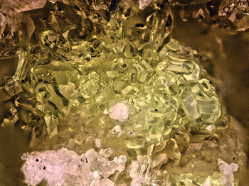
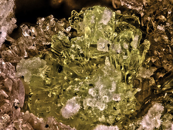
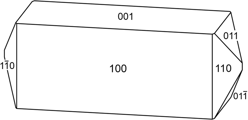
Pseudomeisserite-(NH4) crystals are stout prisms, up to ~0.3 mm long, typically occurring in tightly intergrown aggregates (Figs 1 and 2). Prisms are elongate on [010] and exhibit the forms {100}, {001}, {110} and {011} (Fig. 3). No twinning was observed.
Intergrown short prisms of pseudomeisserite-(NH4) with white ivsite. The field of view is 0.84 mm across.

Intergrown prisms of pseudomeisserite-(NH4) on bladed changoite with white sprays of ferrinatrite. The field of view is 1.0 mm across.

Crystal drawing of pseudomeisserite-(NH4); clinographic projection in standard orientation.

The mineral is light yellow with very pale yellow streak and fluoresces bright lime green under 405 nm (long-wave) ultraviolet (UV) laser illumination (for examining micro-crystals, it is often most effective to use lasers, but they are not readily available in short-wave; however, uranyl minerals that fluoresce under long-wave UV generally fluoresce similarly under short-wave). Crystals are transparent and have a vitreous lustre. The tenacity is brittle, the Mohs hardness is 2½ (based on scratch tests), the fracture is curved or conchoidal and perfect cleavage on {100} was observed. The density measured by flotation in methylene iodide–toluene is 3.22(2) g⋅cm–3. The calculated density is 3.277 g⋅cm–3 for the empirical formula and 3.289 g⋅cm–3 for the ideal formula. At room temperature, the mineral is easily soluble in H2O.
Optically, pseudomeisserite-(NH4) is biaxial (–), with α = 1.536(2), β = 1.559(2) and γ = 1.565(2) (measured in white light). The 2V measured directly on a spindle-stage is 53(1)°; the calculated 2V is 53.5°. Dispersion is r > v, distinct. The mineral is pleochroic: X colourless, Y light yellow and Z pale yellow (X < Z < Y). The optical orientation is Z = b and Y ∧ c = 33° in obtuse β. The Gladstone-Dale compatibility, 1 – (K p/K c), is 0.008 (superior) based on the empirical formula (Mandarino, Reference Mandarino1981).
Raman spectroscopy
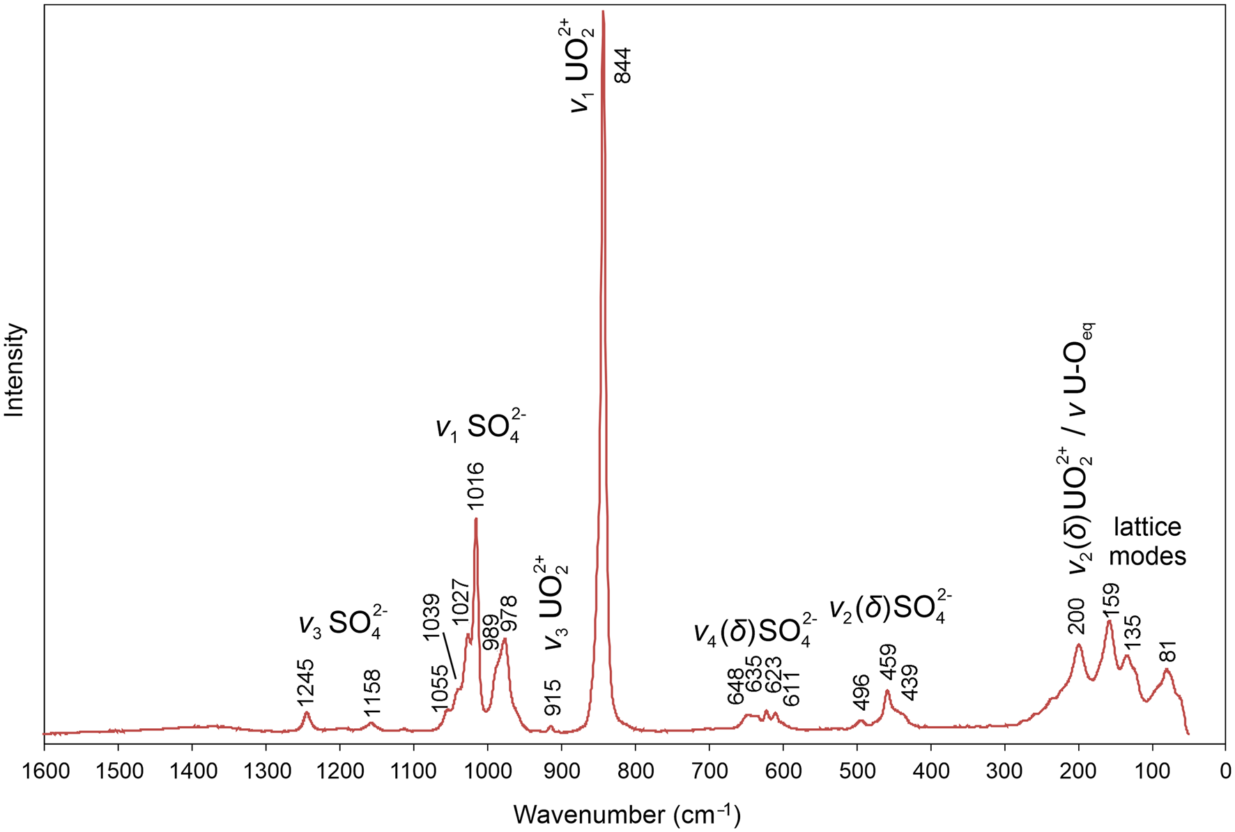
Raman spectroscopy was conducted on a Horiba XploRA PLUS. Pronounced fluorescence was observed using a 532 nm diode laser; consequently, a 785 nm diode laser was utilised. The spectrum, recorded from 1600 to 50 cm–1, is shown in Fig. 4.
The Raman spectrum of pseudomeisserite-(NH4) recorded with a 785 nm laser.

The ν3 (SO4)2– antisymmetric stretching vibrations occur as weak bands at 1245 and 1158 cm–1. Several weak to strong bands/shoulders between 1055 and 978 cm–1 are assignable to the ν1 (SO4)2– symmetric stretching vibrations. The multiple bands observed in this region are consistent with the presence of several symmetrically unique SO4 tetrahedra in the pseudomeisserite-(NH4) structure. The weak band at 915 cm–1 is related to the ν3 (UO2)2+ antisymmetric stretching vibrations. The ν1 (UO2)2+ symmetric stretching vibration is present as a very strong band at 844 cm–1. Bartlett and Cooney (Reference Bartlett and Cooney1989) provided an empirical relationship to derive the approximate U–OUr bond lengths from the band position assigned to the ν1 (UO2)2+ stretching vibration, which gives 1.77 Å, in excellent agreement with U–OUr bond lengths from the X-ray data: 1.763(4), 1.771(4), 1.765(4) and 1.769(4) Å.
Four weak bands between 648 and 611 cm–1 are attributed to the ν4 (δ) (SO4)2– bending vibrations, and those at 496, 459 and 439 cm–1 to the ν2 (δ) (SO4)2– bending vibrations. A band at 200 cm–1 is attributable to the ν2 (δ) (UO2)2+ bending vibrations and/or possibly to ν U–Oeq bending modes. The remaining bands are due to unassigned phonons.
Chemical composition
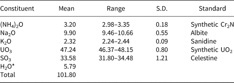
Chemical analyses (four points on four crystals) were performed at the University of Utah on a Cameca SX-50 electron microprobe with four wavelength dispersive spectrometers and using Probe for EPMA software. Analytical conditions were 15 kV accelerating voltage, 10 nA beam current and a beam diameter of 5 μm. Raw X-ray intensities were corrected for matrix effects with a ϕρ(z) algorithm (Pouchou and Pichoir, Reference Pouchou and Pichoir1991). Time-dependent intensity corrections were applied to Na, U and S. Crystals suffered severe beam damage during analyses. Because insufficient material is available for a direct determination of H2O, it has been calculated based upon the structure determination (U = 2 apfu, O = 28 apfu and charge balance). Analytical data are given in Table 1.
Chemical composition of pseudomeisserite-(NH4).

* based on the structure; S.D. – standard deviation.
The empirical formula (calculated on the basis of 28 O apfu) is (NH4)1.49K0.60Na3.87U2.00S5.04O28H7.78. The simplified formula is (NH4,K)2Na4[(UO2)2(SO4)5]⋅4H2O. The ideal formula is (NH4)2Na4[(UO2)2(SO4)5]⋅4H2O, which requires (NH4)2O 4.27, Na2O 10.16, UO3 46.87, SO3 32.80 and H2O 5.90, total 100 wt.%.
X-ray crystallography and structure refinement
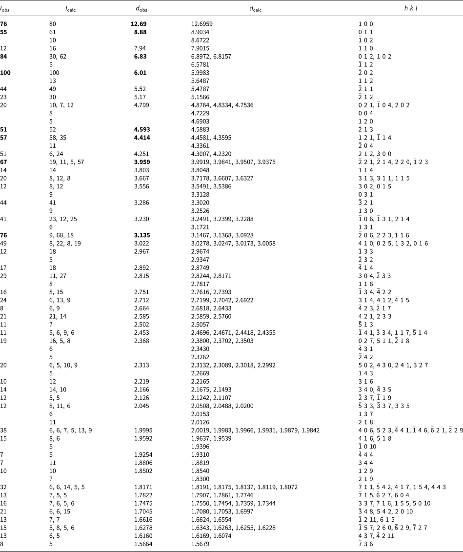
Powder X-ray studies were carried out using a Rigaku R-Axis Rapid II curved imaging plate microdiffractometer, with monochromatic MoKα radiation. A Gandolfi-like motion on the φ and ω axes was used to randomise the samples and observed d values and intensities were derived by profile fitting using JADE 2010 software (Materials Data, Inc.). The observed powder data for pseudomeisserite-(NH4), presented in Table 2, show good agreement with the pattern calculated from the structure refinement. The unit-cell parameters refined from the powder data using JADE 2010 with whole pattern fitting are a = 13.129(7), b = 10.108(7), c = 19.534(7) Å, β = 104.315(12)° and V = 2512(2) Å3.
Powder X-ray data (d in Å) for pseudomeisserite-(NH4)*.

* Only calculated lines with I > 4 are listed. For I > 3 see the supplemental material. The strongest lines are given in bold.
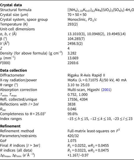
Single-crystal X-ray studies were done using the same diffractometer and radiation used for the powder studies. The Rigaku CrystalClear software package was used for processing the structure data, including the application of an empirical absorption correction using the multi-scan method with ABSCOR (Higashi, Reference Higashi2001). The structure was solved by the charge-flipping method using SHELXT (Sheldrick, Reference Sheldrick2015a). SHELXL-2016 (Sheldrick, Reference Sheldrick2015b) was used for the refinement of the structure. Two large-cation sites were refined with full joint occupancies by K and N (NH4). The N1 site refined to N0.386K0.614(9), while the N2 site refined to N0.767K0.233(10). A difference-Fourier syntheses located all H atom positions associated with the H2O groups, which were then refined with soft restraints of 0.82(3) Å on the O–H distances and 1.30(3) Å on the H–H distances and with the U eq of each H set to 1.2 times that of the donor O atom. However, the H atom positions associated with the NH4 cations were not resolved at this stage of the refinement.
The equivalent isotropic displacement parameters (U eq) of the N1 and N2 sites refined to 0.042 and 0.056, respectively, significantly greater than those of the Na sites (0.021 to 0.037). The total of the refined joint occupancies of the N1 and N2 sites provided a large-cation content of [(NH4)1.15K0.85]Σ2.00, compared with the much more NH4-rich content of [(NH4)1.49K0.60]Σ2.09 obtained from the electron-probe microanalyses (EPMA). These factors suggested that the N1 and N2 sites actually have more N and less K than the refinement indicated. To explore this, we adjusted the occupancies to N0.50K0.50 and N0.91K0.09, respectively, so that these sites refined to U eq values of ~0.031 and provided a total content of [(NH4)1.41K0.59]Σ2.00. The R 1 increased from 0.0251 to 0.0259; however, potential H positions associated with the N sites were revealed. When these were incorporated into the refinement, R 1 decreased to 0.0253. In the final refinement, with H positions included, the joint N/K occupancies of the N1 and N2 sites were again refined, yielding N1: N0.446K0.554(10) and N2: N0.880K0.120(11), with a final total large-cation content of [(NH4)1.33K0.67]Σ2, a final R 1 of 0.0252 and U eq values of 0.037 for the N sites. The H sites associated with the N1 and N2 sites were refined with soft restraints of 0.90(3) Å on the N–H distances and 1.45(3) Å on the H–H distances and with the U eq of each H set to 1.5 times that of the donor N atom; the occupancies of the H sites were tied to those of the corresponding N sites.
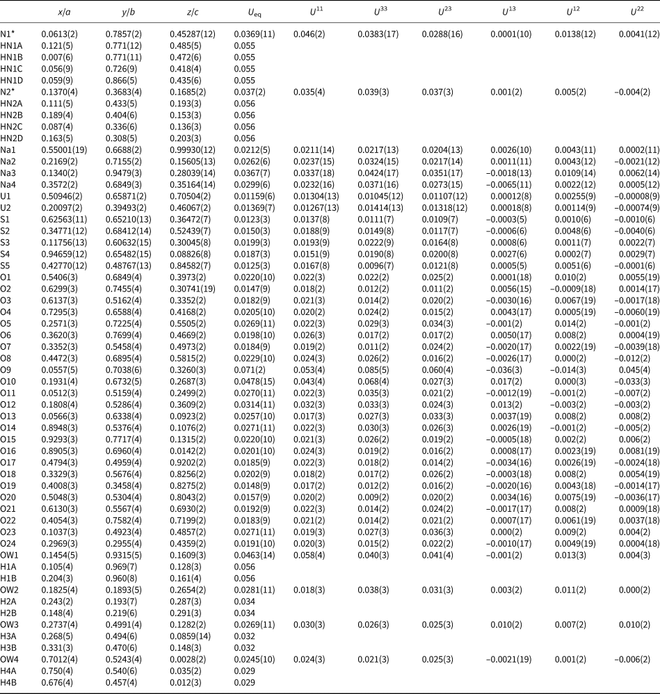
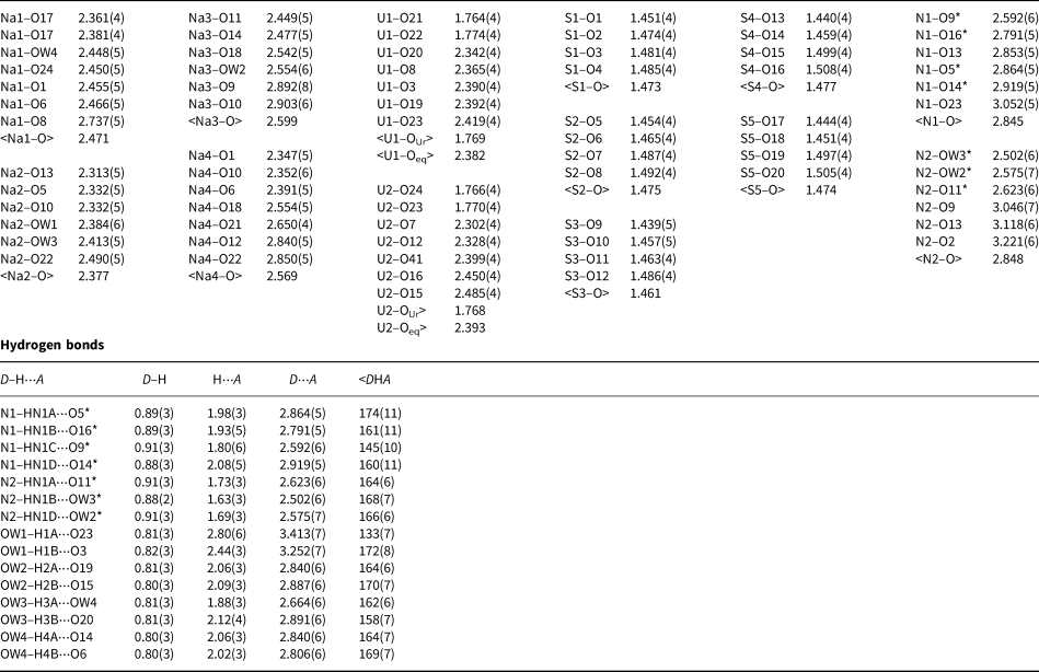
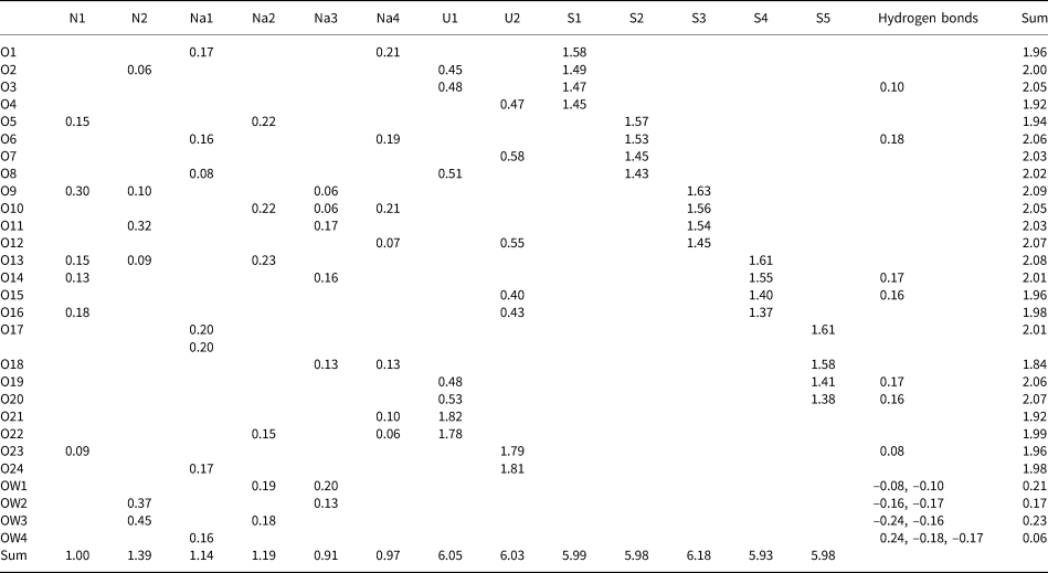
Data collection and refinement details are given in Table 3, atom coordinates and displacement parameters in Table 4, selected bond distances in Table 5 and bond-valence summations (BVS) in Table 6. The crystallographic information files have been deposited with the Principal Editor of Mineralogical Magazine and are available as Supplementary material (see below).
Data collection and structure refinement details for pseudomeisserite-(NH4).

R int = Σ|F o2–F o2(mean)|/Σ[F o2]. GoF = S = {Σ[w(F o2–F c2)2]/(n–p)}½. R 1 = Σ||F o|–|F c||/Σ|F o|. wR 2 = {Σ[w(F o2–F c2)2]/Σ[w(F o2)2]}½; w = 1/[σ2(F o2)+(aP)2+bP] where a is 0.0115, b is 8.0215 and P is [2F c2+Max(F o2,0)]/3.
Atom coordinates and displacement parameters (Å2) for pseudomeisserite-(NH4).

* Refined occupancies: N1: N0.446K0.554(10) and N2: N0.880K0.120(11)

Bond valence analysis for pseudomeisserite-(NH4). Values are expressed in valence units.*

* The N1 and N2 site bond valences are based on refined joint occupancies by K and N. NH4+–O bond valence parameters are from García-Rodríguez et al. (Reference García-Rodríguez, Rute-Pérez, Piñero and González-Silgo2000). All other cation–O bond valence parameters are from Gagné and Hawthorne (Reference Gagné and Hawthorne2015). Hydrogen-bond strengths are based on O–O bond lengths from Ferraris and Ivaldi (Reference Ferraris and Ivaldi1988).
Description of the structure
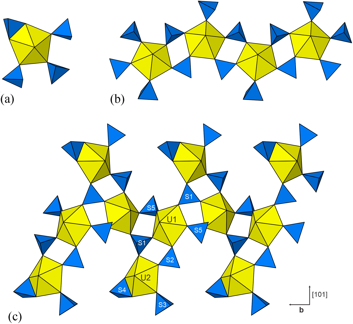
The U1 and U2 sites in the structure of pseudomeisserite-(NH4) are each surrounded by seven O atom sites forming pentagonal bipyramids. This is a most frequent coordination for U6+ in mineral and inorganic structures where the two short apical bonds of the bipyramid constitute the uranyl group (cf. Burns, Reference Burns2005). The two apical O atoms of the bipyramids (OUr) form short bonds with the U, and this unit comprises the UO22+ uranyl group. Five equatorial O atoms (Oeq) complete each of the U coordinations. All Oeq atoms bound to U also participate in SO4 groups. The U1 bipyramid is surrounded by five SO4 groups [centred by S1 (×2), S2 and S5 (×2)], each of which shares one Oeq corner of the U1 bipyramid. The U2 bipyramid is surrounded by four SO4 groups, three of which (centred by S1, S2 and S3) share single Oeq corners and one of which (centred by S4) shares two equatorial O atoms, a polyhedral edge. Such a bidentate linkage between a UO7 pentagonal bipyramid and a SO4 tetrahedron has been previously reported in the Na-uranyl-sulfate minerals klaprothite, péligotite and ottohahnite (Kampf et al., Reference Kampf, Plášil, Kasatkin, Marty and Čejka2017b) and lussierite (Kampf et al., Reference Kampf, Olds, Plášil, Nash and Marty2019; Fig. 5a), all of which occur at the Blue Lizard mine.
(a) The [(UO2)(SO4)4]6– cluster in lussierite. (b) The [(UO2)(SO4)3]4– chain in meisserite. (c) The [(UO2)2(SO4)5]6– band in pseudomeisserite-(NH4).

The U1 bipyramids are linked to one another by pairs of SO4 tetrahedra (centred by S1 and S5) to form a chain along b. This chain is topologically identical to those in meisserite (Plášil et al., Reference Plášil, Kampf, Kasatkin, Marty, Škoda, Silva and Čejka2013); Fig. 5b) and fermiite (Kampf et al., Reference Kampf, Plášil, Kasatkin, Marty and Čejka2015), both of which occur at the Blue Lizard mine. However, in the structure of pseudomeisserite-(NH4), each U1 bipyramid in the chain is also linked to a U2 bipyramid by pairs of SO4 tetrahedra (centred by S1 and S2), thereby yielding a [(UO2)2(SO4)5]6– uranyl-sulfate band (Fig. 5c) having its plane parallel to {10 $\bar{1}$}. This band is the structural unit in the structure of pseudomeisserite-(NH4). To our knowledge, no similar structural unit has been previously reported in the structure of any mineral or synthetic compound. The complexity of U–S polyhedral linkage contributes apparently to the overall complexity of the pseudomeisserite-(NH4) structure. With total complexity of 1329.90 bits/cell (including hydrogen atoms), pseudomeisserite-(NH4) is one of the very complex uranyl minerals (after Krivovichev, Reference Krivovichev2013) and belongs to group of most complex uranyl sulfates (Gurzhiy and Plášil, Reference Gurzhiy and Plášil2019). Based on the topological type of structural unit, it falls in the group of structures that are based on dense chains of polyhedra, such as alwilkinsite-(Y) (Kampf et al., Reference Kampf, Plášil, Čejka, Marty, Škoda and Lapčák2017a) and uranopilite (Burns, Reference Burns2001).
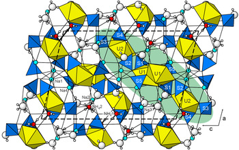
The uranyl sulfate bands are linked through a complex network of bonds involving two large-cation sites (N1 and N2) occupied jointly by NH4 and K, four Na sites (Na1, Na2, Na3 and Na4) and four H2O-group sites (OW1, OW2, OW3 and OW4). The K1, K2 and Na2 sites are six coordinated, while the Na1, Na3 and Na4 sites are seven coordinated. The entire structure is shown in Fig. 6, with one uranyl-sulfate band highlighted.
The crystal structure of pseudomeisserite-(NH4) viewed down b. The Na–O and NH4/K–O bonds are shown as thin black lines. Hydrogen bonds are not shown. The [(UO2)2(SO4)5]6– band is highlighted with a green background. The unit cell is indicated by dashed lines.

The N1 site appears to accommodate roughly equivalent amounts of NH4 and K, while the N2 site exhibits a significantly greater affinity for NH4 over K. There is no obvious explanation for the preference of the N2 site for NH4; in fact, NH4 prominence in the N2 site is counter-indicated by bond-valence considerations (considering NH4 as a spherical cation), as the high BVS for the site (1.39 valence units) would be close to neutral (1.02 vu) with occupancy by K alone. It is worth noting that the bond-valence values reported in Table 6 follow the approach of Garcia-Rodriguez et al. (Reference García-Rodríguez, Rute-Pérez, Piñero and González-Silgo2000), which treats NH4+ strictly as a spherical cation. Other authors (cf. Khan and Baur, Reference Khan and Baur1972) have studied ordered hydrogen bonding exhibited in some NH4 coordinations. Catti and Franchini-Angela (Reference Catti and Franchini-Angela1976) noted that, for short H⋅⋅⋅O bonds with large N–H⋅⋅⋅O angles, the NH4+ group behaves as an ordered hydrogen-bond donor, while long H⋅⋅⋅O distances and relatively small N–H⋅⋅⋅O angles are indicative of behaviour as a strongly electropositive large alkali-like cation. It seems reasonable to assume that NH4–O bonds corresponding to ordered hydrogen bonds provide somewhat greater bond strength than those corresponding to electrostatic bonds, and that this should be particularly true for relatively short N–H⋅⋅⋅O bonds. The N1 and N2 coordinations in the structure of pseudomeisserite each include ordered hydrogen bonds (see Table 6). The N1 site has four such bonds and the N2 site has three. That suggests that the N1 site might be more favourable to NH4 over K; however, the fact that the three ordered N–H⋅⋅⋅O bonds for the N2 site are particularly short may make this site more favourable for NH4 occupancy.
Supplementary material
To view supplementary material for this article, please visit https://doi.org/10.1180/mgm.2020.17
Acknowledgements
Structures Editor Peter Leverett, reviewer Igor Pekov and an anonymous reviewer are thanked for their constructive comments on the manuscript. We are grateful to retired miner Dan Shumway of Blanding, Utah, for advice and assistance in our collecting efforts in Red Canyon. A portion of this study was funded by the John Jago Trelawney Endowment to the Mineral Sciences Department of the Natural History Museum of Los Angeles County. This research was also financially supported by the Czech Science Foundation (project GACR 20-11949S to JP).