Head and neck lipomas are very rare, with most being localised to the posterior neck. A lipoma that extends into the mediastinum is even more uncommon, with only three cases being documented in children. Internal jugular phlebectasia is another atypical condition that occurs in children, often manifested by a lateral neck bulge. Having a cervicomediastinal lipoma with coexisting internal jugular vein phlebectasia is the rarest with only one other case being documented. Reference Tawil and Azizkhan1 In this case report, we demonstrate a cervical–mediastinal lipoma with coexisting internal jugular vein phlebectasia.
Case report
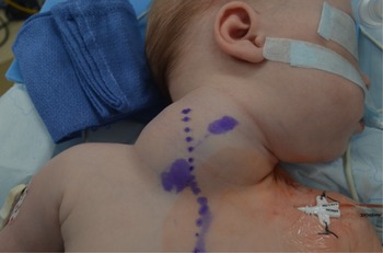
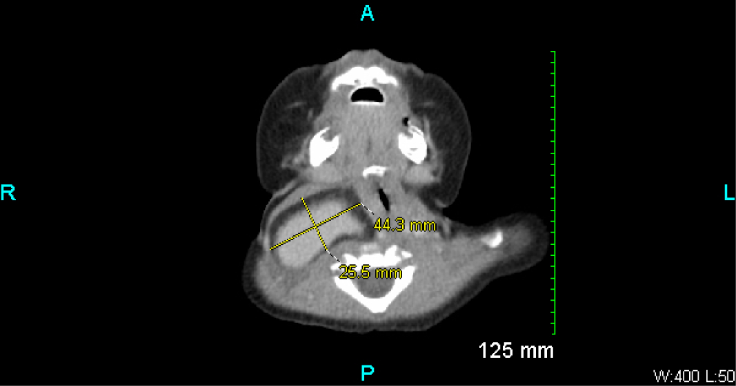
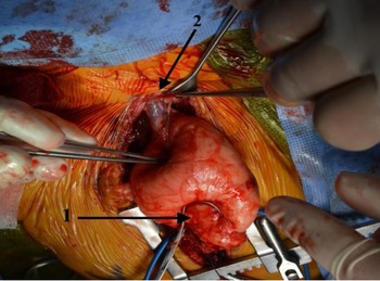
A 6-month-old female, previous 35-week preterm infant, presented with difficulty breathing and stridor that had progressively worsened over the last week with introduction of solid foods. She had no history of stridor, dysphagia, choking or trouble feeding until the start of pureed food. Patient had a 5-week NICU stay for respiratory distress syndrome and had multiple chest X-rays during that time. There was no obvious neck pathology at that time. Patient was brought to the emergency department due to severe retractions with audible noisy breathing and was given racemic epinephrine and Decadron without relief of symptoms. During the transfer of care, it was noticed that patient had a slight bulge on the right side of her neck (Fig 1). Parents had started to notice a subtle difference in the appearance of the right and left side of her neck around her fourth month of life. At this time, a CT was ordered and showed dysplastic dilation of the internal jugular vein surrounded by fat, which was causing significant tracheal deviation with stenosis down to 2 mm at certain points (Fig 2). The surrounding fat measured up to 18 mm thick and extended into the right superior mediastinum. A neck ultrasound confirmed massive dilation of the right internal jugular vein consistent with phlebectasia with large peripheral fat. The radiologist could not determine if there was lipomatous expansion of the vessel wall itself. The right-sided neck mass was increasingly noticeable when the patient cried or tried to sit up. Due to the resulting airway constriction, surgical removal was necessary. Surgery was performed by paediatric cardiothoracic, general and otolaryngology surgeons. A pre-operative bronchoscopy revealed at least 80% compression of the trachea. A median sternotomy was necessary due to the involvement of the distal jugular vein in the mediastinum. First the thymus was dissected and removed followed by dissection of the mass. The mass was not invading any surrounding structures and was well-circumscribed with discrete borders. The internal jugular vein was clamped proximal to the innominate vein and roughly 2 cm below the jugular foramen (Fig 3). The vein was surgically ligated at both ends, and the mass was removed in its entirety. The mass structure and the thymus were sent to pathology. The report revealed unremarkable thymic tissue with no evidence of malignancy. The mass consisted of mature adipose tissue with focal brown fat and intersecting hypocellular bands of fibrosis, consistent with a lipoma with necrosis. At the end of the procedure, a repeat bronchoscopy demonstrated that the trachea compression was relieved. Patient recovered well post-operatively and is followed by ENT and paediatric surgery in the outpatient setting. Focused physical examination revealed no evidence of right-sided cervical venous distension. A neck ultrasound is scheduled during the follow-up period.
Pre-operative right side neck mass.

CT scan of mass measuring 44.3 mm by 25.5 mm, showing the tracheal deviation.

Intra-operative resection of mass with distal (cardiac) portion of right internal jugular vein clamped (arrow 1) and proximal end dissected (arrow 2).

Discussion
A large neck mass in a child presents a diagnostic challenge with a lengthy list of potential diagnoses. Only a few of these diagnoses are associated with a visual increase in size related to increased vagal tone, including superior mediastinal tumours, laryngocele or jugular phlebectasia. Reference Kasim, Hassan, Hassan, Al-Mughairi, Yassin and Rashad2 This patient presented with two of the three possible diagnoses. Jugular vein phlebectasia is described as congenital dilatation of the jugular vein due to deficiencies in the vessel wall. Reference Kasim, Hassan, Hassan, Al-Mughairi, Yassin and Rashad2,Reference Baker, Ingraham, Fine, Iyer and Monroe3 The most common presentation is on the right side. This is thought to be due to the contact between the right innominate vein and the right apical pleura in which any increase in intrathoracic pressure is directly transferred to the right internal jugular vein. Reference Kasim, Hassan, Hassan, Al-Mughairi, Yassin and Rashad2 Another theory is that the right innominate vein travels in the same direction as the superior vena cava leading to higher pressures during inspiration. Conversely, the cause of internal jugular phlebectasia is thought to be unknown with only 206 paediatric cases since 1928. Reference Figueroa-Sanchez, Ferrigno, Benvenutti-Regato, Caro-Osorio and Martinez4 A systemic review of all the current data on internal jugular phlebectasia found no definitive association with previous neck trauma, positive-pressure ventilation or tumours. Reference Figueroa-Sanchez, Ferrigno, Benvenutti-Regato, Caro-Osorio and Martinez4,Reference Sander, Eliçevik, Ünal and Vural5 The most commonly reported venous aneurysms were that of the superior vena cava, however upper extremity aneurysms tend to be underreported due to their mostly indolent nature. Reference Baker, Ingraham, Fine, Iyer and Monroe3 Typically, the treatment for internal jugular phlebectasia is conservative in nature with most surgery occurring for cosmetic benefit.
Lipomas of the head and neck are extremely uncommon with most being localised to the posterior neck or spinal cord. In this case, the lipoma extended into the anterior mediastinum. Mediastinal lipomas are also very rare, and most are found incidentally on chest imaging. There have been two cases of congenital lipomas reported in the literature. Reference Eryılmaz, Yücel, Yücel and Arıcıgil7 Given the size of the patient’s lipoma at the time of discovery, it is plausible that this was a congenital lipoma. This is only the second case of a lipoma with an associated venous aneurysm in a child that has been described. Both cases involved young female patients with compressive neck symptoms due to the increasing size of their lipomas. There was no clear association of the jugular aneurysm and the lipoma in that initial case, but the mass had similar histological features to the case we present here. Tawil and Azizkhan believed in their case, and the two phenomenon were interrelated due to the dilated vascular channels throughout the adipose tissue. Reference Tawil and Azizkhan1 Much like their report, our patient’s jugular vein had channelling into the surrounding fat and filling the lipoma with blood.
There are reports of other vessel abnormalities, such as aneurysm, in conjunction with lipoma growth in various parts of the body. The most commonly reported lipoma with aneurysmal component is in the brain. There are multiple case reports of cerebral arteries with saccular aneurysm, and lipomas occur at the site of aneurysmal tissue. Reference Futami, Kimura and Yamashita8 There are also cases of ventricular aneurysm associated with cardiac lipomas. In the lipomas of cardiac origin, it is most often noted that the lipoma arises from the subpericardium or myocardium. Reference Song, Hickey, Nabi and Chang9 All other reports of lipomas and aneurysms are in arterial or muscular tissue. Here in our case, the aneurysm was venous.
We are proposing that the dilatory change in vascular wall structure may cause fat proliferation to occur. Another hypothesis is that the lipoma caused weakening of the vessel wall and led to dilation. This could occur if the lipoma inhibits or blocks certain growth factors for the smooth muscle causing weakening during haemodynamic stress. It was discussed that the possibility of the lipoma taking on characteristics of a liposarcoma due to the invasive nature it had with the internal jugular vein. However, the lipoma did not invade any other surrounding structure in the patient’s neck. There is a paucity of formal research regarding the association of lipoma and aneurysm due to the scarcity of the condition.
We believe that the main cause of jugular vein phlebectasia in children is due to congenital deficiencies in the vessel wall. If this is the case in our patient, we can conclude that the dilation of the vessel caused a rapid expansion of the surrounding adipose tissue.
Acknowledgements
None.
Financial support
This research received no specific grant form any funding agency, commercial or not-for-profit sectors.
Conflicts of interest
None.