Introduction
Diodontus Curtis, 1834 (Hymenoptera: Pemphredonidae) is a genus of terricolous, solitary, predatory wasps (family status according to Sann et al. Reference Sann, Niehuis, Peters, Mayer, Kozlov and Podsiadlowski2018), represented by 76 described species (Pulawski Reference Pulawski2018). The highest species diversity is observed in Palaearctic grasslands and deserts. The genus is less diverse in the Nearctic and Afrotropical regions. It is only marginally represented in northwestern Indomalaya and, to the current knowledge, missing from the Australasian and Neotropical regions.
Recent revisions of the European representatives of Diodontus (Dollfuss et al. Reference Dollfuss, Bouček, Bitsch and Bitsch2001; Olszewski et al. Reference Olszewski, Ljubomirov, Wiśniowski, Kowalczyk and Krzyżyński2016) do not include species described from North Africa and western Asia, therefore, the status of multiple Mediterranean species is not completely clear. An identification key for Diodontus from this region is not available. A review of DNA barcodes available from the Barcode of Life Data System (www.boldsystems.org) Barcode Index Number (BIN) database (Ratnasingham and Hebert Reference Ratnasingham and Hebert2007, Reference Ratnasingham and Hebert2013) and new unpublished data of the authors demonstrate that several widespread species of Diodontus actually represent complexes of morphologically cryptic taxa that need a revision with the application of molecular characters.
The goals of this publication are description of two new western Mediterranean species from the species-rich D. minutus (Fabricius, 1793) species group, reconstruction of their phylogenetic relationships, assessment of selected DNA markers for their use in species recognition, and presentation of a provisional identification key to the described Mediterranean species of this group.
Material and methods
Specimens
The studied material and the type specimens of the new species described here are deposited in the following collections:
CCSE – Collection of Christian Schmid-Egger, Berlin, Germany
CJG – Collection of Joseph Gusenleitner, Linz, Austria
CKS – Collection of Konrad Schmidt, Heidelberg, Germany
COB – Collection of Øistein Berg, Haslum, Norway
CSG – Collection of Severiano F. Gayubo, Salamanca, Spain
CUP – Charles University, Department of Zoology, Prague, Czech Republic
MNHN – Muséum National d’Histoire Naturelle, Paris, France
MSNG – Civic Museum of Natural History Giacomo Doria, Genova, Italy
MZH – Finnish Museum of Natural History, Helsinki, Finland
NHMUK – Natural History Museum, London, United Kingdom
NHMW – Natural History Museum, Vienna, Austria
NRCV – Nature Research Centre, Vilnius, Lithuania
OLML – Biologiezentrum-Oberösterreichisches Landesmuseum, Linz, Austria
OUMNH – Oxford University Museum of Natural History, Oxford, United Kingdom
RMNH – Naturalis Biodiversity Centre, Leiden, the Netherlands
TUZ – Natural History Museum, University of Tartu, Estonia
ZMAN – Zoological Museum Amsterdam, the Netherlands
ZMUC – Copenhagen Zoological Museum, Denmark
Morphological characters
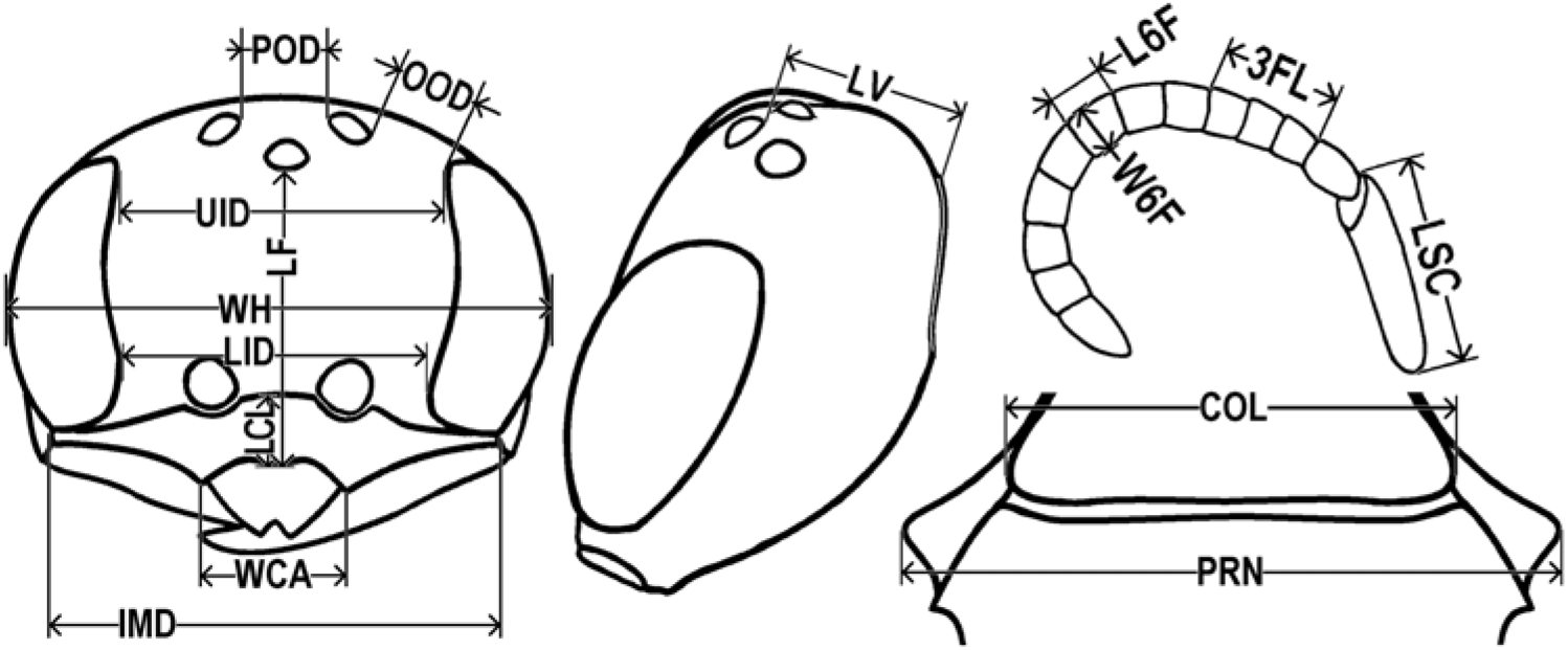
We used morphometric measurements (Fig. 1) based on Budrys (Reference Budrys and Jonaitis1996). Width of head is defined as the maximum visible width in dorsal or frontal view; length of face is defined as the distance between fore margin of mid-ocellus and midpoint of the lower margin of the clypeus when viewed at a right angle; lower interocular distance is defined as the shortest distance between inner margins of eyes approximately at the level of antennal sockets in frontal view; upper interocular distance is defined as the shortest distance between inner margins of eyes approximately at the level of mid-ocellus in frontal view; length of vertex is defined as the shortest distance between hind margin of mid-ocellus and midpoint of occipital carina when viewed at a right angle; post-ocellar distance is defined as the shortest visible distance between inner margins of hindocelli when measured at a right angle; oculo-ocellar distance is defined as the shortest visible distance between outer margin of hindocellus and margin of eye when measured at a right angle; intermandibular distance is defined as the distance between outer margins of genal swellings just above the fore mandibular condyles in anterodorsal view; length of clypeus is defined as the distance between midpoint of frontoclypeal suture and midpoint of the lower margin of the clypeus when viewed at a right angle; width of clypeal apex is defined as the distance between the tips of lateral teeth on the lower margin of clypeus; length of scape is the maximum visible length of the scape; length of flagellomeres 1–3 is at maximum visible combined length of first three flagellomeres, without pedicel; length of flagellomere 6 is at maximum visible length; width of pronotum is defined as the distance between tips of pronotal lobes in dorsal view; width of flagellomere 6 is at maximum visible width without the layer of trichoid sensilla; width of pronotum is defined as the distance between tips of pronotal lobes, dorsal view; width of pronotal collar is defined as the distance between lateral edges of transverse carina of pronotal collar, anterodorsal view.
Morphometric measurements used in Diodontus species descriptions. COL, width of pronotal collar; IMD, inter-mandibular distance; LCL, length of clypeus; LF, length of face; LID, lower interocular distance; LSC, length of scape; LV, length of vertex; L6F, length of flagellomere 6; OOD, oculo-ocellar distance; POD, post-ocellar distance; PRN, width of pronotum; UID, upper interocular distance; WCA, width of clypeal apex; WH, width of head; W6F, width of flagellomere 6; 3FL, length of flagellomeres 1–3.

The measurement was done using an ocular micrometer in a Nikon SMZ1000 binocular microscope (www.nikon.com). Images were obtained using digital camera (Nikon DS-Fi2) connected to the microscope. The images were stacked using the software CombineZP (Alan Hadley; https://alan-hadley.software.informer.com).
Kolmogorov–Smirnov test for normality of measurement distribution, analysis of variance, and canonical discriminant analysis of the measurements were performed using Statistica, version 8 (Statsoft, Tulsa, Oklahoma, United States of America). The subsets of measurements significantly contributing to the discriminant models for the separation of males and females were selected using backward stepwise procedure.
In the descriptions, the morphological terminology mostly follows Bohart and Menke (Reference Bohart and Menke1976), with the following exceptions: the term “orbital gland” is used instead of “orbital fovea.” This structure of female face is often convex in apoid wasps (Hymenoptera: Apoidea); therefore, the term “fovea” is misleading. The hypothesis of glandular nature of the orbital gland needs a focussed investigation, though it is supported by occasional presence of a layer of wax-like excrete on this structure in specimens of Diodontus and other wasp and bee genera (unpublished observations). Instead of “propodeal enclosure” we use the morphological term “metapostnotum.”
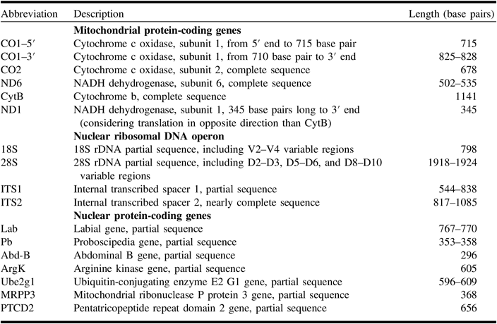
Selection of molecular characters
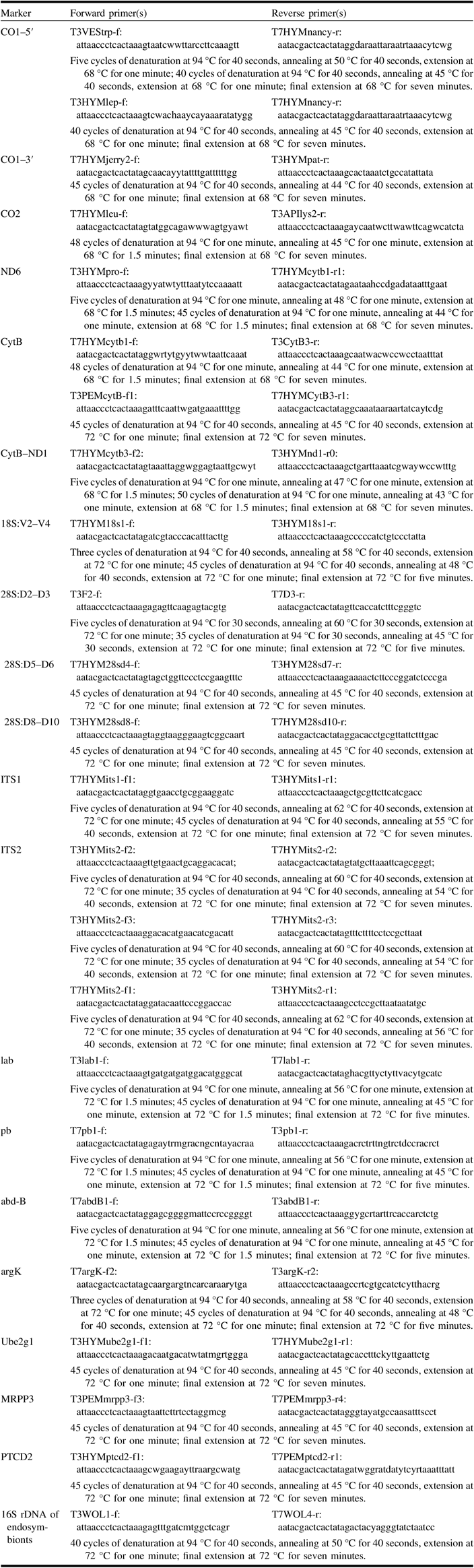
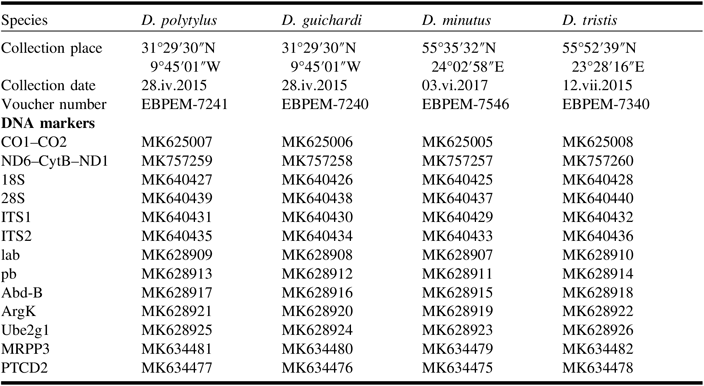
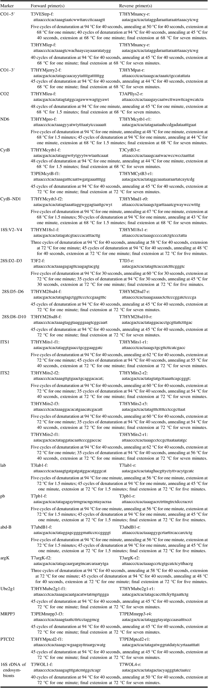
We used the mitochondrial DNA markers, namely cytochrome c oxidase subunits 1 and 2 (CO1 and CO2), NADH dehydrogenase subunits 1 and 6 (ND1 and ND6), and cytochrome b (CytB) genes; the ribosomal DNA (rDNA) operon; and selected protein-coding genes (Table 1). Among the latter, we studied the Hox genes that control early development of morphological character-rich anterior and posterior body segments, namely labial (lab), proboscipedia (pb), and abdominal B (Abd-B). The other nuclear markers were chosen from recently published sequences of 3256 protein-coding genes, obtained from the whole-body transcriptomes of 167 species of Hymenoptera (Peters et al. Reference Peters, Krogmann, Mayer, Donath, Gunkel and Meusemann2017, supplementary materials). After a preliminary assessment of the evolutionary rate by overall mean distance among the sequences, using MEGA, version 7 (Kumar et al. Reference Kumar, Stecher and Tamura2016), we selected two highly conserved genes, namely ubiquitin-conjugating enzyme E2 G1 (Ube2g1) and arginine kinase (ArgK), and two highly variable genes, namely mitochondrial ribonuclease P protein 3 (MRPP3) and pentatricopeptide repeat domain 2 (PTCD2). Pairs of newly constructed and tested T7-tailed and T3-tailed primers that were used for amplification of the studied DNA markers and their polymerase chain reaction conditions are listed in Table 2. In case of ITS2, which was difficult to amplify, we used different combinations of the listed forward and reverse primers, trying to obtain the best sequencing results. The exon-intron structure of the nuclear protein-coding genes was reconstructed comparing them with the structure of orthologous genes of Apis mellifera Linnaeus, 1758 (Hymenoptera: Apidae), available from GenBank (https://www.ncbi.nlm.nih.gov/genbank) (Benson et al. Reference Benson, Karsch-Mizrachi, Lipman, Ostell and Wheeler2005).
DNA markers used as molecular characters for Diodontus species identification and phylogeny reconstruction.

T7-tailed and T3-tailed primers used for the amplification of DNA markers and assessment of the presence of endosymbionts: name and sequence (5′–3′) and polymerase chain reaction conditions (all include an initial denaturation at 95 °C for five minutes).
Fresh material of most European Diodontus species suitable for the analysis of multiple genes was not available to us; therefore, the phylogenetic relationships of the newly described species with them were assessed using the CO1–5′ barcode sequences publicly available from the Barcode of Life Data System (Ratnasingham and Hebert Reference Ratnasingham and Hebert2007, Reference Ratnasingham and Hebert2013).
We also checked the samples for maternally inherited cytoplasmic incompatibility inducing endosymbionts. These intracellular bacteria may stimulate a divergence of mitochondrial clades, modify their evolutionary rates, and even act as speciation agents (Rokas Reference Rokas2000). We used original primers T3WOL1-f and T7WOL4-r (Table 2), which were successful in amplification of partial 16S rDNA of Wolbachia Hertig, 1936 (Rickettsiales: Anaplasmataceae) and Spiroplasma Saglio et al., 1973 (Entomoplasmatales: Spiroplasmataceae) from flight muscles of several apoid and vespoid wasps.
DNA extraction, polymerase chain reaction, and sequencing
DNA was extracted from thoracic muscle tissues of specimens preserved in 96% ethanol, using either the protocol of the GeneJet Genomic DNA Purification Kit (Thermo Fisher Scientific Baltics, Vilnius, Lithuania), or the method described by Stunžėnas et al. (Reference Stunžėnas, Petkevičiūtė and Stanevičiūtė2011). In the latter case, a piece of muscle tissue was placed in 50 µL 1× Tris-borate-EDTA (TBE) buffer (Thermo Fisher Scientific Baltics) on a sterile microscope glass slide with matt edge, homogenised using the matt edge of another slide, collected and incubated at 95 °C for 10 minutes, then at −20 °C for three minutes, and centrifuged at 12 000× g for five minutes. Afterwards, 25 µL of the supernatant buffer with the total DNA was transferred to a fresh tube and stored at −20 °C. The polymerase chain reaction mixture in a volume of 25 μL consisted of 12.5 μL of 2× polymerase chain reaction buffer (Thermo Fisher Scientific Baltics), 2.5 μL of 10 pmol of each primer, 7.5 μL of deionised water, and 1 μL of genomic DNA. Original T7-tailed and T3-tailed forward and reverse polymerase chain reaction primers were constructed and tested, preliminarily assessing their features with online BiSearch Primer Design and Search Tool (Tusnády et al. Reference Tusnády, Simon, Váradi and Arányi2005). Synthesis of the primers was accomplished at Macrogen (Seoul, South Korea). The primers and polymerase chain reaction conditions are presented in Table 2. The success of the polymerase chain reaction was assessed by electrophoresis on 1.5% agarose gel (Thermo Fisher Scientific Baltics). The polymerase chain reaction products were purified following the protocol of Exonuclease I/FastAP Thermosensitive Alkaline Phosphatase (Thermo Fisher Scientific Baltics) or using electrophoresis and the GeneJet Gel Extraction Kit (Thermo Fisher Scientific Baltics). They were sequenced at Macrogen (Seoul, South Korea) with the BigDye Terminator version 3.1 Cycle Sequencing Kit (Applied Biosystems, Foster City, California, United States of America). For sequencing, universal primers T7 (aatacgactcactatag) and T3 (attaaccctcactaaag) were applied.
Phylogeny reconstruction
The obtained nucleotide sequences were aligned using the BioEdit 7.2.5 software (Hall Reference Hall1999), either manually or with application of the ClustalW multiple alignment algorithm. The nucleotide substitution model most likely for the studied sequences was selected from the list of 24 models available in MEGA version 7 (Kumar et al. Reference Kumar, Stecher and Tamura2016), applying the maximum likelihood method and considering the lowest Bayesian Information Criterion score. The substitution patterns were best described by the general time reversible model with gamma distribution evolutionary rates (we used four discrete categories) and a fraction of invariable sites (GTR+G+I). We applied this model in reconstructions of phylogenetic relationships using the maximum likelihood algorithm, implemented in MEGA 7, and the method of Bayesian inference, using MrBayes, version 3.2.3 (Ronquist and Huelsenbeck Reference Ronquist and Huelsenbeck2003). For the latter analysis, the sequences were converted from Fasta to Nexus format using Mesquite 3.51 (Maddison and Maddison Reference Maddison and Maddison2015). We treated each gene and, where present, the combined sequence of exons and the combined sequence of introns of a gene, as separate sections. The GTR+G+I nucleotide substitution model was applied and the nucmodel parameter was set to “codon” for the sections of mitochondrial and nuclear protein-coding genes; the GTR+G nucleotide substitution model was applied and nucmodel parameter set to “4 × 4” for sections of tRNA, rDNA, internal transcribed spacers, and introns; the number of substitution types (nst) was set to “mixed”. For the reconstruction of phylogenetic relationships between two newly described species of Diodontus and D. minutus, Markov Chain Monte Carlo (MCMC) analyses were run for 500 000 generations, with tree sampling every 1000 generations; D. tristis (Vander Linden, 1829) was used as an outgroup. Reconstructing the phylogenetic tree of European Diodontus on the base of the available barcoding CO1–5′ sequences, we ran the Markov Chain Monte Carlo analyses for 3 000 000 generations, with tree sampling every 5000 generations; Passaloecus corniger Shuckard, 1837 (Hymenoptera: Pemphredonidae) was used as an outgroup. In both cases, the convergence between runs was considered sufficient after the standard deviation of split frequencies reaching a value 0.01.
MEGA 7 was used for the comparison of pairwise evolutionary divergence among the studied DNA markers, estimated as a percentage of base differences per site (p-distance); standard error estimates were obtained by a bootstrap procedure (10 000 replicates).
Results
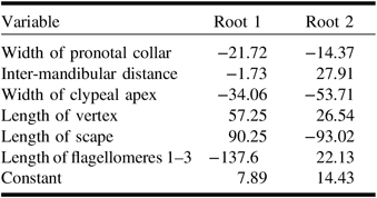
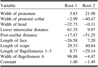
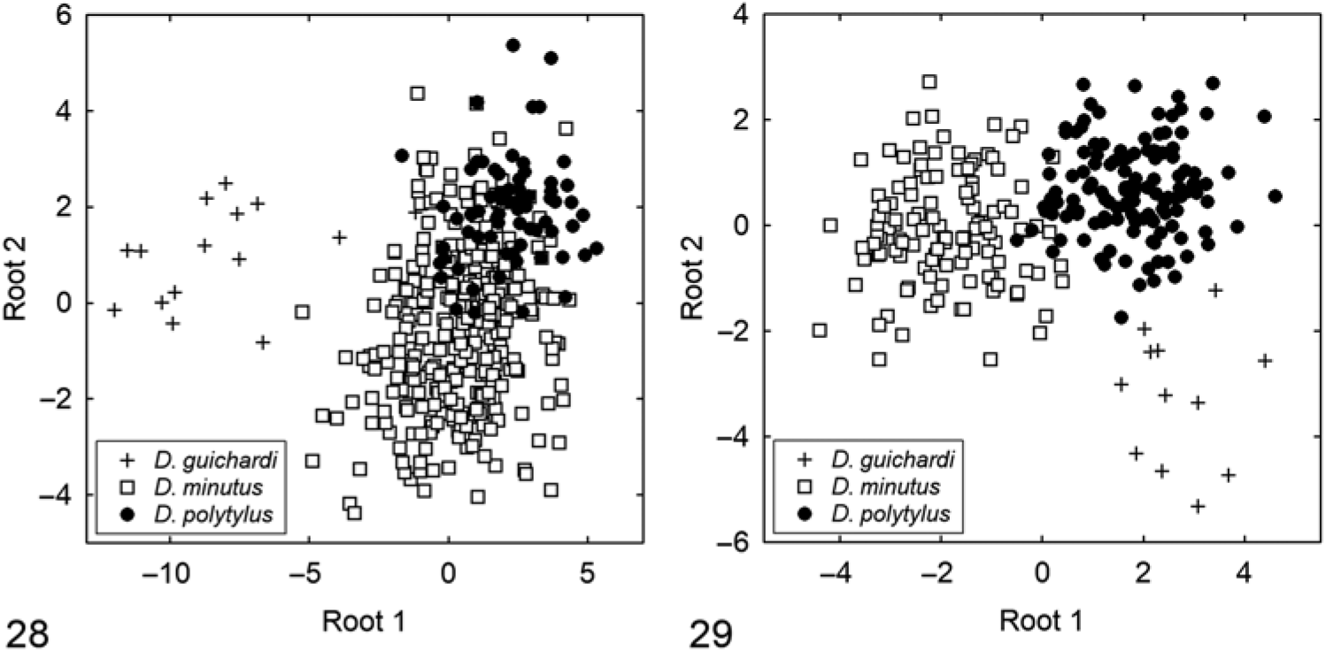
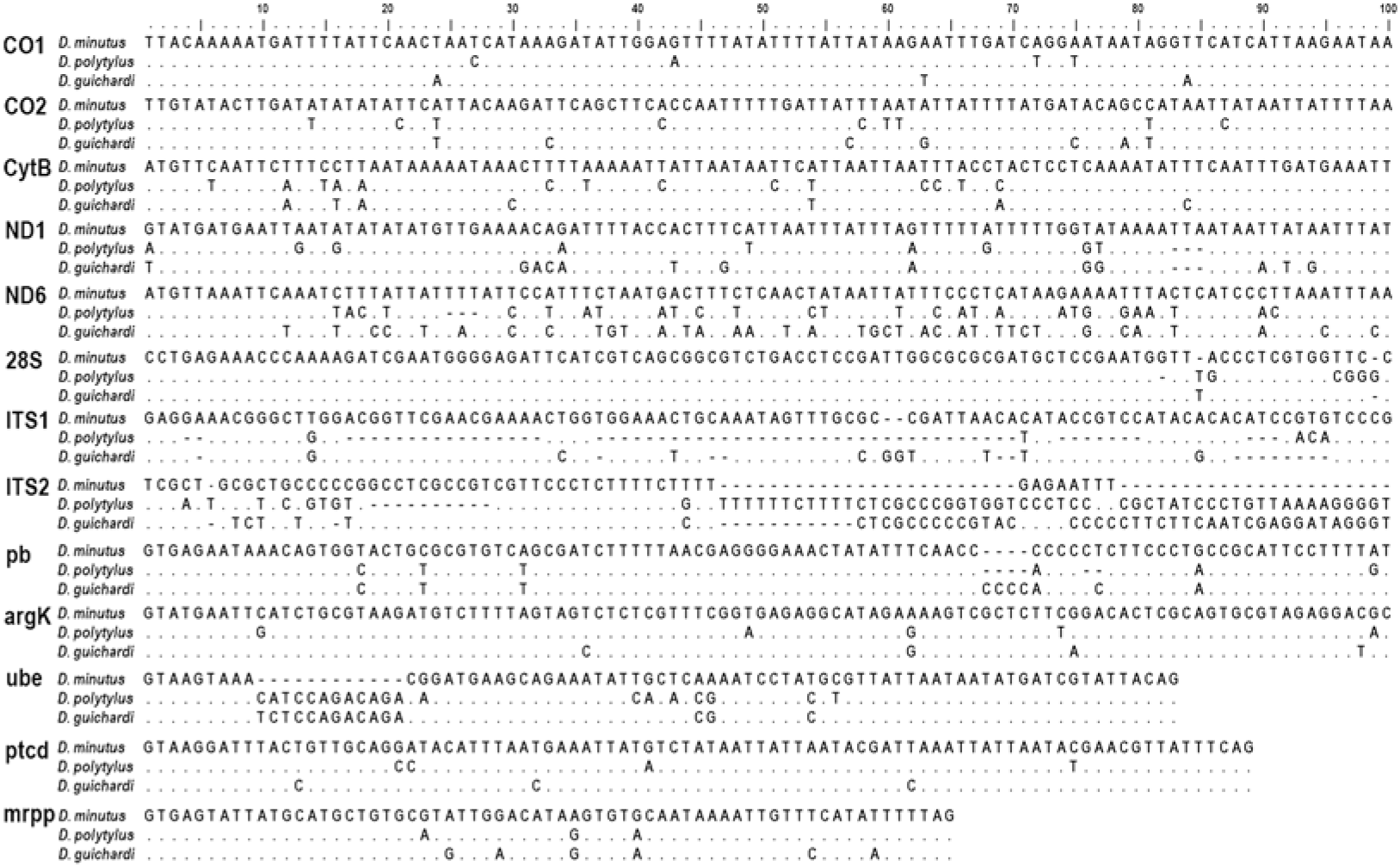
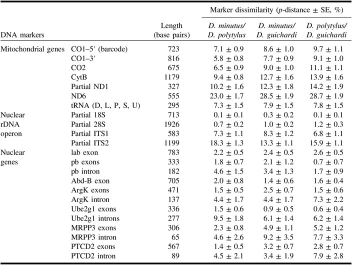
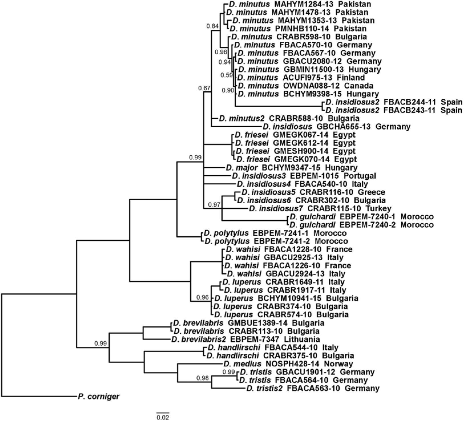
The two species described here as new were morphologically close to Diodontus minutus. Discriminant functions developed for morphometric measurements (Tables 3–4) allowed a correct identification of 87% of females (Fig. 28) and 97% of males (Fig. 29) of the three species. However, they could be reliably separated by multiple discrete molecular characters (Fig. 30). Pairwise evolutionary divergence of the DNA markers among the species is presented in Table 5. Reconstruction of phylogeny of European Diodontus species using the CO1–5′ barcode sequences available from the Barcode of Life Data System confirmed the evolutionary differentiation and genetic distinctness of the new species (Figs. 31–32). The obtained new DNA sequences were deposited in GenBank (Table 6).


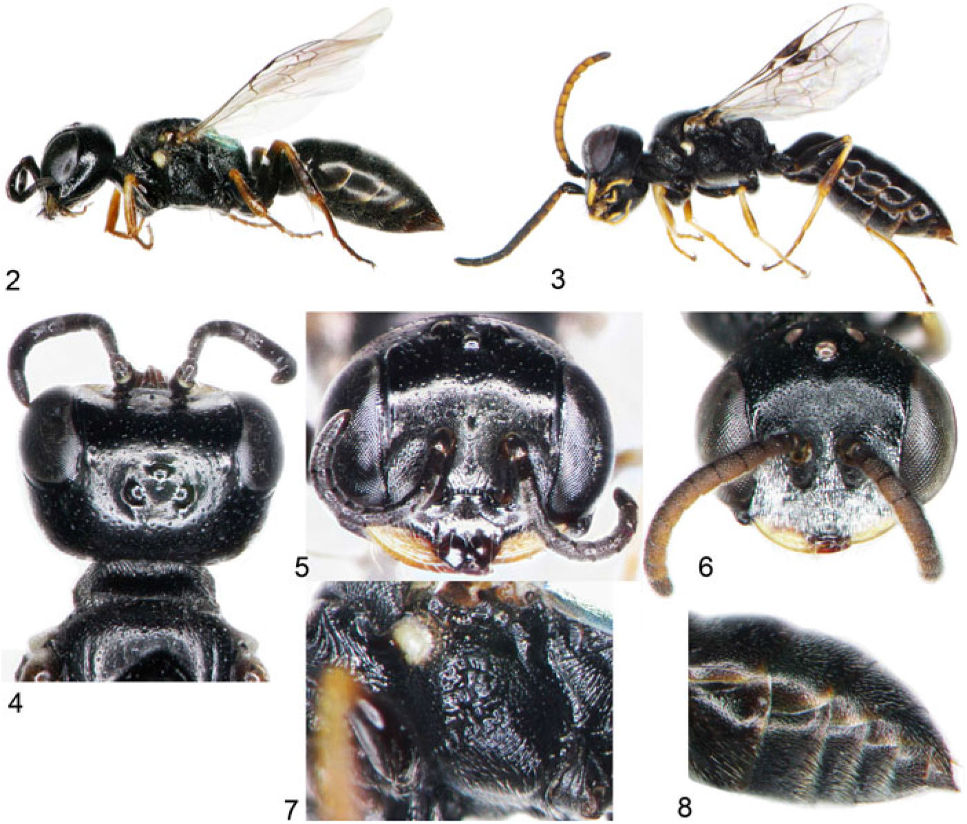
Diodontus polytylus new species. 2, Lateral habitus of female; 3, lateral habitus of male; 4, head, pronotum, and scutum of female; 5, face of female; 6, face of male; 7, mesopleuron of female; 8, pilosity of abdominal sterna in male.

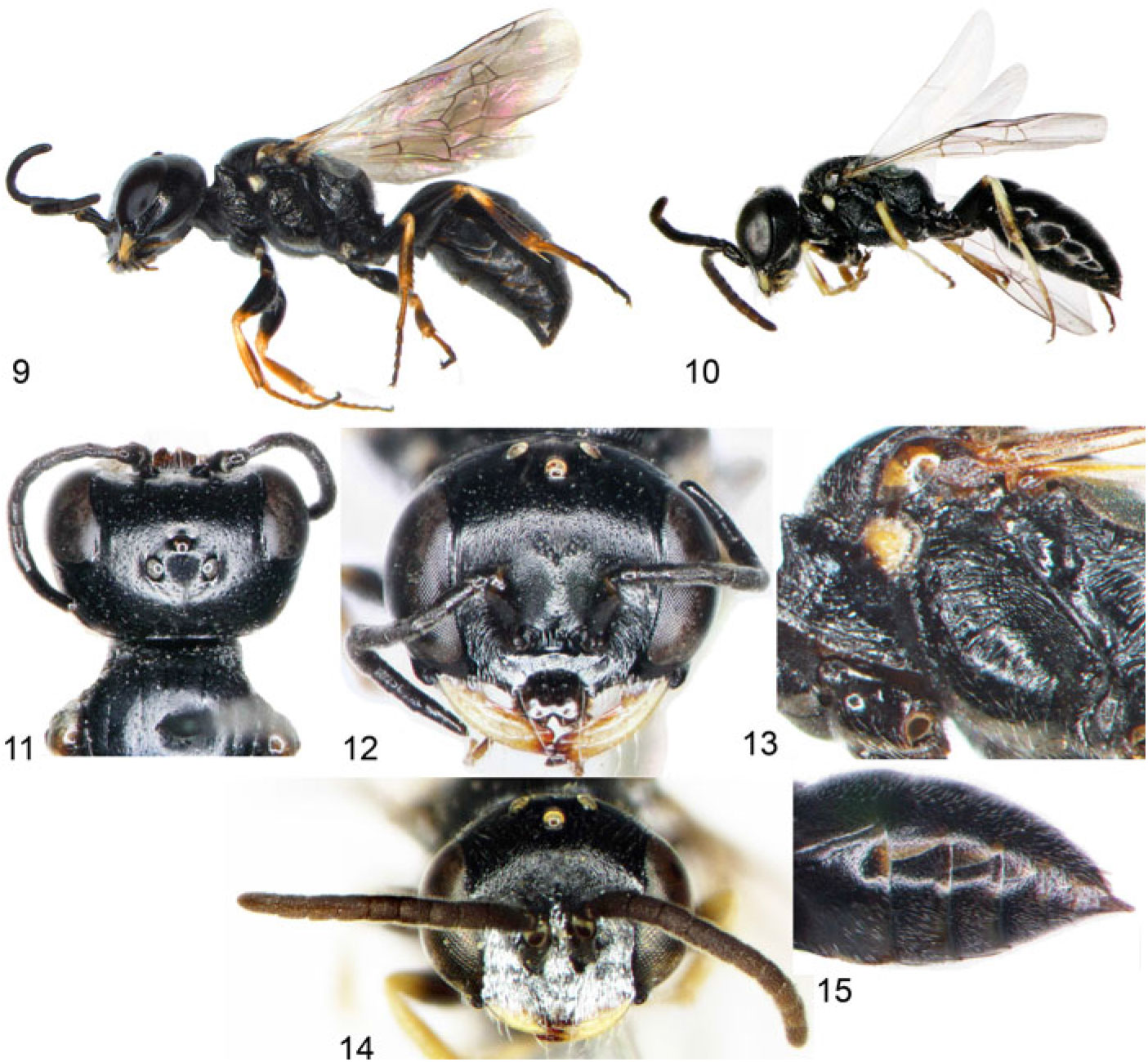
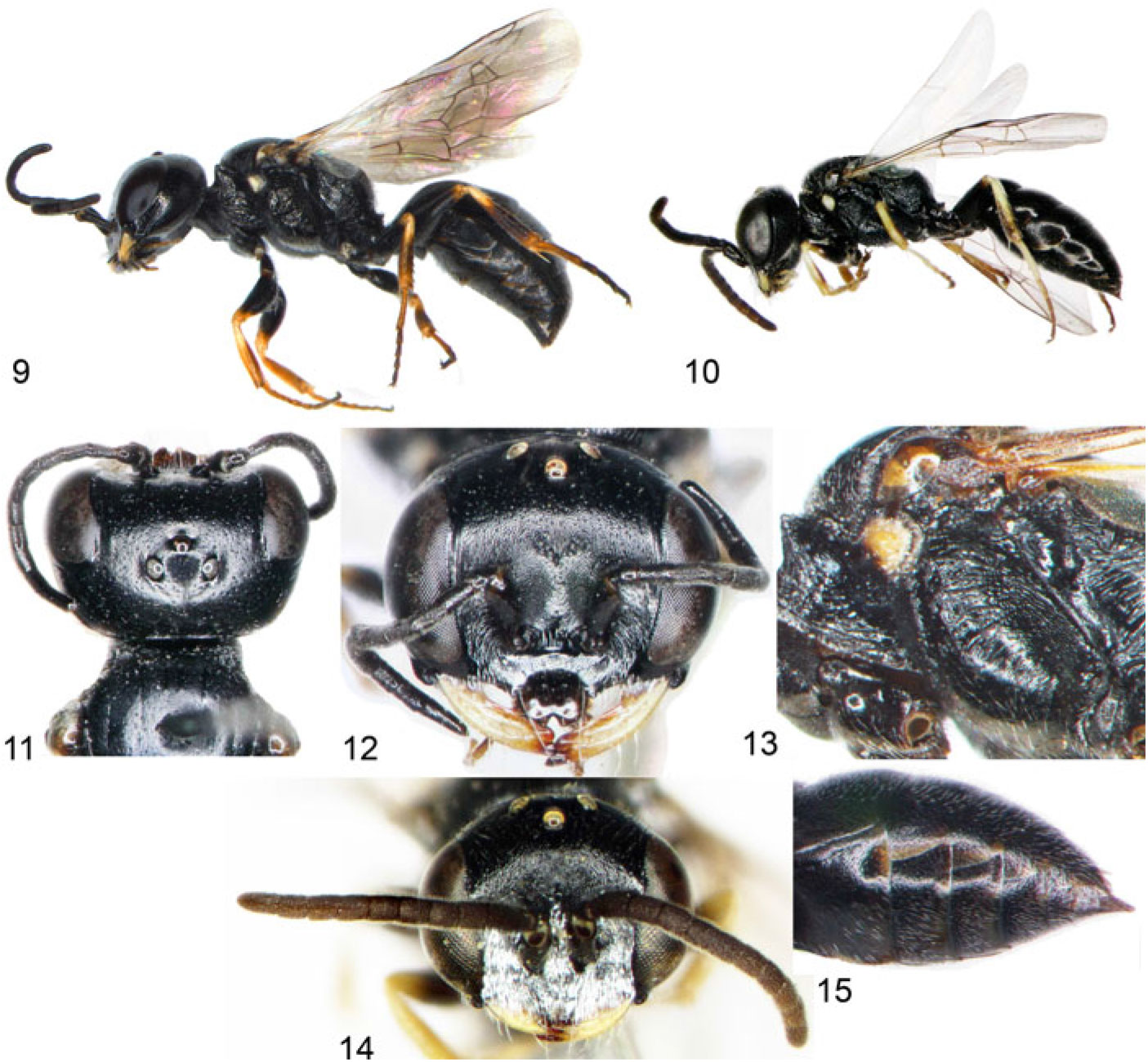
Diodontus guichardi new species. 9, Lateral habitus of female; 10, lateral habitus of male; 11, head, pronotum, and scutum of female; 12, face of female; 13, mesopleuron of female; 14, face of male; 15, pilosity of abdominal sterna of male.
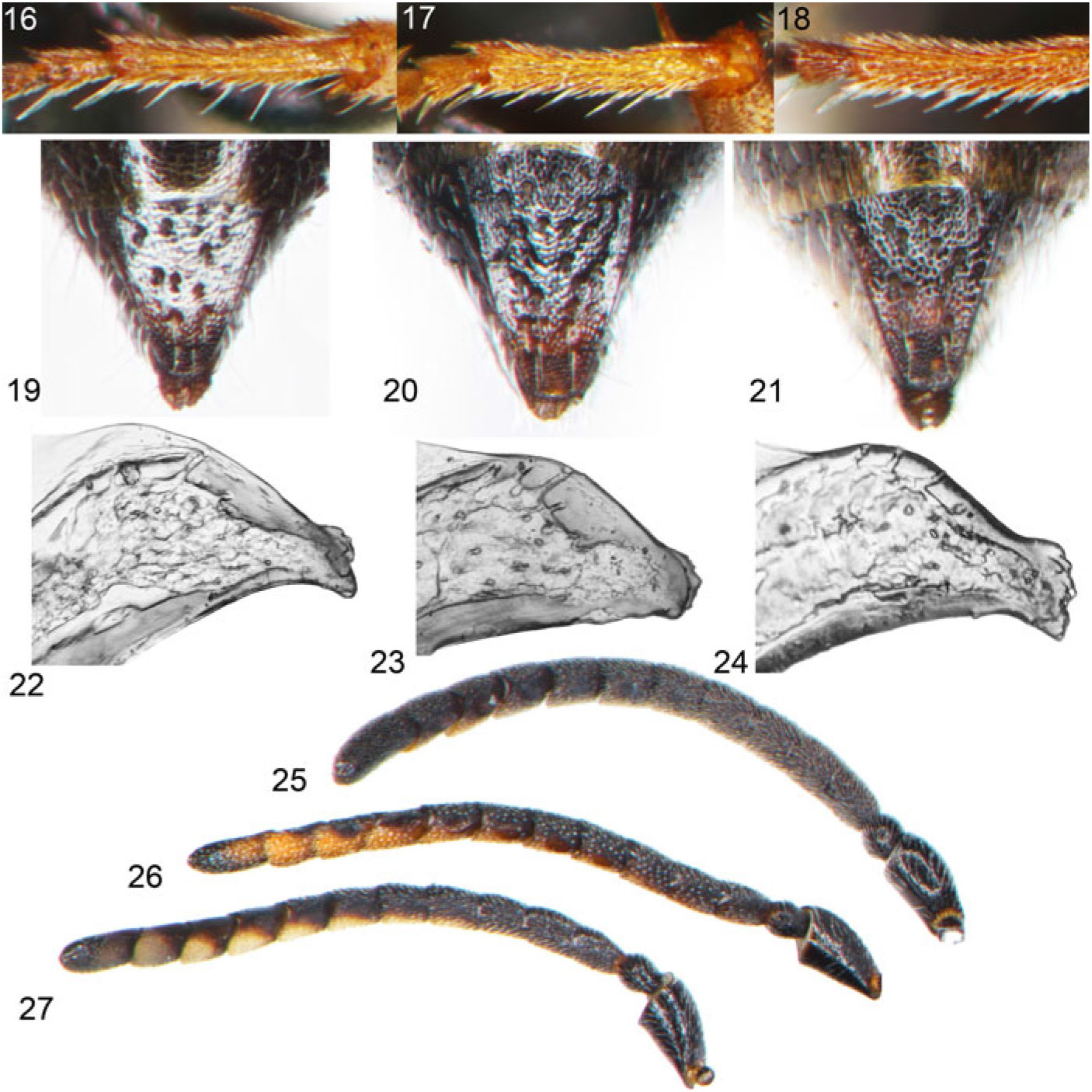
Diodontus comparison characters. 16, Diodontus guichardi probasitarsal rake of female; 17, D. polytylus probasitarsal rake of female; 18, D. minutus probasitarsal rake of female; 19, D. guichardi pygidial plate of female; 20, D. polytylus pygidial plate of female; 21, D. minutus pygidial plate of female; 22, D. guichardi apex of penial valve of male; 23, D. polytylus apex of penial valve of male; 24, D. minutus apex of penial valve of male; 25, D. guichardi antenna of male; 26, D. polytylus antenna of male; 27, D. minutus antenna of male.


Comparison of selected molecular characters of Diodontus polytylus and D. guichardi with those of D. minutus. CO1, 5′ end of mitochondrial cytochrome c oxidase, subunit 1; CO2, 5′ end of mitochondrial cytochrome c oxidase, subunit 2; CytB, 5′ end of mitochondrial cytochrome b; ND1, 3′ end of mitochondrial NADH dehydrogenase, subunit 1; ND6, 5′ end of mitochondrial NADH dehydrogenase, subunit 6; 28S, partial sequence of variable region D2 of 28S rDNA; ITS1, partial sequence of internal transcribed spacer 1 of nuclear rDNA operon; ITS2, partial sequence of internal transcribed spacer 2 of nuclear rDNA operon; pb, the lesser intron of proboscipedia gene; argK, intron of arginine kinase gene; ube, intron of ubiquitin-conjugating enzyme E2 G1 gene; ptcd, intron of pentatricopeptide repeat domain 2 gene; mrpp, intron of mitochondrial ribonuclease P protein 3 gene. Dots – identical nucleotides; dashes – deletions.

Pairwise evolutionary divergence between Diodontus minutus, D. guichardi, and D. polytylus, estimated as a percentage of base differences per site (p-distance) in selected DNA markers. Standard error estimates obtained by a bootstrap procedure (10 000 replicates).

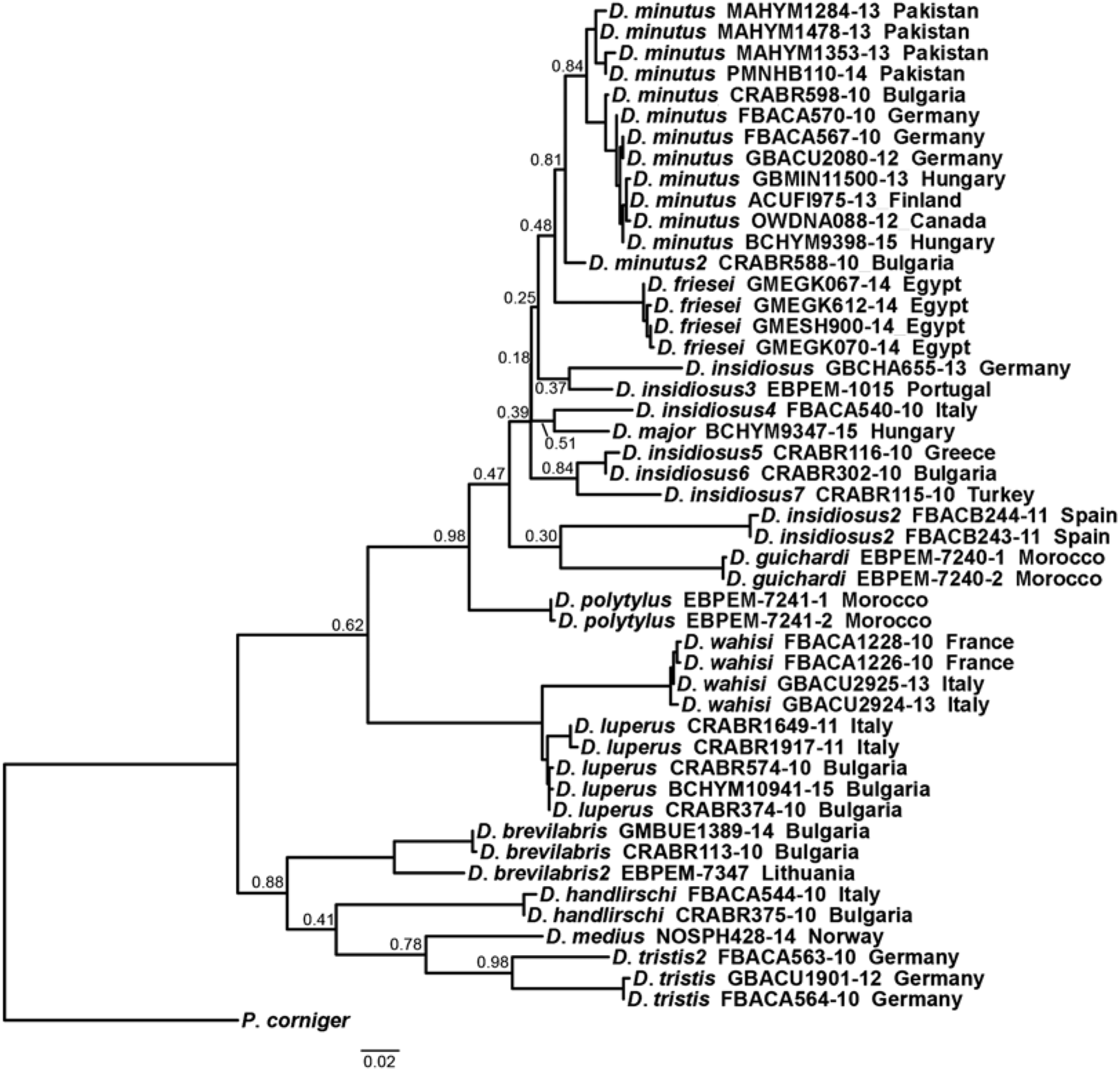
Maximum likelihood tree of Diodontus guichardi and D. polytylus with European species of Diodontus, reconstructed using the 658 base-pair-long molecular CO1 barcode sequence. Only mitotypes of species with available barcode sequence included; mitotype labels include the specimen process identification codes of the Barcode of Life Data System or (labels including EBPEM) the codes of specimens with unpublished barcode sequences from the database of the authors, and the collection country. Only probabilities of branches lower than 1.00 are presented. Passaloecus corniger was used as an outgroup.

Bayesian inference tree (see Fig. 31 title for details).

Accession numbers to DNA sequences of Diodontus specimens (vouchers in NRCV) deposited in GenBank (www.ncbi.nlm.nih.gov/genbank).

Diodontus polytylusBudrys, new species
http://zoobank.org/urn:lsid:zoobank.org:act:38F4DEC6-E993-47E9-9E1E-D157898E637A.
Type material. Holotype: ♂, Tunisia, Tozeur (33°55′N 08°08′E), 22.iv.1955, P.M.F. Verhoeff (RMNH).
Paratypes: Algeria: no exact locality data, 4.iv.1893, 4♂, 26.vi.1893, 1♂, 27.vi.1893, 2♂, Eaton (NHMUK); Ain Franine, 26.iv.1959, 1♀, J. Barbier (NHMW); Algiers, 6.iv.1893, 1♂, Eaton (NHMUK), 19.iv.1898, 1♂, Morice (OUMNH), 25.v.1947, 3♂, Rombout (RMNH); Annaba, 28.xi.1893, 1♀, Eaton, 10.viii.1897, 1♂ (no collector data) (NHMUK); Biskra, 27.iii.1895, 1♂, 7.iv.1897, 3♂, 20.iv.1897, 1♂ (no collector data) (NHMUK); El Harrach, iv.1943, 1♂, K.M. Guichard (NHMUK); Les Andalouses, 27.iv.1958, 2♂, J. Barbier (MNHN), 8.vi.1958, 3♀1♂, 22.vi.1958, 2♀1♂, J. Barbier (NHMW); 10 km E Remchi, 18.iv.1983, 10♂, R. Leys, P. van der Hurk (RMNH).
Chad: Zouar, 1.iv.1953, 1♀ (NHMUK).
Libya: Gharyan, 2.v.1951, 1♀, K.M. Guichard (NHMUK); Hun, 7.ii.1952, 1♀, K.M. Guichard (NHMUK); Querqarish, 6.iii.1952, 4♂, K.M. Guichard, 17.v.1952, 1♀, K.M. Guichard (NHMUK); Tarhunah, 22.vii.1955, 2♂, K.M. Guichard (NHMUK).
Morocco: Agadir, 23–26.v.1950, 3♂, P.M.F. Verhoeff (RMNH), 9.iii.1974, 5♀8♂ (4♀6♂ NHMUK, 1♀2♂ OUMNH), 4.iv.1974, 1♀, K.M. Guichard, G.R. Else (NHMUK), 10.ii.1988, 1♀, W. Perraudin (CCSE), 6.iii.1988, 1♀, W. Perraudin (cKS), 8.ii.1988, 1♂, K.M. Guichard (NHMUK), 11.iii.1989, 1♀, K.M. Guichard (NHMUK), 11.iv.2000, 1♀, Øistein Berg (COB); 12 km N Agadir, 21.iii.1987, 2♂, J. Gusenleitner (CJG); 30 km E Agadir, 29.iv.1980, 1♂, W. Perraudin (CCSE); Amouguer, 23.v.1995, 1♀4♂, M. Halada (OLML); Aoufouss, 24.iv.1987, 1♀1♂, M. Edwards (NHMUK); Aoulouz, 26.v.1995, 15♀18♂, C. Schmid-Egger (CCSE); Assafid, 20.iv.1988, 1♂, J. Gusenleitner (CJG); Azemmour, 28–30.v.1950, 3♀4♂, P.M.F. Verhoeff (RMNH); Boumalne, 21.iv.1987, 4♂, M. Edwards (NHMUK); El Gfifate, 28.iv.1987, 1♂, M. Edwards (NHMUK); Erfoud, 27.iv.1987, 8♂, K.M. Guichard, M. Edwards (NHMUK); 30 km N Errachidia (“Ksar-es-Souk”), 5.iv.1986, 1♂, K. Warncke (CKS); Essaouira, (no date), 3♂, R.E. Turner (NHMUK), 11.iv.1987, 1♀4♂, K.M. Guichard (NHMUK), 27.iv.1985, 1♂, LaRoche (CKS), 28.iv.2015, 12♂, yellow pan trap (fixed in ethanol for DNA study; voucher EBPEM-7241), V. Soon (4♂ NRCV, 8♂ TUZ); Goulimine, 20.iii.1989, 1♂ (NHMUK), 23.iii.1989, 1♂, K.M. Guichard (NHMUK); 10 km N Imilchil, 30.viii.1992, 1♀, K. Warncke (CKS); Imlil, 23.v.1995, 1♂, M. Hauser (CCSE); Laayoune, 13.ii.1988, 1♂, K.M. Guichard (NHMUK); Marrakech, 18–20.v.1950, 1♂ P.M.F. Verhoeff (RMNH), 19–21.vi.1968, 1♀, P.M.F. Verhoeff (RMNH); Mhamidas, 29.v.1995, 1♂, C. Schmid-Egger (CCSE); Mogador, (no date), 3♀1♂, R.E. Turner (NHMUK); Oued R’dat, 6.vi.1985, 4♀, K.M. Guichard (2♀ NHMUK, 2♀ OUMNH); Oued Sous, 31.iii.1983, 1♂, G.R. Else (NHMUK), 26.v.1995, 3♀1♂, M. Hauser (CCSE); Oulad Teima, 20.v.1995, 3♀1♂, M. Hauser (CCSE); Ounara, 10.iv.1987, 4♂, K.M. Guichard (NHMUK); Qued Issene, 23.iii.1987, 1♂, J. Gusenleitner (CJG); environs Sidi Abdeljelil, 8.v.1997, 3♀6♂, P. Prudek (CUP); Sidi-Ifni–Goulimine road, 6 km, 15.iii–1.iv.1974, 11♂, K.M. Guichard, G.R. Else (1♂ RMNH, 9♂ NHMUK, 1♂ NRCV); Sidi Moussa, 19.iv.1988, 1♀, J. Gusenleitner (CJG); Souk-el-Arba-du-Sahel, 26.iii.1987, 1♀, J. Gusenleitner (CJG); Tadouarte, 12.iv.1988, 1♀1♂, J. Gusenleitner (CJG); Tafraouten, 7.iv.1983, 1♂, G.R. Else (NHMUK); Taliouine, 26.v.1995, 2♀2♂, C. Schmid-Egger (CCSE); 10 km E Tan-Tan, 18.iii.1974, 1♂, K.M. Guichard, G.R. Else (NHMUK); Tansikht, 17.iv.1987, 1♀2♂, M. Edwards (NHMUK); 10 km SW Tazenakht, 30.v.1995, 2♀, C. Schmid-Egger (CCSE); Tinghir, 29.iv.1987, 1♀, M. Edwards (NHMUK); Tinzouline, 27.v.1995, 1♀, F. Brechtel (CCSE); 20 km N Tiznit, 24.iii.1987, 1♀, J. Gusenleitner (CJG); Zagora environs, 15.v.1997, 7♂, P. Prudek (CUP).
Portugal: Acoteias, 28.vii.1983, 1♀3♂, S.F. Gayubo (CSG); Aldeia da Tor, 27.vii.1983, 7♂, S.F. Gayubo (CSG); Freixedas, 11.viii.1994, 1♀, J. Garcia (CSG); Izeda, 2.viii.1993, 1♀, J. Garcia (CSG); Junqueira, 6.viii.1993, 1♀, J. Garcia (CSG); Lagoa de Albufeira, 1.viii.1967, 2♂, P.M.F. Verhoeff (RMNH); Lisbon environs, 20–30.v.1949, 1♂, P.M.F. Verhoeff (RMNH); Odeleite, 22.vii.1983, 2♀, S.F. Gayubo (CSG); Paderne, 30.vii.1983, 6♂, S.F. Gayubo (CSG); Resende, 16–19.vii.1953, 2♀, P.M.F. Verhoeff (RMNH); Ribabelida, 1.viii.1995, 1♂, J. Garcia (CSG); São Bartolomeu de Messines, 1.viii.1983, 1♀, S.F. Gayubo (CSG); São Miguel de Machede, 3.vii.1995, 1♂, J. Garcia (CSG); Silves, 2.viii.1983, 2♀, S.F. Gayubo (CSG); Trafaria, 17.vi.1954, 1♂, N.F. d’Andrade (RMNH), 21.vii.1950, 1♂, P.M.F. Verhoeff (RMNH); Vale Pereiro, 1.viii.1993, 1♀, J. Garcia (CSG).
Spain: Abadia, 2.v.1982, 1♂, S.F. Gayubo (CSG); Alora, 22.v.1967, 1♂, M.J. and J.P. Duffels (ZMAN); Azuaga, 23.vi.1985, 1♀, S.F. Gayubo (CSG); Castiejon, 10.ix.1988, 1♀, C. Heras (CSG); Cenes de la Vega, 14.vi.1989, 1♀1♂, S.F. Gayubo (CSG); Cuevas del Valle, 21.vii.1987, 2♀, S.F. Gayubo (CSG); Encinas de Abajo, 10.viii.1980, 1♀1♂, S.F. Gayubo (CSG); Gavilanes, 22.vii.1987, 2♂, S.F. Gayubo (CSG); Hoyos, 19.vi.1987, 1♂, F. Torres (CSG); Hornillos, 18.vii.1988, 1♂, C. Heras (CSG); La Bazana, 22.vi.1985, 1♂, S.F. Gayubo (CSG), 25.vi.1985, 1♂, S.F. Gayubo (CSG); Lastras de Cuellar, 4.ix.1986, 1♀, S.F. Gayubo (CSG); Miranda del Castañar, 12.viii.1987, 1♀, F. Torres (CSG); Monesterio, 15.vi.1985, 1♂, S.F. Gayubo (CSG); Montemolin, 14.vi.1985, 2♂, S.F. Gayubo (CSG), 15.vi.1985, 1♀3♂, S.F. Gayubo (CSG); Moron de la F., 11.vi.1989, 2♀, S.F. Gayubo (CSG); Pallares, 24.vi.1985, 5♂, S.F. Gayubo (CSG); Pedroso de la Carballeda, 29.vii.1984, 1♂, S.F. Gayubo (CSG); Penanegra Pass, 4.viii.1987, 1♀, S.F. Gayubo (CSG); Penaparda, 7.viii.1980, 2♂, S.F. Gayubo (CSG); El Puente de Sanabria, 1.viii.1984, 3♀1♂, S.F. Gayubo (CSG); Riomalo de Abajo, 13.viii.1987, 1♀, F. Torres (CSG); San Ciprian, 1.viii.1984, 1♀1♂, S.F. Gayubo (CSG); Saucelle, 25.iv.1980, 1♀, S.F. Gayubo (CSG), 27.iii.1981, 1♀1♂, S.F. Gayubo (CSG); Segura de Leon, 16.vi.1985, 2♂, S.F. Gayubo (CSG); Toledo, 25–27.vii.1967, 3♀ (RMNH), 16–18.viii.1967, 1♂ (RMNH), 2–3.vii.1968, 1♂, P.M.F. Verhoeff (RMNH), 26.v–5.vi.1979, 1♂, K.M. Guichard (NHMUK), 28.v.1979, 1♂, K.M. Guichard (NHMUK); Valdefuentes de Sangusin, 1.ix.1978, 5♀, S.F. Gayubo (CSG).
Tunisia: 3♀3♂, Tozeur (33°55′N 08°08′E), 22.iv.1955, P.M.F. Verhoeff (RMNH); Akarit, 19.v.1993, 1♀1♂, M. Hauser (CCSE), 18.vi.1994, 1♀, C. Schmid-Egger (CCSE); 5 km N Al Kaf, 22.vi.1994, 1♀, C. Schmid-Egger (CCSE); Bordj Toumi, 17.v.1992, 8♂, J. Gusenleitner (CJG); Bou Hedma National Park, 15–17.iii.1993, 1♂, M. Hauser (CCSE); Chenini, 15.iv.1988, 1♂, R.T.A. Schouten (RMNH); 5 km W Douz, 21.iii.2001, 1♂, C. Saure (CCSE); Fernana, 10.vii.1979, 2♂, J. Gusenleitner (CJG); Gabes, 21.iv.1971, 1♀, J. Gusenleitner (CSG), iv.1930, 4♂, R. Meyer (RMNH 2♂, NHMW 2♂); 20 km S Gabes, 18–19.ii.1986, 1♂ (ZMUC), 18–19.iii.1986, 1♂ (ZMUC); Gafsa, 21.vi.1994, 1♀1♂, C. Schmid-Egger, M. Hauser (CCSE); Hammamet, 26.iv.1955, 4♂, P.M.F. Verhoeff (RMNH); Houmt Souk, 29.iii.1994, 1♀, U. Willand (CCSE); 14 km SE Houmt Souk, 3.iv.1992, 1♀, J. Gusenleitner (CJG); Kasserine, 11.vi.1980, 1♂, K.M. Guichard (NHMUK); E of Kasserine, 9.v.2001, 3♀, P. Rosa (CCSE); Kebili, 10.v.1992, 5♂, J. Gusenleitner (CJG), 5.iv.1994, 5♂, U. Willand (CCSE); 10 km N Korba, 29.iii.1986, 2♀5♂ (ZMUC); 7 km W Korba, 27.v.1999, 1♂, O. and M. Niehuis (CCSE); Ksar Jouamaa, 26.iii.2001, 1♂, C. Schmid-Egger; Metouia, 20.vi.1994, 8♀2♂, C. Schmid-Egger, M. Hauser (CCSE); Nefta, 5.v.1973, 1♂, J. Gusenleitner (CSG), 14.iv.1981, 1♀, J. Gusenleitner (CJG), 13.v.1913, 1♂, Morice (OUMNH); Sousse, 30.iii.1989, 1♂, Hohmann (CKS); Tabarka, 5.viii.1978, 1♂, K.M. Guichard (NHMUK); Tamerza, 23.iii.2001, 1♂, C. Schmid-Egger (CCSE); 10 km NNW Thelepte, 24.iii.2001, 1♀1♂, C. Saure (CCSE); Tozeur, 8.v.1913, 2♂, Morice (OUMNH), 24–28.iii.1978, 2♀9♂, K. Guichard (OUMNH), 23.iii.2001, 1♀, C. Schmid-Egger (CCSE); Tunis, 1898, 2♂, Schmiedeknecht (RMNH, NHMW); Tunis environs, xi.1881, 1♀1♂ (MSNG), iv.1882, 2♂, G. and L. Doria (MSNG); Zarzis, 18.iii.1986, 3♀2♂ (ZMUC).
Diagnosis. The female of D. polytylus closely resembles females of D. minutus, D. longicornis Beaumont, 1960, D. oraniensis (LePeletier, 1845), and D. guichardi new species in the body sculpture, colouration, and the distance between the lateral teeth of the lower edge of clypeus being approximately equal to the distance between this tooth and the eye. Diodontus polytylus differs from these species by a weaker alutaceous microsculpture, thus a more shiny appearance of the upper frons; the punctures of the upper frons are sparser and a bit larger than in the listed species, though smaller than, e.g., in D. friesei Kohl, 1901, and most of the specimens currently considered to be D. insidiosus Spooner, 1938. The molecular CO1–5′ barcodes from the Barcode of Life Data System ascribed to D. insidiosus demonstrate a barcode index number divergence (Schmid-Egger et al. Reference Schmid-Egger, Straka, Ljubomirov, Blagoev, Morinière and Schmidt2018) implying that this nominal species represents a complex of several cryptic undescribed species requiring a revision. The female of D. polytylus can be differentiated from D. longicornis by a smaller distance between the hindocelli and a distinctly convex dorsal part of the orbital gland, which is nearly flat in D. longicornis.
The male of D. polytylus differs from all other described species of Diodontus in the largest number of tyloidea present on flagellomeres 4–11 (Fig. 26). It differs from D. guichardi,D. longicornis, and D. oraniensis by a paler, yellowish-brown ventral side of flagellum and by the presence of dense pilosity on posterior margins of sterna 3–5 (Fig. 8), similar to that in D. minutus. The male of D. polytylus can be separated from D. minutus by a straight, not dilated mesobasitarsus, and from D. friesei by a finer punctation of the upper frons. In comparison to D. polytylus, males of most D. insidiosus specimens have a more rounded, less transverse head, a coarser punctation of upper frons, and a thicker flagellum with flagellomeres not longer than wide.
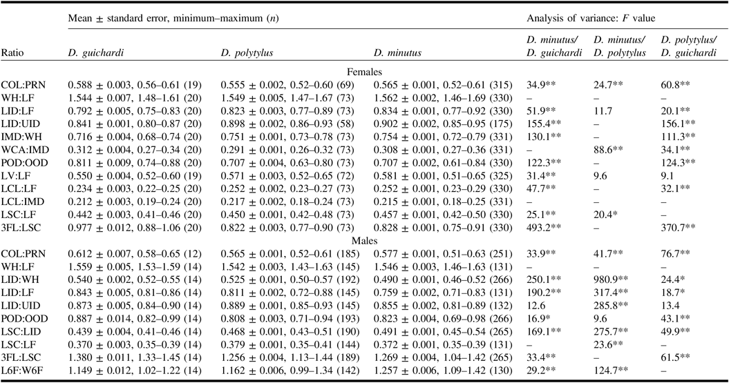
Comparing the body proportions of the described species to morphometrically similar D. minutus, D. polytylus has a slightly smaller pronotal collar in both sexes, a smaller distance between the lateral teeth of the lower edge of clypeus of female, shorter flagellomeres, inner orbits less converging ventrad, and a wider lower face of male (Table 7).
Selected morphometric ratios of Diodontus guichardi, D. polytylus, and D. minutus.

For the ratios that are different in pairwise comparisons (analysis of variance: Tukey’s honest significance test for unequal n; P < 0.05), F values are presented and marked by * if difference significant at P < 0.01, or ** if difference significant at P < 0.001.
See Fig. 1 for definitions of character abbreviations.
Female (Fig. 2). Body length 3.8–5.5 mm. Body black; spiracular lobe of pronotum and spot on tegula pale yellow; basal half of mandible, protibia anteriorly and mesotibia basally bright to dark yellow; distal half of mandible dark yellow to reddish brown; tip of mandible dark red to nearly black; tibia posteriorly and mesally brown to nearly black; lateral side of mesotibia brown to nearly black with more-or-less distinct brown line or a row of brown spots; metatibia basally pale yellow to dark yellow, laterally nearly black or sometimes with more-or-less distinct brown line, or a row of brown spots; tarsi pale brown to dark brown; pterostigma nearly black with dark brown strip along the edge of wing.
Head weakly transverse (frontal view; Fig. 5), width of head:length of face = 1.55 (here and hereafter the average value; a more detailed morphometric summary in Table 7). Face comparatively wide, lower interocular distance:length of face = 0.82. Inner orbits weakly converging ventrad, lower interocular distance:upper interocular distance = 0.90. Vertex strongly developed, length of vertex:length of face = 0.57. Ocelli forming obtuse triangle, post-ocellar distance:oculo-ocellar distance = 0.71 (Fig. 4). Frontal line in front of fore ocellus not impressed. Area of interantennal process nearly flat. Dorsal part of orbital gland moderately convex, prominence not wider than 0.5 width of hindocellus; mid-part narrower than distance between it and inner orbit. Mandibular condyles relatively widely separated, intermandibular distance:width of head = 0.75. Clypeus relatively long, length of clypeus:length of face = 0.25. Clypeal apex tridentate, moderately broad, width of clypeal apex:inter-mandibular distance = 0.29; mid-tooth slightly shorter than lateral teeth. Labrum subtriangular, with subelliptical apical notch. Scape moderately long, length of scape:length of face = 0.45. Flagellum comparatively short, length of flagellomeres 1–3:length of scape = 0.8. Upper frons smooth, moderately shiny, with distinct shallow irregularly scattered punctation; interspaces alutaceous, 2–6 times as wide as punctures. Pilosity of upper frons sparse, shorter than 0.5 width of ocellus. Pilosity of lower frons between eye and antennal socket pale grey, relatively sparse, not concealing an underlying microsculpture. Upper part of clypeus with relatively sparse short pilosity; shiny impunctate area above lower edge covers approximately 0.5 of the height of the clypeus.
Pronotal collar of moderate width, width of pronotal collar:width of pronotum = 0.55, with narrowly rounded sides and weakly convex dorsal carina (frontal view). Lateral surface of pronotal collar weakly shiny, rugulose. Probasitarsal rake weak (Fig. 17), with thinner preapical spines than in D. minutus (Fig. 18). Scutum smooth, moderately shiny, alutaceous, with sparse, irregularly scattered, fine but distinct punctation. Posteroventral part of mesopleuron smooth, coriaceous, weakly shiny, with distinct fine punctation, posteriorly more shiny, finely strigose (Fig. 7). Metapostnotum with fine but more-or-less distinct transverse wrinkle or carina between anterior horizontal surface and posterior pit. Propodeum partly shiny, moderately rugose, with small but distinct lateral angles (dorsal view), with thin carina separating lateral and posterior surfaces.
Gastral terga smooth, minutely punctate; tergum 1 strongly shiny, without microsculpture between punctures; remaining terga shiny, with fine alutaceous microsculpture. Pygidial plate subtriangular, coarsely punctate, moderately shiny, with distinct granulose microsculpture (Fig. 20).
Male (Fig. 3). Body length 2.7–4.3 mm (holotype 4.0 mm). Colouration similar to that of female, except of ventral side of flagellum yellowish brown and legs paler, with more extended pale-yellow markings on tibiae.
Proportions of head and pronotum similar to those of female: width of head:length of face = 1.54, lower interocular distance:length of face = 0.81, lower interocular distance:upper interocular distance = 0.89, width of pronotal collar:width of pronotum = 0.57. Ocelli forming wider, more obtuse triangle than in female, post-ocellar distance:oculo-ocellar distance = 0.81. Clypeal apex bidentate; labrum subtrapezoidal, with wide subtriangular emargination of apical margin. Scape moderately long, length of scape:length of face = 0.38. Flagellum comparatively short, length of flagellomeres 1–3:length of scape = 1.26; flagellomeres longer than wide, length of flagellomere 6:width of flagellomere 6 = 1.16; flagellomeres 4–11 with smooth, flat, ovoid tyloidea; flagellomeres 8–10 with weakly protruding apicoventral edge (Fig. 26). Microsculpture and pilosity similar to those in female, with following exceptions: upper frons with slightly stronger and denser punctation and more distinct microsculpture; pilosity of lower frons and clypeus dense and silver, concealing the underlying microsculpture (Fig. 6).
Sculpture and pilosity of thorax and gaster similar to those of female, with following exceptions: punctation of scutum denser, interspaces 0.8–2.5 times as wide as punctures. Probasitarsi and mesobasitarsi weakly bent, mesobasitarsus not dilated. Tergum 6 with two lateral groups of 3–4 small pale-brown spines; tergum 7 with subtrapezoidal shiny, punctate pygidial plate, delimited by distinct carina. Sterna 3–5 with denser and longer pilosity medio-posteriorly than remaining sterna (Fig. 8). Penial valve with short, narrowly rounded apex and small dorsal preapical tubercle (Fig. 23).
Distribution. Southern Spain, Portugal, Morocco, Algeria, Tunisia, Libya, and Chad.
Etymology. Species name refers to the unique morphological character of this species, the largest number of tyloidea on the male flagellum of any Diodontus.
Diodontus guichardi Budrys, new species
http://zoobank.org/urn:lsid:zoobank.org:act:F3330D99-8BB7-4566-9B98-52BF84955503.
Type material. Holotype: ♀, Morocco, Sidi-Moussa-d’Aglou, 10 km W of Tiznit (29°43′N 9°50′W), 12.iii.1974, K.M. Guichard, G.R. Else (NHMUK).
Paratypes. Morocco: 1♂, Sidi-Moussa-d’Aglou, 10 km W of Tiznit (29°43′N 9°50′W), 12.iii.1974, K.M. Guichard, G.R. Else (NHMUK); Agadir, 9.iii.1974, 5♂, K.M. Guichard (NHMUK); Douar Laarab, 29.iv.2015, 1♀1♂, V. Soon (TUZ); Essaouira, 27.iv.1985, 1♀, LaRoche (CKS), 11.iv.1987, 1♀1♂, K.M. Guichard (NHMUK), 28.iv.2015, 20♀70♂, yellow pan trap (fixed in ethanol for DNA study; voucher EBPEM-7240), V. Soon (4♀12♂ NRCV, 16♀58♂ TUZ), 29.iv.2015, 1♀, V. Soon (TUZ); 6 km E Essaouira, 29.iv.2015, 1♀1♂, V. Soon (TUZ); 1 km E Essaouira, 22.iii.1997, 2♀, S.M. Blank (CCSE); 10 km E Essaouira, 22.iii.1997, 1♀, M. Hauser (CCSE); Mehdya, 31.v–1.vi.1950, 1♀, P.M.F. Verhoeff (RMNH); Mogador, 9–14.v.1926, 1♀, Lindberg (MZH); Ounara, 22.v.1985, 2♀, K.M. Guichard (RMNH), 21–22.v.1985, 3♀3♂, K.M. Guichard (OUMNH); Sidi-Moussa-d’Aglou, 2.iv.1974, 1♀, K.M. Guichard, G.R. Else (NHMUK); Sidi R’bat, 31.v.1995, 1♂, M. Hauser (CCSE); Tizi n’Tichka, 19.iii.1997, 4♀5♂, S.M. Blank (CCSE); 20 km N Tiznit, 24.iii.1987, 1♀, J. Gusenleitner (CJG).
Diagnosis. The female of D. guichardi resembles the females of D. minutus, D. longicornis, D. oraniensis, and D. polytylus by the body sculpture, colouration, and the distance between the lateral teeth of the lower edge of clypeus approximately equal to the distance between this tooth and the eye. It differs from D. minutus and D. polytylus by a finer punctation of the upper frons, which is hardly discernible on the underlying alutaceous microsculpture; the latter is slightly stronger than in D. polytylus, making the upper frons less shiny, though it is weaker than coriaceous or granulose microsculpture of the upper frons in D. longicornis and D. oraniensis.
The male of D. guichardi can be separated from D. minutus in having a straight, slender mesobasitarsus (which is bent and dilated in D. minutus) and lacking a longer pilosity on the margins of sterna 3–5; it also differs from D. minutus, D. polytylus, D. friesei, and D. insidiosus (see a note on this nominal species in the diagnosis of D. polytylus) by a sparser and at least slightly finer punctation of the upper frons, and a darker (dark brown to pale brown, but not yellow ventrally) flagellum. Diodontus oraniensis and D. longicornis differ from this new species by a markedly longer and darker flagellum.
Comparing to the morphometrically similar D. minutus and D. polytylus, D. guichardi has a wider pronotal collar, more widely separated hindocelli and longer flagellomeres 1–3 in both sexes, a shorter clypeus, a lesser distance between mandibular condyles, a slightly shorter vertex, inner orbits more converging ventrad, thus a narrower lower face of female, and slightly thicker and shorter mid-flagellomeres of male (Table 7).
Female (Fig. 9). Body length 4.1–5.2 mm (holotype 4.4 mm). Body black; spiracular lobe of pronotum and spot on tegula yellowish white; mandible and protibia anteriorly, mesotibia and metatibia basally pale yellow; tip of mandible dark red to black; mesotibia laterally brown; metatibia laterally nearly black, sometimes with brown spots (indistinct on holotype); tarsi pale brown to dark brown; pterostigma nearly black with brown strip along the edge of wing.
Head weakly transverse (Fig. 12), width of head:length of face = 1.54. Face moderately narrow, lower interocular distance:length of face = 0.79. Inner orbits appreciably converging ventrad, lower interocular distance:upper interocular distance = 0.84. Vertex moderately developed, length of vertex:length of face = 0.55. Ocelli forming obtuse triangle, post-ocellar distance:oculo-ocellar distance = 0.81 (Fig. 11). Frontal line in front of fore ocellus slightly impressed. Area of interantennal process nearly flat. Dorsal part of orbital gland weakly convex, prominence narrower than 0.5 width of hindocellus; mid-part twice narrower than distance between it and inner orbit. Mandibular condyles relatively weakly distanced, intermandibular distance:width of head = 0.72. Clypeus relatively short, length of clypeus:length of face = 0.23. Clypeal apex tridentate, comparatively broad, width of clypeal apex:intermandibular distance = 0.31; mid-tooth shorter than lateral ones. Labrum semicircular, with narrow subtriangular apical notch. Scape moderately long, length of scape:length of face = 0.44. Flagellum comparatively long, length of flagellomeres 1–3:length of scape = 1.0. Upper frons smooth, diffusedly shiny, with very fine, superficial, scattered punctation; interspaces finely coriaceous, 1.5–4.0 times as wide as punctures. Pilosity of upper frons sparse, shorter than 0.5 width of ocellus. Pilosity of lower frons between eye and antennal socket pale grey, moderately dense, not concealing underlying microsculpture. Upper part of clypeus with moderately dense, short pilosity; shiny impunctate area above lower edge comparatively broad, covering approximately 0.7 of the height of the clypeus.
Pronotal collar of moderate width, width of pronotal collar:width of pronotum = 0.59, with angulate sides and nearly straight dorsal carina (frontal view); lateral surfaces shiny, rugose. Probasitarsal rake weak (Fig. 16), with thinner preapical spines than in D. minutus (Fig. 18). Scutum smooth, weakly shiny, alutaceous, with very fine, irregularly scattered punctation. Posteroventral part of mesopleuron smooth, alutaceous, shiny, with scattered fine punctation, without distinct strigosity posteriorly (Fig. 13). Metapostnotum posteriorly shiny, nearly smooth, without transverse carina separating anterior horizontal and posterior declining surfaces. Propodeum shiny, with comparatively weak rugosity, indistinct lateral angles and barely discernible carina separating lateral and posterior surfaces.
Gastral terga smooth, minutely punctate; tergum 1 strongly shiny, without microsculpture between punctures, tergum 2 with very weak alutaceous microsculpture; remaining terga with more distinctly coriaceous interspaces between punctures. Pygidial plate subtriangular, strongly punctate, shiny, with more smoothened coriaceous microsculpture (Fig. 19) than in D. minutus (Fig. 21).
Male (Fig. 10). Body length 3.4–3.9 mm. Colouration similar to that of female, except of ventral side of flagellum dark brown to pale brown, getting paler toward apex, and paler tibiae with more extended pale yellow markings.
Proportions of head and pronotum similar to those of female: width of head:length of face = 1.56, lower interocular distance:length of face = 0.84, lower interocular distance:upper interocular distance = 0.87, width of pronotal collar:width of pronotum = 0.61. Ocelli forming wider, more obtuse triangle, post-ocellar distance:oculo-ocellar distance = 0.89. Clypeal apex bidentate; labrum subtrapezoidal, with shallow subtriangular emargination of apex. Scape comparatively short, length of scape:length of face = 0.37. Flagellum moderately short, length of flagellomeres 1–3:length of scape = 1.38; mid-flagellomeres slightly longer than wide, length of flagellomere 6:width of flagellomere 6 = 1.15; flagellomeres 6–10 with smooth ovoid tyloidea; flagellomeres 7–10 with weakly protruding apicoventral edge (Fig. 25). Microsculpture and pilosity similar to those in female, except of pilosity of lower frons and clypeus that is dense and silvery, concealing the underlying microsculpture (Fig. 14).
Sculpture and pilosity of thorax and gaster similar to those in female, with following exceptions: punctation of scutum fine but relatively dense, interspaces 0.5–3.0 times as wide as punctures; only centre of scutum sparsely punctate. Probasitarsi and mesobasitarsi weakly bent, mesobasitarsus not dilated. Tergum 6 with two lateral groups of 3–4 small pale-brown spines; tergum 7 with weakly delimited widely subelliptic pygidial plate. Pilosity of sterna 3–5 short and even, without denser and longer pilosity at posterior edge (Fig. 15). Penial valve with acute apex and smaller dorsal preapical tubercle (Fig. 22), compared to that of D. minutus (Fig. 24).
Distribution. Morocco.
Etymology. The species is named in honour of Kenneth M. Guichard, whose prominent efforts of collecting in little studied areas allowed the discovery of the here-described and multiple other wasp species.
Phylogenetic relationships
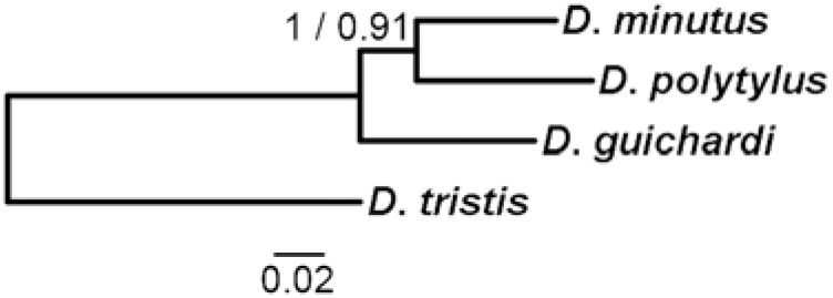
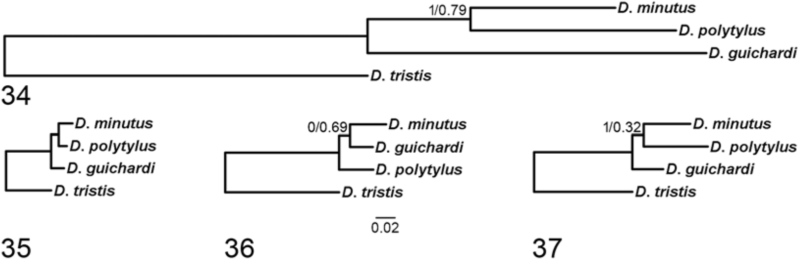
Phylogeny reconstruction based on the barcoding CO1–5′ DNA sequence places D. polytylus as the sister taxon to the clade that includes D. minutus, D. friesei, D. major Kohl, 1901, the D. insidiosus species complex, and D. guichardi, thus considering the latter species phylogenetically closer to D. minutus than D. polytylus (Figs. 31–32). However, a reconstruction based on all studied DNA markers, using D. tristis as an outgroup, demonstrates a closer relatedness of D. polytylus to D. minutus, with D. guichardi being a sister taxon of these two species (Fig. 33). Such relationships are supported by the reconstructions based on combined mitochondrial markers (Fig. 34), exons of nuclear protein-coding genes (Fig. 35), and combined markers of the nuclear rDNA operon (Fig. 37). In contrast, phylogeny reconstruction based on the studied introns of nuclear protein-coding genes supports a closer relatedness of D. minutus and D. guichardi, similarly to the CO1–5′-based reconstruction (Fig. 36). In the latter tree, however, the common node of D. minutus and D. guichardi has a relatively low probability (0.69), when using the likelihood algorithm, or the two possibilities of bifurcation of the tree, including D. guichardi, D. minutus and D. polytylus, are equiprobable, when using the Bayesian inference.
Our attempts to confirm the presence of bacterial endosymbionts, which might induce cytoplasmic incompatibility, in the specimens used for DNA studies were not successful. Therefore, we consider that these specimens have not been infected by Wolbachia or other endosymbionts; thus, the observed relative evolutionary rates of their mitochondrial DNA markers are not biased in relation to the nuclear DNA markers.
Provisional key to the Mediterranean species of Diodontus minutus species group (species distribution information includes unpublished data from the collections available with the authors)

Discussion
Dry grasslands and deserts of Mediterranean area, western Asia, and central Asia seem to be a region of intense speciation for the genus Diodontus, where multiple highly variable species with doubtful taxonomic status occur. At least a part of them may represent complexes of phylogenetically young cryptic species or species in statu nascendi. An earlier morphometric study (Budrys Reference Budrys and Jonaitis1996) revealed wide intraspecific variation ranges of body proportions in multiple species of this genus. The current research confirms that even an application of discriminant analysis, based on multiple measurements, may not allow a reliable morphometric delimitation of closely related species.
Using morphological characters and keys to European species of Diodontus (Dollfuss et al. Reference Dollfuss, Bouček, Bitsch and Bitsch2001; Olszewski et al. Reference Olszewski, Ljubomirov, Wiśniowski, Kowalczyk and Krzyżyński2016), the females of the two species, described here as new, would be misidentified as D. minutus due to similar colouration and the distance between lateral teeth of clypeal apex being approximately equal to the distance between the lateral tooth and the eye. Males of both new species would be misidentified as D. insidiosus due to similar colouration and nearly straight, narrow mesobasitarsus. In collections, D. polytylus has been also often misidentified as D. friesei.
The application of molecular characters is much more fruitful in species recognition (Fig. 30). The barcoding CO1–5′ region allows a reliable separation of both new species from the remaining D. minutus species group, including D. friesei and the D. insidiosus species complex. The latter complex is represented by multiple geographically restricted and relatively highly divergent haplotypes forming multiple barcode index numbers (Schmid-Egger et al. Reference Schmid-Egger, Straka, Ljubomirov, Blagoev, Morinière and Schmidt2018, unpublished data). We consider it as a complex of cryptic species, possibly some of them in a state of allopatric speciation, requiring a detailed revision with the application of molecular characters. Diodontus friesei forms a well-defined barcode index number (Figs. 31–32); thus, it must be considered as a full species. The oldest name of this complex, D. hyalipennis Kohl, 1892, must be attributed to a species from the southern Caucasus that is most probably different from D. friesei and D. insidiosus; however, its status needs a clarification.
Phylogenetic relationships of Diodontus guichardi, D. minutus, and D. polytylus, reconstructed using algorithms of Bayesian inference and maximum likelihood from all studied molecular markers (Table 1). Only probability of branch lower than 1.00 is presented; the probability estimated using the Bayesian inference is before slash, the probability using maximum likelihood is after slash. Diodontus tristis was used as an outgroup.

Phylogenetic relationships of Diodontus guichardi, D. minutus, and D. polytylus, reconstructed using algorithms of Bayesian inference and maximum likelihood from four groups of molecular markers (dendrograms at the same scale). 34, Mitochondrial DNA; 35, exons of nuclear protein-coding genes; 36, introns of nuclear protein-coding genes; 37, markers of nuclear rDNA operon. Only probabilities of branches lower than 1.00 are presented; the probability estimated using the Bayesian inference is before slash, the probability estimated using maximum likelihood is after slash. Diodontus tristis was used as an outgroup.

An adequate reconstruction of phylogenetic relationships among these phylogenetically young species may be problematic, particularly if a single DNA marker, e.g., the CO1–5′ barcode, is used. Varying results of phylogeny reconstructions based on different groups of molecular characters (Figs. 34–37) reflect the differences in evolutionary rates of these markers. We consider the most coherent tree with probability of all nodes equal to 1.00 using both the likelihood algorithm and the Bayesian inference, based on the exons of nuclear protein-coding genes (Fig. 35) as the most probable reconstruction of phylogenetic relationships among D. guichardi, D. polytylus, and D. minutus.
A comparison of the studied molecular characters of Diodontus demonstrates that
(1) The evolutionary rate of the “variable” regions of nuclear 18S and 28S rDNA is too low for the separation of closely related species or reconstruction of phylogeny in genera with high ongoing speciation rate.
(2) The internal transcribed spacers ITS1 and particularly ITS2 allow a perfect separation of closely related species due to a high evolutionary rate; however, they may be difficult to sequence due to large and variable length, exceeding 1000 base pairs in some species of Diodontus. A large part of the variability of these markers is represented by extensive indels, which are problematic for evolutionary interpretation; therefore, ITS1 and ITS2 seem to be less applicable for phylogeny reconstructions than for species identification.
(3) Exons of protein-coding genes have a low evolutionary rate, which may be insufficient for the separation of closely related species. However, their evolutionary coherence seemingly allows the most consistent reconstruction of phylogeny (Fig. 35), in comparison to the other groups of markers (Figs. 34, 36–37).
(4) Introns of protein-coding genes may have a high evolutionary rate, comparable to that of mitochondrial markers (Table 5). They may be easily amplified by selecting the conserved primer-binding sites on the exons nearby; thus, they may be perfectly applicable for species identification. However, they often include indels, which are problematic for evolutionary interpretation. Therefore, they seem to be less suitable for phylogeny reconstruction than exons.
(5) Some mitochondrial markers may have a considerably higher evolutionary rate than the well-studied CO1–5′ barcode (Table 5; Fig. 30) and they can be easily amplified due to the availability of conserved regions nearby for the selection of primer-binding sites. An example of such marker in Diodontus is ND6; another example is mitochondrial ATP synthase subunit 8 gene in Chrysis Linnaeus, 1761 (Hymenoptera: Chrysididae) (Orlovskytė et al. Reference Orlovskytė, Budrys, Budrienė, Radzevičiūtė and Soon2016). The exploration of mitochondrial markers with a considerably higher evolutionary rate than that of CO1, specific for particular taxa, may be useful for the separation of closely related species as well as reconstruction of their phylogeny or at least in the clarification of their microevolutionary or phylogeographic patterns.
We consider these conclusions as hypotheses for testing in other taxa rich in closely related and cryptic species.
Acknowledgements
This study was partly supported by a grant from the Research Council of Lithuania (contract MIP-042/2014) and by the European Union Framework Programme 7 Project SCALES (Securing the Conservation of Biodiversity across Administrative Levels and Spatial, Temporal and Ecological Scales) (contract 226852). The research was carried out using the research infrastructure of the Nature Research Centre under Lithuanian open access network initiative. The authors thank the Doctoral School of Earth Sciences and Ecology (financed by the European Union Development Foundation) for funding, and Mohamed Ghamizi (Marrakesh, Morocco) for helping to arrange the expedition to Morocco, and are grateful to two anonymous reviewers whose comments and suggestions greatly improved the manuscript.