Mr President, Mr President Elect, Fellows and Members, Ladies and Gentleman, it is a pleasure to address the Section of Laryngology this morning and deliver the 94th Semon Lecture. I would like to thank the Semon Committee for their kind invitation. My lecture will discuss Sir Felix Semon (the man himself), highlight the history of head and neck surgery, and then discuss the requirements of a modern-day thyroid surgeon. I have no conflict of interest and nothing to declare.
There can be no doubt that an autobiography is an unrivalled vehicle for telling the truth about other people, whilst never is a writer so rarely well inspired as when he/she talks about themselves! Sir Felix Semon's autobiography was published posthumously by his son, Henry, in 1926,Reference Semon and McIntyre1 with redacted comments about the treatment of Emperor Frederick III. Many would admit a more balanced view is that given by Professor Donald Harrison, who states at the beginning of his book ‘Every man nowadays has his disciples, and it is usually Judas who writes the biography’ – perhaps preparing us for his conclusions later on.Reference Harrison2
Sir Felix Semon (Figure 1) was born in 1849, and his autobiography begins ‘It was not sung at my cradle that my life would be an eventful one. I was born at Danzig (then in Western Prussia) as the eldest son of the merchant, Simon Joseph Semon (8 December 1849). On both sides my parents sprang from prolific Jewish families, and my father was the kindest man I have ever known. My mother's character differed considerably with kindness and affection being more latent in her soul!’
Sir Felix Semon (1849–1919).

The front page of the autobiography begins, ‘He is Physician Extraordinary to HM King Edward VII along with President of the Laryngological Society of London, and Physician of Diseases of the Throat at St Thomas's Hospital’. Semon qualified as Doctor of Medicine Member of Royal College of Physicians (‘MD MRCP’) (later Fellowship of the Royal College of Physicians of London (‘FRCP’)), and studied medicine in Berlin. He then travelled to Vienna, Paris and then London, where he worked for Sir Morell McKenzie and chose a career in otolaryngology. Together, they founded the specialty of laryngology and contributed to the first text book on ENT. Both individuals were prominent in founding the laryngological section of the Royal Society of Medicine, in 1907, when the British Rhinolaryngological Association (founded by McKenzie) amalgamated with the Laryngological Society of London (founded by Semon).Reference Weir and Mudry3
Semon was keen to progress within ENT; he was a self-publicist (he enjoyed self-aggrandisement), and often quoted his private patients, who included William Gladstone, Winston Churchill, Lillie Langtry, as well as royalty, naming Crown Prince Frederick and Princess Victoria, King Edward VII and Queen Victoria amongst others. He had married Lady Augusta Semon, who enjoyed with him an interest in music, and together they were able to afford a somewhat middle-class existence in London. They were often guests of the Royal Family, both in London and Scotland.
In Vienna, Semon was exposed to concepts of integration between clinical care and basic research (the beginnings of translational medicine). He published widely, gave formal lectures, and was interested in upper respiratory tract infections including syphilis, tuberculosis, as well as benign and malignant diseases of the larynx, working closely with Sir Henry Butlin. His contributions to thyroid surgery relate more to his abilities as a physician rather than a surgeon, drawing our attention within ENT to hypothyroidism, Semon's law, music and the singing voice, along with conservation surgery.
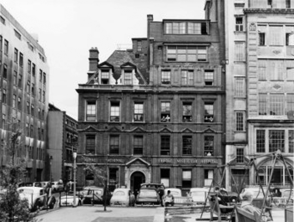
Sir Morrell Mackenzie had founded The Journal of Laryngology & Otology in 1887 (with Norris Wolfendon). What followed was the first ENT hospital in this country (at Golden Square, London; Figure 2), which later was incorporated within Gray's Inn Road.
The ENT Hospital, Golden Square, London, circa 1950s. Courtesy of Mr Navnit Shah.

Semon retired in 1909, and was given a luxurious dinner hosted by Sir Henry Butlin, followed by an around-the-world tour with Thomas Cook in 299 days! The legacy of his lectures lives on today, promoting ongoing education within laryngology.
The history of the thyroid gland dates back to the seventh century, when goitre (derived from the Latin ‘guttur’ for ‘throat’) was described by Paul of Aegina, and then later by Marco Polo in Turkistan (in 1127).Reference Welbourn4 The normal thyroid (Greek for ‘shield’) was not described until the Renaissance, when Leonardo Da Vinci produced his anatomical studies. At this time, a goitre was considered a thing of beauty, with many portraits depicting women with thyroid swellings indicating maturity, and therefore ripeness for both marriage and childbirth.
The first thyroid operation dates back to the seventeenth century, when Willhelm Fabricus (1646) described an operation performed by a second doctor who was then imprisoned for manslaughter as the child subsequently died. In 1791, Pierre Joseph Desault (head and neck and ENT surgeon) carried out the first successful partial thyroidectomy. The majority of the operations at this time were either non-cutting or cutting without completely removing the thyroid, often using procedures such as enucleation and ligation.Reference Welbourn4
By the 1850s, mortality was 40 per cent, haemorrhage and infection were major problems, and surgery was condemned. The introduction of anaesthesia by William Moreton (1842), anti-sepsis by Joseph Lister (1867) and haemostasis in 1872 (when Spencer Wells invented his haemostat) all helped to drastically reduce complication rates.Reference Welbourn4–Reference Watkinson and Clarke6 What followed then was a prolific expansion in the world of general surgery, involving Theodor Billroth, Emil Theodor Kocher, Charles Mayo, George Crile, William Halstead and Hayes Martin.Reference Welbourn4 Indeed, Kocher became the first surgeon to be awarded the Nobel Prize in Physiology (in 1909), which related to ‘his work on the physiology, pathology and surgery of the thyroid gland’. Billroth and Kocher inspired Halstead, Crile, Mayo and Lahey, along with Sir James Berry at the Royal Free Hospital, London (he of Berry's ligament), as well as Sir Cecil Joll who also worked at the Royal Free Hospital (as well as the Royal Marsden Hospital). Further contributions came from Arthur Dunhill and later on from Oliver Beahrs at the Mayo Clinic.Reference Welbourn4
At this time, thyroid surgery was in the domain of the general surgeon. Later, Sir Arthur Dunhill became senior surgeon at St Bartholomew's Hospital, developing a major interest in thyroid surgery. He went on to become senior surgeon to the Royal Household, and operated on Sir Winston Churchill's inguinal hernia on 11 June 1947. However, some thirty years ago, otolaryngologists began to develop an interest in the thyroid. Subsequently, Mr Omar Shaheen became the first UK ENT surgeon to publish a book on thyroid surgeryReference Shaheen7 and give a Semon Lecture on that topic. By 2015, ENT surgeons had become the principal specialty involved in thyroid and parathyroid surgery in the UK. Currently, the modern-day thyroid surgeon will have undergone appropriate specialist training, and often a specialist fellowship. Most will be involved in teaching and training (some in laboratory research), but all will be attending meetings and courses, performing audits, and publishing their results.Reference Watkinson8,Reference Watkinson, Street and Harrison9,Reference Watkinson10
Thyroid disease is common; thyroid cancer is uncommon. The incidence of malignancy is rising, due in part to over-investigation, and the identification and treatment of low-grade cancers, but survival remains the same. Surgery is often recommended for benign disease (often those with obstructive symptoms), thyrotoxicosis, suspected or proven malignancy, as well as cosmesis and patient choice. Thyroidectomy is an operation that varies day by day, with no one procedure being the same. Many would describe routine surgery as a biopsy, isthmusectomy, a diagnostic lobectomy or a straightforward total thyroidectomy on a normal to moderately enlarged euthyroid gland. Currently in the UK, about 12 000 thyroidectomies (and rising) are performed each year. There will be times when thyroid surgery is not straightforward, and I will use my experience of ‘the difficult thyroid’ to illustrate this (Table 1).
Difficult thyroid

In order to be a good thyroid surgeon, one should first be a good thyroid physician. Thyroid cancer is different to squamous cell carcinoma; diagnosis pertains to both structure and function, and blood tests do not lie. Sometimes the history can be difficult; there may be language problems, as well as not enough time or information. Thyroids can be difficult to assess with regard to upper airway obstruction (often patients cannot ie flat), and clinical assessment is a poor predictor of upper airway obstruction, which is best assessed by a respiratory flow loop.Reference Gittoes, Miller, Kaykin, Sheppard and Franklyn11
Sometimes, the diagnosis may be difficult (i.e. lymphangioma), or the surgery may be problematic because of large and retrosternal benign goitres, thyrotoxicosis, or invasive malignancy for both differentiated thyroid cancer and medullary thyroid cancer. Often these cancers necessitate nodal surgery (either primary or revision operations). Primary site surgery really now consists of two operations: a lobectomy (or extended lobectomy) and a total thyroidectomy.Reference Watkinson, Street and Harrison9,Reference Hobbs and Watkinson12 Occasionally, it may be prudent to do a lobectomy on one side and a subtotal lobectomy on the other side (near-total thyroidectomy). This can help preserve vital structures (but not usually function). The old-fashioned subtotal thyroidectomy (Dunhill procedure), which aimed to preserve tissue in both sides of the neck, should be avoided. Historically, this procedure was conducted to reduce complications and preserve function, but it meant tissue could grow back, making further revision surgery hazardous.
Patients with thyrotoxicosis (Graves’ disease, a toxic solitary nodule (Plummer's disease) or a toxic multinodular goitre) can be challenging regarding their biochemical state, vascularity and gland size. One should operate (where possible) in the euthyroid state. Beta-blockers, lithium and sometimes Lugol's iodine may be required. Surgery involves meticulous haemostasis; use extracapsular dissection to preserve four parathyroids and thank God for the fifth parathyroid! Many patients (because of metabolic hungry bone syndrome) have been started on vitamin D, which can reduce the incidence of post-operative hypocalcaemia.
The operation of thyroid lobectomy is a set manoeuvre, and proceeds (a bit like Strictly Come Dancing) in a quick-slow-quick-quick-slow pattern.Reference Watkinson, Street and Harrison9,Reference Watkinson10,Reference Hobbs and Watkinson12 Sometimes, surgery is difficult because of gland size. Often, it is extensive and retrosternal, giving rise to obstructive symptoms (the thyroid cork). Treatment with surgery is usually by extended lobectomy or total thyroidectomy, without recourse to a sternal split. Appropriate access is crucial. Use a correctly sized incision; often some strap muscles are divided. Do the easy side first, and begin with the upper pole. Stay within the pseudocapsule, and work medial to lateral. Make sure you get into the right plane for lower pole vessels. Always get control inferiorly, and try to have a good assistant. The anatomy can be complicated; the course of the nerve (often splayed by a tubercle of Zuckerkandl) may vary. It is important to remember, ‘never deliver the baby until the cervix is fully dilated’. The key to the posterior mediastinum is the lateral neck, and the lower the goitre, the higher the cut. But the bigger the lesion, the bigger the risk. It is better to have a large scar than a small tombstone!
One of the most feared complications following thyroidectomy is a recurrent laryngeal nerve palsy. Semon's law comments on vocal fold position in both health and disease (1881), and states that ‘in all progressive organic lesions, the abductor fibres are more susceptible and thus first to be paralysed compared to the adductor fibres’.Reference Semon and McIntyre1–Reference Weir and Mudry3,Reference Semon13 This is an unsatisfactory theory, because, if it were true, the paralysed fold would lie in the median position. It also predicts that the affected fold would shift laterally over time, which is opposite to the gradual adduction widely observed over time. Another theory (the Wagner and Grossman Hypothesis, 1897),Reference Wagner14,Reference Grossman15 states that ‘complete paralysis of the recurrent laryngeal nerve results in the vocal cord lying in a paramedian position because of an intact cricothyroid muscle which adducts the vocal cord’. The problem with this theory is that the vocal fold palsy caused by a left apical bronchial carcinoma produces a fold in the cadaveric position.
Current knowledge states that four nerves supply the larynx, and that the only motor fibres within the superior laryngeal nerve are to the cricothyroid muscle. This means that normal voice should equate to no neuronal injury, but recent anatomical studies have shown communications from above with that below. The internal branch of the superior laryngeal nerve anastomoses with the recurrent laryngeal nerve on the posterior cricoid muscle (Galen's anastomosis). This can be either single, double or a plexus. There can also be a cross-anastomosis with the other side (cricoid communication or plexus; Figure 3).Reference Henry, Pękala, Sanna, Vikse, Sanna and Saganiak16 There is sometimes also an arytenoids plexus connecting the anterior branch of the recurrent laryngeal nerve with the arytenoids branch of the internal branch of the superior laryngeal nerve. This would explain the discrepancies in outcomes when nerves are either damaged or suffer severe trauma, and yet the fold moves normally as well as when a palsy is not expected and one then occurs. Future studies will help map out the normal physiology of neuronal vocal fold control.Reference Henry, Pękala, Sanna, Vikse, Sanna and Saganiak16
The posterior relations of laryngeal anatomy, showing the anastomotic connections between the internal branch of the superior laryngeal nerve (ILN) above and the recurrent laryngeal nerve (RLN) below on the cricoid muscle (Galen's anastomosis). Also shown are the cross-anastomotic cricoid connections (CA) and the arytenoid plexus (AP). The letters (a) and (p) refer to the anterior and posterior branches, respectively. Courtesy of Elsevier.

Surgery can be challenging for locally advanced disease in relation to blood vessels, the pharynx, larynx and trachea, the recurrent laryngeal nerve, the mediastinum, as well as strap muscles and skin, but major extirpative surgery may be indicated in some cases.Reference Watkinson10,17–Reference Ishii, Kim, Watkinson, Mallick, Harmer, Mazzaferri and Kendall-Taylor19 However, it is not always possible to remove the entire tumour without damaging both recurrent laryngeal nerves. A small residue of tumour may be left behind to be dealt with by radioactive iodine ablation and subsequent thyroid-stimulating hormone suppression with levothyroxine (T4) (with or without external beam radiotherapy). Quality of life issues are important, such that alternatives should always be considered, with treatment being tailored to the individual patient.17
Some necks can be difficult to examine and assess because of obesity or previous treatment. Others are difficult to operate on because of extensive disease, mediastinal extension, difficult histology or recurrence. There is usually no role for either ‘berry picking’ or prophylactic lateral neck dissection. Selective neck dissection is based on the natural patterns of drainage in the untreated neck (Figure 4), and is more difficult than radical surgery. The surgery should take into account the ‘corners of consternation’ (Figure 5).Reference Watkinson and Gilbert5,Reference Watkinson and Clarke6,Reference Warner, Jennings, Watkinson, Watkinson and Clarke20 Current guidelines outline the indications for both central and lateral neck dissection.17 Keep your options open, maintain the ability to go medial to lateral, as well as lateral to medial, and don't be afraid to divide the sternomastoid muscle for access. Level IIB can be a problem (Suarez's triangle). Remember the contralateral neck and, as a general rule when in the zone, dissect the zone. An exception to this would be recurrent neck disease, which should always be restaged, and treatment should be surgical whenever possible. The basis of selective neck dissection alters as patterns of drainage change following treatment, and so local excision often prevails.
A schematic diagram showing the extent of primary (‘A’ & ‘D’) and secondary (‘B’ & ‘C’) surgery for differentiated thyroid cancer. The operation is usually a total thyroidectomy for localised disease, dissecting levels VI (‘A’) and sometimes VII (‘D’) for high-risk cases. For lateral disease (‘B’ & ‘C’) in the untreated neck, selective surgery targets levels II, III and IV (and sometimes Vb), staying below the accessory nerve (‘xi’). RLN = recurrent laryngeal nerve. Courtesy of Hodder Arnold Press.

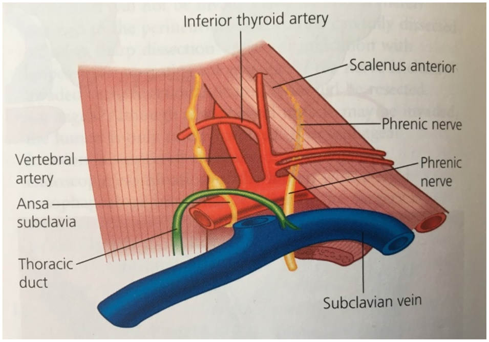
The minor ‘corners of consternation’ include Chassaignac's triangle (above), as well as the retropharyngeal and parapharyngeal spaces. This triangle is bounded medially and laterally by the longus colli and scalenus anterior muscles, and inferiorly by the subclavian vein. The floor is the prevertebral fascia, and anteroinferiorly within the triangle are the scalene (and some level IV) nodes covered anteriorly by the internal jugular vein and the sternomastoid muscle. The major ‘corners of consternation’ form the boundaries of a rectangle created by the fusion of the anterior and posterior triangles. Courtesy of Hodder Arnold Press.

Neck aesthetics and the siting of the scar are important. Sit the patient up in the anaesthetic room, and confirm both the level and shape of the scar. Incisions change based on neck shape, sternomastoid tendon prominence and the access required. A standard Kocher incision is often used, which lies within the suprasternal notch. Further access to the neck can be achieved by extending the cut into the lateral utility incision (Figure 6), or alternatively using a midline transverse incision (popularised by Shah).Reference Watkinson and Gilbert5,Reference Watkinson and Clarke6 Symmetry is important (size less so), as is meticulous closure, but poor scars do occur. After 6 or 12 months, sometimes scar revision is required. Unsightly scars can be hypertrophic or keloid, and poorly sited in both level and direction. Scar revision can be difficult, and, like the cheek, within the lower neck big is beautiful. There is no room for small flaps, and scar revision usually involves incision, mobilisation, and advancement with sometimes either a neck lift or drop. Occasionally, if there is excess neck skin, this can be excised and closed by advancement when excellent results are possible.
This patient underwent total thyroidectomy and left selective neck dissection (levels II–V) for differentiated thyroid cancer. Access was obtained using traditional midline access (a), extended laterally as a thyroid utility incision to allow neck surgery below (b). This heals well, as it involves a Kocher and lazy ‘S’ incision back to back, giving good access to the five major ‘corners of consternation’.

Thyroid cancer in children is rare; it can be difficult to manage and is emotionally demanding. Nodules are more likely to be malignant and previous neck irradiation is a significant risk factor. Children aged under 10 years tend to have more aggressive disease; the general principles of management are similar to adults, but must include a paediatric endocrinologist and oncologist. Usually, total thyroidectomy is recommended; some patients require selective neck dissection. Decisions regarding radioactive iodine I-131 therapy should be individualised. Life-long follow up is advised.17
Patients can be difficult to follow up both clinically and biochemically. Multidisciplinary team (MDT) protocols exist for monitoring thyroid hormone levels, serum thyroglobulin and calcitonin, along with investigation pathways for possible recurrent disease. Those cases that are extremely difficult to manage relate to tumour and physician factors. Early detection is important to improve cure and survival, and local recurrence is often treated with further surgery. Metastatic disease involving the lungs and other soft tissues that are not amenable to surgery should be treated with radiotherapy. Isolated bone metastases can be treated with a combination of orthopaedic intervention, radioactive iodine I-131 therapy and external beam radiotherapy.17
It is important that you consent for success, but be prepared for less. If it is not written down, it did not happen. Complications should be discussed based on your own experience (combined with the literature). Common complications should be listed, but nowadays (following the Montgomery case21) the more uncommon ones (such as ptosis) should also be included. Hence, for total thyroidectomy, consider bilateral vocal fold palsy and therefore consent for tracheostomy.
How can we make the difficult thyroid case easier? Train well, build your pyramid, do a fellowship and subspecialise. Work within guidelines,17 treasure the MDT, audit your data, keep up to date and publish one's results. The operation can be made easier with technology. Loupes and headlights help, and the nerve stimulator is now becoming mandatory, with benefits for teaching and training, re-do cases, difficult cases, and use in a medicolegal setting. The harmonic scalpel and LigaSure™ devices can speed up surgery, but there is no evidence that they improve outcomes. Access to rapid parathyroid hormone measurements is now the norm. Minimally invasive and robotic surgical procedures will have their place in selected cases. Currently, many surgeons continue to use fine ties and bipolar diathermy.
Who should do the surgery? It is not your specialty that matters, but your training and expertise, together with one's working environment.Reference Watkinson8 So what about the future? British otolaryngology has lately contributed significantly to thyroid research. Studies have looked at the aetiology and pathogenesis of goitre in both benign and malignant disease. A number of growth factors have been identified,Reference Susarla, Watkinson and Eggo22,Reference Kim, Franklyn, Boelaert, Eggo, Watkinson and McCabe23 and some goitres may be susceptible to gene therapy.Reference Ramsden, Buchanan, Egginton, Watkinson, Mautner and Eggo24 Other studies have looked at reversing the deactivation of the sodium/iodide symporter receptor so that radioactive iodine uptake can be restored.Reference Smith, Read, Turnell, Watkins, Watkinson and Lewy25 Such techniques could then be used in combination with near-complete surgery, so that we can tailor adjuvant radiotherapy and novel systemic treatments (such as tyrosine kinase inhibitors) to improve outcomes.Reference Wang, Zafereo, Dadu, Ferrarotto, Busaidy and Lu26
My lecture started with Felix, and finishes with the helix! Personalised medicine can detect familial non-RET medullary thyroid cancer mutations. Families who have in the past required prophylactic therapy no longer need an operation if they are gene-negative.Reference Smith, Read, Hoffman, Brown, Bradshaw and Campbell27 Over the next decade, there will be advances in translational medicine because of molecular profiling, along with developments in nerve regeneration or repair, and transplantation. Clinical trials are also important to safely introduce new treatments such as minimally invasive techniques with robotics, along with individualised surgery regimens. The management of the difficult thyroid may involve gene switching and antigenic therapy, gene therapy with molecular profiling of thyroid cancer, and, again, personalised surgery with adjuvant systemic therapy.Reference Read, Seed, Fong, Modasia, Ryan and Watkins28–Reference Almubarak, Qassem, Alghofaili, Alzahrani and Karakas30 There will be improvements in follow up (including measuring circulating DNAReference Allin, Shaikh, Carter, Thway, Sharabiai and Gonzales-de-Castro31), and the question regarding ‘lobectomy versus total thyroidectomy’ outcomes in low-grade differentiated thyroid cancer will be answered by the recently approved ‘HoT’ (hemi- vs total thyroidectomy) trial.
My advice to those in training is be patient and stay focused, listen to and care about your patients. Be intellectually curious and strive to consistently innovate over your entire career. Be technically gifted, develop the art of thyroid surgery and learn to love it. Have a life mission to make a difference. Plan a subspecialty and do a fellowship if you can. Work as a team and have fun along the way. Don't be overcritical of yourself, and remember ‘Every surgeon carries about him a little cemetery, in which from time to time he goes to pray, a cemetery of bitterness and regret of which he sees the reason for certain of his failures’ (René Leriche, 1951).Reference Watkinson and Gilbert5
We started with the cradle and finish with the grave. Sir Felix Semon wrote in February 1921 ‘that during the last few weeks, I have been better apart from shortness of breath after small efforts but except my own family, I have nothing left that interests me and if I knew that to-morrow morning I should not awake, I should go as quietly to bed as if a long life were in store for me’. Sir Felix Semon died a few days later from angina, on 1 March 1921. His memorial tablet in Golders Green reads ‘Monumentum Ejus Incrementum Scientiae’, which translates as ‘Tribute to a Great Scientific Career’. Sir Donald Harrison mentioned Judas at the beginning of his biography on Semon. He concluded that ‘Semon was highly intelligent, industrious but also self-opinionated, probably not warm-hearted; a complex character, a mixture of sainthood but perhaps with a touch of sin!’
This lecture has been about some of the greats in general and endocrine surgery, as well as in ENT and plastic surgery. Modern-day thyroid surgery is now truly multidisciplinary, and whilst historically the thyroid belonged to the general surgeon, now, with increased specialisation, more and more cases are being treated by otolaryngologists and dedicated endocrine surgeons. The surgery demands appropriate training and subspecialisation, with dedication to teaching, audits and research. I believe the future is bright for ENT and endocrine surgery, and for that we have to thank Sir Felix Semon's lecture series, together with Mr Omar Shaheen and Mr Tony Bull (Semon Lecture, 2000),32 for bringing thyroid and facial plastic surgery into the domain of otolaryngology.