INTRODUCTION
Infection of sheep with the abomasal-dwelling helminth parasite Teladorsagia circumcincta is the main cause of ovine parasitic gastroenteritis in temperate climates, leading to significant production losses and increased costs for prevention and control of the disease (Nieuwhof and Bishop, Reference Nieuwhof and Bishop2005). While anthelmintics remain the principal control measure, the problem of anthelmintic resistance (Gilleard, Reference Gilleard2006) has led to increased interest in harnessing the sheep's own native immunity to control the parasite, primarily by vaccination. Gradual exposure to low levels of the parasite in the field results in an acquired immunity, which can be modelled in an experimental setting by trickle infection (Smith et al. Reference Smith, Jackson, Jackson and Williams1983). The resulting immunity is manifest in immune exclusion and expulsion of parasites following subsequent challenge with large doses of infective T. circumcincta L3 larvae (Smith et al. Reference Smith, Jackson, Jackson and Williams1983). The cytokine expression profile in immune sheep has been characterized as predominantly Th2-like (Craig et al. Reference Craig, Miller, Smith and Knight2007), with strong mast cell and eosinophil responses in the abomasum (Stevenson et al. Reference Stevenson, Huntley, Smith and Jones1994). However, immunity is not long lived following cessation of exposure, and it effectively wanes after as little as 6 weeks following anthelmintic clearance of the worms (Jackson et al. Reference Jackson, Greer, Huntley, McAnulty, Bartley, Stanley, Stenhouse, Stankiewicz and Sykes2004).
It has been shown through ex vivo analysis of immune abomasal biopsies, that factors present in the immune abomasum inhibit the penetration of infective larvae into the abomasal crypts (Jackson et al. Reference Jackson, Greer, Huntley, McAnulty, Bartley, Stanley, Stenhouse, Stankiewicz and Sykes2004). In order to identify factors that may be responsible for protection of the immune abomasum, and which may become less abundant in the immune waning abomasum, we adopted a proteomic approach to compare abomasal mucosa from immune, immune waning and naïve sheep.
MATERIALS AND METHODS
Antibodies
Rabbit polyclonal antibodies to ATP synthase beta, calreticulin, desmin, GRP78, GRP94, PDI and thioredoxin, and chicken anti-bovine serum albumin were supplied by Abcam (Cambridge, UK). Donkey anti-ovine serum albumin was supplied by AbD Serotec (Kidlington, UK). A rabbit antibody to galectin-15 was kindly donated by Thomas Spencer (Texas A&M University, USA). Horseradish peroxidase (HRPO)-conjugated secondary anti-rabbit and anti-donkey antibodies were supplied by Stratech Scientific (Newmarket, UK).
Sheep and trickle infection with T. circumcincta
Experiments were performed using yearling sheep housed under worm-free conditions. The T. circumcincta strain used was fully benzimidazole-sensitive and was originally derived in the UK and maintained at Moredun Research Institute for approximately 30 years. Infection procedures, worm harvesting, counting and staging was carried out as previously described (Halliday et al. Reference Halliday, Routledge, Smith, Matthews and Smith2007, Reference Halliday, Morrison and Smith2009). Briefly, sheep (n=12) were subjected to trickle infection with 2000 T. circumcinta L3 larvae 3 times per week for 6 weeks. A further 6 worm-naïve sheep were not infected. All sheep were then given an oral dose of fenbendazole (day 0) according to the manufacturer's recommendations. Seven days later, the 6 uninfected sheep (naïve) and 6 of the immune sheep were stunned with a captive bolt, exsanguinated, and the abomasum removed and opened along its lesser curvature. The abomasum was washed 3 times in warm saline, and then abomasal fold samples (∼2cm2) were removed for protein extraction, paraformaldehyde-fixation and RNA purification.
After a further 5 weeks, the remaining 6 trickle-infected sheep (immune-waning) were necropsied and samples taken in the same way.
Sample preparation
Samples of abomasal fold (approx 250 mg) were homogenized in 500 μl of extraction buffer (8 m urea; 2% CHAPS; 0 4% dithiothreitol (DTT), containing 1 Roche complete-Mini™ EDTA-free protease inhibitor tablet per 5 ml) using a FastPrep-FP120 disrupter (Qbiogene, Cambridge, UK). The supernatants were processed through QIAshredder tubes (Qiagen, Crawley, UK), centrifuged at 4°C for 15 min at 15 000 g and then diluted with extraction buffer to the equivalent of 200 mg wet weight tissue per ml, aliquotted and frozen (−80°C).
Two-dimensional gel electrophoresis (2DGE)
For each sample, an Immobiline DryStrip pH 4–7 (18 cm; GE Healthcare, Chalfont St Giles, UK) was rehydrated for 12 h in the presence of a mixture of 450 μl of 8 m urea; 2% CHAPS; 0 4% DTT containing IPG buffer, pH 4–7 (1 in 200 v/v; GE Healthcare) and tissue extract (40 μl, equivalent to 5 mg wet weight tissue). Isoelectric focussing followed at 8000 V for 35 000 Vh (Protean IEF Cell, Bio-Rad, Hemel Hempstead, UK). Focussed strips were then stored at −80°C until required for the 2nd dimension separation. After thawing, strips were incubated in equilibration buffer (6 m urea; 2% SDS; 20% glycerol; 0 375 m Tris-HCl, pH 8 8) containing 130 mm DTT for 15 min, then in equilibration buffer containing iodoacetamide (135 mm) for a further 15 min. The strips were then placed on top of SDS-PAGE gels (12% T; Protean II; Bio-Rad) and electrophoresis was carried out at 24 mA per gel until complete. Finally, gels were stained with colloidal Coomassie blue (Imperial Protein Stain, Thermo Scientific, Cramlington, UK), according to manufacturer's instructions.
Image analysis and data processing
Gels were imaged at 600 dpi using a flatbed scanner (Imagescanner, GE Healthcare). Images were imported into Dymension software (Version 3.0.1.2, Syngene, Cambridge, UK) as 3 sample groups (naïve, immune and immune waning), with n=6 in each group. Spot detection, gel warping, spot matching and densitometry were then carried out automatically by the software. Consensus spot volumes were exported as normalized values relative to total integrated spot volume (set to 100 for each gel). Within each sample group, the mean normalized spot volume was calculated for each consensus spot. Mean values were then used to calculate fold-changes for each spot in comparisons between the different groups. Group-wise statistical comparisons were also made for each spot using Student's t-test (Microsoft Excel). Following these comparisons, individual consensus spots were flagged as potentially differentially expressed where P<0 05 (Student's t-test), fold change >2 00 and the mean volume for the spot in at least one of the groups was above a set threshold value (0 1).
Cluster analysis
Cluster analysis was performed as recommended by Meunier et al. (Reference Meunier, Dumas, Piec, Bechet, Hebraud and Hocquette2007) for proteomic data. Briefly, the complete data set was normalized by row then log transformed and imported into Permutmatrix hierarchical clustering software (Caraux and Pinloche, Reference Caraux and Pinloche2005). The data were then processed using the Pearson distance setting for dissimilarity, and clustered on both rows and columns using Ward's minimum variance method, as recommended by Meunier et al. (Reference Meunier, Dumas, Piec, Bechet, Hebraud and Hocquette2007).
Representative gels
Pools were created for each of the naïve and immune sample groups by combining an equal quantity of each of the 6 biological replicates. 2D gels were then run on the pooled samples, as above, and a composite gel was created for reference purposes by merging the 3 images.
Spot identification by peptide mass fingerprinting
Selected spots were excised from gels, digested with trypsin, and peptide fingerprinting was performed by MALDI-TOF mass spectrometry (Ultraflex™ II, Bruker Daltonics). Spot identity was determined by database searching with Mascot software (Perkins et al. Reference Perkins, Pappin, Creasy and Cottrell1999) (http://www.matrixscience.com/), using NCBInr database, mass tolerance of ±0 15 Da, species set to ‘mammalia’ and fixed modification= Cys-carbamidomethylation. As an exception, spot 66 was identified using SwissProt database, species=‘other mammalia’ and ±0 5 Da.
Western blotting
Pooled samples from each of the 3 groups (i.e. naïve, immune and immune-waning) were separated by SDS-PAGE (12%T), alongside molecular weight standards (broad range, unstained, Bio-Rad) then transferred by semi-dry blotting (Trans-Blot SD, Bio-Rad) to PVDF membrane (Immobilon-P, Millipore, Watford, UK). The blots were incubated in blocking buffer (2% skimmed milk powder in 10 mm Tris-HCl, 150 mm NaCl, 0 5% Tween-80) for 1 h, then incubated overnight at 4°C with primary antibodies, as appropriate, diluted to 1 μg/ml in blocking buffer. Blots were washed, then incubated with the appropriate HRPO-conjugated secondary antibodies and imaged using Chemiluminescent Peroxidase Substrate (Sigma) and an Image Station IS440CF (Kodak). After Western blotting was complete, the blots were post-stained with Coomassie Blue (Imperial Protein Stain, Pierce) to visualize the molecular weight standards.
Anti-serum albumin immunohistochemistry
Samples of abomasal fold taken during post-mortem of experimental sheep were fixed in 4% paraformaldehyde in PBS, processed into paraffin blocks, then 4 μm sections were cut onto coated slides (Surgipath, Peterborough, UK). Antigen retrieval was performed by microwaving the sections for 4 min with 10 mm sodium citrate buffer, pH 6 0. Sections were incubated overnight at 4°C with chicken anti-bovine serum albumin or control chicken IgY (both at 2 μg/ml) in PBS containing 0 5 m NaCl and 0 5% Tween 80. Slides were then washed and incubated with rabbit anti-chicken IgY-horseradish peroxidise (Stratech Scientific; 1/500 dilution). Immunoperoxidase labelling was revealed using NovaRed (Vector Labs, Peterborough, UK) and slides counterstained with Meyer's haematoxylin.
RT-PCR detection of ovine serum albumin expression
RNA was purified from abomasal samples and reverse transcribed as previously described (Knight et al. Reference Knight, Pate, Smith and Miller2007). Primers specific for serum albumin and 3 housekeeping genes were designed using the Primer 3 program: http://frodo.wi.mit.edu/cgi-bin/primer3/primer3_www.cgi. The housekeeping genes (ATPase, RW1 and TM57) were chosen on the basis of transcriptomic profiling of naïve and T. circumcincta immune abomasum (Knight et al. Reference Knight, Griffith, Pemberton, Pate, Guarneri, Anderson, Talbot, Smith, Waddington, Fell, Archibald, Burgess, Smith, Miller and Morrison2011) to show no significant change. Primers and semi-quantitative PCR conditions were as follows. Serum albumin (X17055, EMBL) 5′-CCCCAAGTGTCAACTCCAAC, 3′-AAACATGGCCGTCTGTTCAC, annealing temp: 52°C; 32 cycles. ATPase (X02813, EMBL) 5′-GCTGACTTGGTCATCTGC, 3′-CAGGTAGGTTTGAGGGGATAC, annealing temp: 60°C; 31 cycles. RW1 (FE026635, GenBank) 5′-CCACTCCCCACCCTCCTCAT, 3′-TGCAGATGGTCTCAGCCGAAT, annealing temp: 58 °C; 30 cycles. TM57 (FE030125, GenBank) 5′-GCTGAAAGTTCGGGAGCTTCG, 3′-GTCCAGAGGGGCTGGTCTCC, annealing temp: 58°C; 30 cycles.
Gels were imaged using a Bio-Rad FXPro Imager and gel densitometry performed using Kodak 1D software. The serum albumin PCR product from naïve abomasum was purified using a PCR product purification kit (QIAgen) and sequenced by DNA Sequencing & Services, University of Dundee, Scotland. Sequence analysis was carried out using multiple alignment and sequence similarity search programs available on http://www.ebi.ac.uk/Tools/sequence.html, http://www.ncbi.nlm.nih.gov and http://www.ensembl.org/.
RESULTS
2DGE analysis of sheep abomasal extracts
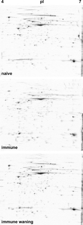
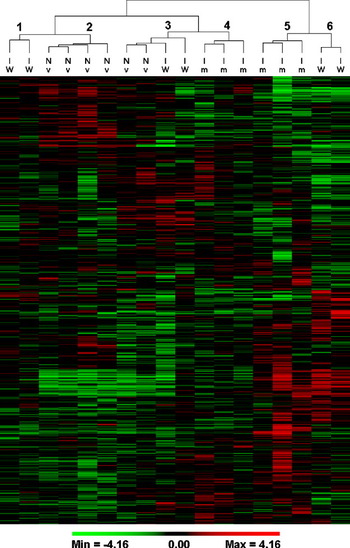
Two-dimensional gel images of abomasal extracts from naïve, immune and immune waning sheep (total of 18 gels) were subjected to computational analysis in Dymension software, which yielded a total of 1103 spots matched across the gels. Example gels are shown in (Fig. 1). Cluster analysis (Permutmatrix) of the exported spot expression data resolved the expression patterns of individual gels into 6 groups (Fig. 2). Samples from naïve sheep clustered into groups 2 and 3; immune samples into groups 4 and 5, and immune waning samples clustered into groups 1, 3 and 6.
Effect of Teladorsagia circumcincta immune status on protein expression in the sheep abomasal mucosa. Mucosal tissue extracts from naïve (n=6), immune (n=6) and immune-waning (n=6) groups were separated by 2DGE, stained with Coomassie Blue and imaged as described in the Materials and Methods section. Image analysis was performed using Dymension software (Syngene). The image shows the reference warped gel image for each of the 3 sample groups following automated sample alignment.

Hierarchical clustering of sheep abomasal extract 2DGE spot densitometry data. Gel densitometry data for 1103 matched spots across samples originating from Teladorsagia circumcincta naïve (Nv, n=6), immune (Im, n=6) and immune waning (IW, n=6) sheep was exported from Dymension software then normalized and log transformed as described in the Materials and Methods section. The data were then imported into Permutmatrix software, and clustering was performed on both rows and columns. The samples segregated into 6 distinct groups, with naïve and immune samples clustering separately.

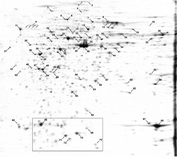
For reference purposes, a further 3 2D gels from pooled naïve, immune and immune-waning samples were overlaid to create a single gel image showing all the spots selected for identification (Fig. 3). To illustrate the level of variability between gels, the rectangular section of the gel highlighted in Fig. 3 is shown online in Suppl. Fig. 1 for all 18 gels.
Reference 2DGE image for sheep abomasum. Pooled naïve, immune and immune-waning abomasum extracts were subjected to 2DGE as described in the Materials and Methods section. Gel images were aligned (Dymension) and merged to create a single composite containing all the spots identified. The annotation shows those spots that were successfully identified by tryptic peptide mass fingerprinting (identities of differentially expressed spots are shown in Table 1 and a full listing is given online in Suppl. Table 1). The highlighted area is shown online for each of the 18 gels in Suppl. Fig. 1.

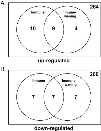
In total, 287 spots were expressed at a mean level above the arbitrary threshold of 0 1 in at least one of the groups. The numbers of spots differentially expressed (P<0 05 and fold change >2) in immune and/or immune-waning groups compared to naïve are shown in Venn diagrams (Fig. 4).
Summary of differentially regulated spots. (A and B) Number of spots in the immune and immune-waning groups that are significantly up- or down-regulated, respectively, compared to the naïve group. Criteria for selection of the spots are explained in the Materials and Methods section.

Identities of differentially regulated proteins
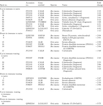
In order to investigate spots of interest, they were excised and processed for peptide mass fingerprinting, which identified a total of 73 spots. Differentially expressed spots are listed in Table 1 (a full listing of all 73 spots arrowed in Fig. 3 is given online in Suppl. Table 1). Of the 73 definitively identified spots, 18 different spots were significantly differentially expressed in immune vs naïve and/or immune-waning vs naïve comparisons (P<0 05 and fold change >2). The most highly up-regulated protein in immune compared to naïve gels was galectin-15 (6 9-fold up-regulated in immune abomasal extracts, not significantly up-regulated in immune-waning compared to naïve and significantly down-regulated −5 75 fold in immune-waning vs immune; Table 1). Thioredoxin (spot 73) was the only other spot, apart from proteolytic fragments, to be significantly up-regulated only in the immune vs naïve comparison, although the level of up-regulation (1 3-fold) was below the arbitrary cut-off. Up-regulated in immune vs naive samples were fragments of actin (spots 41 and 64), tubulin-beta 2C chain (spot 50) and serum albumin (spot 44). Other serum albumin fragments were clearly up-regulated in immune and immune-waning samples but did not reach statistical significance, such as spot 66, a ∼14 kDa N-terminal fragment. Intact serum albumin spots 13 and 15 were significantly down-regulated in the immune and immune-waning group, when compared to naïve.
Sheep abomasal proteins identified by peptide mass fingerprinting

Molecular chaperones, such as calreticulin and protein disulphide isomerase (PDI) were prominent in the differentially expressed list (Table 1). Fragments of calreticulin were significantly up-regulated in both immune and immune-waning samples (spots 30, 37 and 38), while the parent spot (11) was down-regulated (P<0 05, fold change: −1 7 and −1 8, respectively). Additionally, protein disulphide isomerase (PDI; spot 16), stress-70 protein (HSPA9; spot 8), and protein disulphide isomerase A3 (GRP58; spot 19) were down-regulated in immune samples, while endoplasmin (GRP94; spot 3) and GRP58 were significantly down-regulated in immune-waning. Fragments of PDI (spots 22 and 27) were up-regulated in immune-waning samples, suggesting that PDI was the subject of proteolytic processing.
In addition, an ATP synthase beta-chain fragment (ATP5B; spot 49) was up-regulated in immune-waning vs naïve samples. Intact ATP5B (spot 21) was slightly but significantly down-regulated in immune samples (−1 3-fold), as was desmin (−1 79-fold).
Western blotting confirmation
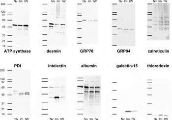
Where suitable antibodies were available, proteins flagged as significantly up- or down-regulated in the 2DGE analysis were confirmed by Western blotting using pooled samples from the naïve, immune and immune-waning groups (Fig. 5). ATP synthase beta exhibited a single band at approximately 50 kDa in the naïve sample, but was found to exhibit a lower MW fragment (approx 40 kDa) in both immune and immune-waning samples. The desmin signal was reduced in immune and immune-waning samples compared to naïve, and lower MW fragments (approximately 32–34 kDa) were also observed. Four antibodies to molecular chaperones were investigated, including GRP94, calreticulin, PDI and GRP78; the latter as an example of a molecular chaperone that did not apparently change significantly on the 2DGE analysis. In fact, the GRP78 Western blot showed a strong single band at approximately 75 kDa in the naïve sample, but was present as a finely separated doublet in immune and immune-waning samples, suggesting a potential post-translational modification, such as proteolytic cleavage of a small fragment from the molecule. Since the antibody used is specific for the N-terminus of GRP78, this suggests that any putative cleavage must occur at the C-terminus. The GRP94 (endoplasmin) Western blot detected a band at approximately 40 kDa, with evidence of an additional fragment in immune and immune-waning samples. GRP94 was detected in naïve samples by 2DGE at a much higher MW (∼90 kDa), but the reason for the lack of detection of the intact protein in the naïve pool is unknown. The calreticulin Western blot contained a number of bands of 60 kDa and below. The immune and immune-waning samples contained a greater number of bands than the naïve samples. The naïve sample run on the PDI Western blot contained a band at approximately 60 kDa, which was missing in immune and immune-waning samples, and which in turn contained larger amounts of apparent fragments of approximately 50 and 45 kDa. The positive control intelectin Western blot showed a band at approximately 37 kDa, as expected, and this was much more intense in the immune sample than in the naïve or immune-waning samples. In the serum albumin Western blot, a band at 67 kDa, was detected, alongside the bovine serum albumin MW standard. In the immune and immune-waning samples, this band was less intense, and an approximately 50 kDa fragment was more intense than in the naïve sample. Further fragments were detected in the immune and immune-waning samples, but the 14 kDa N-terminal fragment was not detected, suggesting that the antibody preferentially detected the C-terminus of serum albumin. Galectin-15 was not detected in the naïve sample but was strongly expressed in the immune sample and less so in the immune-waning sample. Finally, thioredoxin Western blotting showed an increased expression in the immune and immune-waning samples compared to the naïve, again with the latter being rather less intense than in the immune. With the exception of GRP78 and GRP94, these Western blots were consistent with the protein expression changes detected by 2DGE.
Confirmatory Western blots on spots of interest identified by proteomic analysis. Pooled extracts from naïve (Nv), immune (Im) and immune-waning (IW) sheep were analysed by Western blot using antibodies against ATP synthase beta, desmin, GRP78, GRP94, calreticulin, PDI, intelectin, albumin, galectin-15 and thioredoxin.

Further investigation of serum albumin degradation
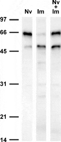
In order to investigate whether or not the observed degradation of serum albumin in immune and immune-waning samples was artifactual, a series of mixed extractions was carried out (Fig. 6). Samples of naïve and immune abomasum were either extracted separately, or the naïve and immune tissues were mixed prior to extraction. Thus, if albumin degradation was due to the liberation during extraction and action on albumin of an immune-expressed proteinase that was refractory to the proteinase inhibitor cocktail used, then the mixed sample would contain more degraded albumin and less intact albumin than would be expected by simple mixing of the extracts (following denaturation by heating in SDS-PAGE reducing buffer). The results obtained (Fig. 6) are consistent with the degraded albumin in immune samples being formed prior to extraction.
Generation of albumin fragments during tissue extraction. Abomasum samples from naïve and immune abomasum were extracted with urea buffer, either alone or together, then analysed by anti-serum albumin Western blotting. The naïve sample (Nv) contained mainly intact serum albumin (67 kDa). Apparent proteolytic degradation fragments of albumin were detected in the immune sample (Im), with a prominent 50 kDa fragment and loss of the parent 67 kDa band. The composite extract (Im + Nv), appeared to contain a higher ratio of intact to degraded albumin than the immune sample, showing that presence of the immune sample did not lead to artifactual degradation of albumin from the naïve sample. This was confirmed using 3 separate combinations of naive and immune extracts (not shown).

Sheep serum albumin immunohistochemistry
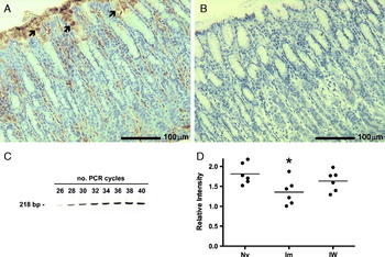
The localization of serum albumin was investigated by immunohistochemistry of naïve, immune and immune-waning abomasal folds using a chicken anti-albumin antibody. Most samples showed intercellular labelling within the lamina propria (Fig. 7A), as would be expected of an abundant plasma protein. This labelling appeared to be strongest towards the base of the abomasal fold. However, additional intracellular labelling was detected in epithelial cells at or close to the surface of crypts (Fig. 7A). Although this labelling was somewhat patchy and difficult to quantify, it was suggestive of local albumin expression. Control IgY was negative (Fig. 7B).
Local expression of serum albumin in the sheep abomasum. Paraformaldehyde-fixed tissue sections from naïve sheep abomasum were immunoperoxidase labelled with chicken anti-bovine serum albumin (A) and control chicken IgY (B). Note the presence of immunopositive mucus cells at, and close to (arrowed) the epithelial surface. RT-PCR of naïve abomasal mRNA using primers specific for sheep serum albumin (C) gave a product of the expected size and sequence (not shown). Semi-quantitative PCR (D), relative to a combination of 3 housekeeping genes (see Materials and Methods section, showed a significant decrease in serum albumin mRNA expression in immune compared to naïve sheep (* Mann-Whitney, P<0 05).

Analysis of serum albumin mRNA transcription
Control abomasal RNA gave a positive RT-PCR signal of the expected size (218 bp) with albumin-specific primers (Fig. 7C). The PCR product was sequenced and found to be exactly as expected from the published sequence (Brown et al. Reference Brown, Dziegielewska, Foreman and Saunders1989). Samples of mRNA from naïve, immune and immune-waning abomasum were subjected to semi-quantitative RT-PCR analysis, and expressed relative to 3 housekeeping genes. This indicated (Fig. 7D) that serum albumin mRNA expression was significantly down-regulated in tissues of the immune phenotype.
DISCUSSION
This study has shown that the protein expression profile in the abomasum of sheep with no prior exposure to T. circumcincta is distinct to that in sheep that have been previously exposed to a trickle infection with the parasite. The expression profiles for naïve sheep and immune sheep clustered separately from each other indicating that T. circumcincta trickle infection produced a detectable alteration in the host abomasal proteome. Furthermore, differences were discovered between recently exposed animals (immune) and those examined 6 weeks after exposure had ceased (immune-waning). The immune sheep resolved into 2 separate groups, and while 2 of the immune-waning sheep clustered with immune sheep, a further 2 clustered with naïve sheep, and the final 2 clustered separately. This suggests a degree of variability in the development and/or maintenance of immune responses between different animals following cessation of exposure.
Proteins that are up-regulated in the immune abomasum, but are less highly expressed in immune-waning are potential candidates for effectors of the immune exclusion phenomenon, which occurs by the development of a mucosal microenvironment that discourages larval establishment in the abomasal crypts (Jackson et al. Reference Jackson, Greer, Huntley, McAnulty, Bartley, Stanley, Stenhouse, Stankiewicz and Sykes2004). The 2DGE analysis highlighted galectin-15, thioredoxin and certain fragments of cytoplasmic actin and calreticulin as possessing these characteristics. Western blotting for galectin-15, thioredoxin and calreticulin was consistent with this pattern, as was that for intelectin.
Expression of galectin-15 (also known as ovgal11) was induced in immune tissues but was minimal in immune-waning samples. This lectin was previously described by Dunphy et al. (Reference Dunphy, Balic, Barcham, Horvath, Nash and Meeusen2000) to be induced in the abomasal mucosa of sheep infected with H. contortus, and was immunolocalized to the upper epithelial layer of the abomasal mucosa (Dunphy et al. Reference Dunphy, Balic, Barcham, Horvath, Nash and Meeusen2000). It is likely that this molecule plays a role in modifying mucus properties as a response to H. contortus infections (Meeusen et al. Reference Meeusen, Balic and Bowles2005), and indeed, it was recently detected during a proteomic analysis of changes to the abomasal mucus layer following T. circumcincta infection (Athanasiadou et al., Reference Athanasiadou, Pemberton, Jackson, Inglis, Miller, Thevenod, Mackellar and Huntley2008).
Intelectin was included as a positive control in our Western blot analysis, since we have previously found that sheep intelectins are up-regulated following T. circumcincta infection (French et al. Reference French, Knight, Smith, Brown, Craig, Pate, Miller and Pemberton2008, Reference French, Knight, Smith, Pate, Miller and Pemberton2009). Here, immunoreactive intelectin (which could be a mixture of intelectin-1, -2 or -3) was most highly expressed in pooled immune abomasum, and was much less abundant in immune-waning. It was surprising that this protein did not make a prominent appearance in the proteomic analysis, but we have found that intelectin-2 co-purifies with sheep gastric mucins (Pemberton et al. Reference Pemberton, Verdon, Inglis and Pearson2011), and this intelectin-mucin interaction, which required SDS and reducing agent treatment to disrupt, may have resulted in its exclusion from the pores of the isoelectric focussing gels. The striking up-regulation of intelectin and galectin-15 in immune mucosa, their decrease in expression in immune-waning mucosa, and their potential for interaction with mucins make them good candidates as effectors of immune exclusion of parasites such as T. circumcincta.
Additionally, a number of proteins were strongly down-regulated in immune compared to naïve mucosa. These include calreticulin, GRP94, HSPA9 and PDI, which are molecular chaperones involved with protein expression, folding and packaging. These data may be indicative of reduced mRNA expression, increased degradation, or artifactual proteolytic cleavage. Alternatively, the proteins may be actively exported, such as occurs during the formation and release of exosomes (Conde-Vancells et al. Reference Conde-Vancells, Rodriguez-Suarez, Embade, Gil, Matthiesen, Valle, Elortza, Lu, Mato and Falcon-Perez2008). The increased abundance of lower MW forms of calreticulin in immune versus naïve samples suggests that this protein undergoes proteolytic degradation. Western blotting analysis also indicated proteolytic processing of GRP78 and PDI. Selective cleavage of endoplasmic reticulum proteins calreticulin, GRP78 and PDI was previously noted in hepatocellular carcinoma, which was suggested as a possible mechanism for promoting protein secretion efficiency (Chignard et al. Reference Chignard, Shang, Wang, Marrero, Brechot, Hanash and Beretta2006). However, artifactual proteolysis cannot be ruled out as an explanation.
The local expression of substantial levels of serum albumin in control abomasa (Fig. 7, cf. A and B) was unexpected and noteworthy. This was confirmed by RT-PCR, and a possible site of expression by mucus-producing epithelial cells was suggested by the immunohistochemistry results. Extra-hepatic expression of serum albumin was previously shown in bovine mammary gland (Shamay et al. Reference Shamay, Homans, Fuerman, Levin, Barash, Silanikove and Mabjeesh2005) and human brain microglial cells (Ahn et al. Reference Ahn, Byun, Cho, Kim, Yoo, Kim, Paek, Kim, Simpson and Lee2008) and therefore may be more widespread than is generally acknowledged. At the protein level, serum albumin was found to be down-regulated in immune and immune-waning samples compared to naïve abomasum. It is not possible to distinguish local and plasma-derived albumin in this analysis, but at the mRNA level, a significant reduction in albumin expression was determined in immune compared to naïve samples. The proposed site of local albumin expression would indicate that it is constitutively released into the gastric mucus, suggesting a possible innate protective role.
The most abundant serum albumin fragment detected in this study appeared to be the ∼14 kDa spot 66, corresponding to the N-terminal lobe of albumin (Asp1 – Lys114; calculated MW=13 0 kDa; pI=4 7), which contains 3 internal disulphide bridges but is not linked to the rest of the molecule by disulphide bridging and would therefore be free to diffuse away following cleavage. We have previously found the homologous fragment to be generated by incubating bovine serum albumin with sheep mast cell proteinase-1 (sMCP-1, Pemberton et al. Reference Pemberton, Huntley and Miller1997). SMCP-1 is known to be highly up-regulated in the abomasal mucosa in response to T. circumcincta infection (Huntley et al. Reference Huntley, Gibson, Brown, Smith, Jackson and Miller1987), but was not directly detected in this analysis since its isoelectric point is too high (pI=8 9).
The N-terminal albumin fragment contains the single free cysteine residue (Cys34) of the molecule. This cysteine has been estimated to make up more than 70% of the free-radical neutralizing capacity of serum (Bourdon and Blanche, Reference Bourdon and Blanche2001), and is important in binding nitric oxide (Stamler et al. Reference Stamler, Jaraki, Osborne, Simon, Keaney, Vita, Singel, Valeri and Loscalzo1992). However, it is unusually unreactive in disulphide exchange reactions, due to an interaction with Tyr84 (Stewart et al. Reference Stewart, Blindauer, Berezenko, Sleep, Tooth and Sadler2005). It is interesting to note that the canonical substrate for human mast cell chymase has been determined to be Tyr84 of human serum albumin (Raymond et al. Reference Raymond, Ruggles, Craik and Caughey2003). Stewart and colleagues have shown how this cleavage would release the structural constraint on Cys34, thus enhancing the thiol reactivity of serum albumin (Stewart et al. Reference Stewart, Blindauer, Berezenko, Sleep, Tooth and Sadler2005). It is less clear how generation of the ∼14 kDa albumin N-terminal fragment might affect the reactivity of Cys34. It may possibly result in a structural rearrangement that activates Cys34 or, alternatively, it may simply generate a form which diffuses more easily to where it is required, for example as an antioxidant or NO-binding protein. Alternative roles for albumin within the abomasal mucosa could include small molecule binding/transport and effects on mucus viscosity via interactions with mucins (List et al. Reference List, Findlay, Forstner and Forstner1978).
Thioredoxin, which was selectively up-regulated to a modest degree in immune sheep, is also an antioxidant. There is expected to be an increased requirement for antioxidant capacity at the mucosal surface during nematode infection, in order to prevent excessive host tissue damage from host-derived anti-parasite oxidants. For example, abomasal expression of the powerful oxidant, dual oxidase-2 (Duox2) was shown to be markedly elevated following infection with H. contortus (Menzies et al. Reference Menzies, Reverter, Andronicos, Hunt, Windon and Ingham2010). Increased expression of the antioxidant glutathione peroxidase-2 was shown to occur in a concerted manner (Menzies et al. Reference Menzies, Reverter, Andronicos, Hunt, Windon and Ingham2010), and it seems likely that thioredoxin and serum albumin also contribute to the host-protective antioxidant screen during anti-parasite responses.
In conclusion, this study has indicated that galectin-15 and intelectin are selectively up-regulated in immune but not immune-waning abomasum following T. circumcinta trickle infection. Further studies are required to determine whether these proteins are effectors of (or simply markers of) the immune exclusion phenomenon. Additionally, the local abomasal expression of albumin and its proteolytic degradation pattern suggests that further investigation into the functional significance of albumin modifications by mast cell proteinases is warranted.
ACKNOWLEDGEMENTS
This work was supported with funding from the Biotechnology and Biological Sciences Research Council (BBSRC grant no: BB/E01867X/1). D.P.K. was also partly supported by a Programme Grant from Scottish Government (RERAD).