Introduction
During expeditions to Costa Rica in 2008, M.P. Mier Durante and three of the authors (N.N., P.H., V.M.) collected several viviparous female aphids on Blechnum buchtienii Rosenstock (Blechnaceae) in Cerro Buenavista or Cerro de la Muerte (Talamanca mountain range). These were identified as belonging to the tribe Macrosiphini (Hemiptera: Aphididae: Aphidinae) sensu the “Remaudière, Quednau, and Stroyan extended classification” (Nieto Nafría and Favret Reference Nieto Nafría and Favret2011). Given their peculiar characteristics we believe they represent a new species. Subsequently, one of the authors (V.M.) visited the capture locality several times to monitor the population and verifying that the aphids developed and reproduced on the host plant.
Specimens caught on ferns in Mexico belonging to the collection génerale d'aphids of the Muséum national d'Histoire naturelle, Paris, France were also examined.
Material and methods
A comparative morphological study was conducted on species in genera (i) whose apterae possess swollen siphunculi and have been recorded in North America or (ii) are known to feed on ferns. Along with the new material from Costa Rica, we also examined aphid specimens in the collections of the Muséum national d'Histoire naturelle (Paris, France) and the Universidad de León (Leon, Spain). We consulted the works of Robinson (Reference Robinson1966), Miyazaki (Reference Miyazaki1968, Reference Miyazaki1971), Hille Ris Lambers (Reference Hille Ris Lambers1969), Ghosh (Reference Ghosh1974), Ghosh et al. (Reference Ghosh, Basu and Raychaudhuri1977), Moritsu (Reference Moritsu1983), Remaudière (Reference Remaudière1983), Cook (Reference Cook1984), Chakrabarti and Banerjee (Reference Chakrabarti and Banerjee1989), Heie (Reference Heie1992, Reference Heie1994, Reference Heie1995), Remaudière and Muñoz Viveros (Reference Remaudière and Muñoz Viveros1992), Foottit and Richards (Reference Foottit and Richards1993), Remaudière and Remaudière (Reference Remaudière and Remaudière1997), Lee et al. (Reference Lee, Holman and Havelka2002), Sorin and Arakawa (Reference Sorin and Arakawa2005), Blackman and Eastop (Reference Blackman and Eastop2006), and Blackman (Reference Blackman2010).
The type specimens are preserved in microscopic slides with a water-soluble mounting medium (Nieto Nafría and Mier Durante Reference Nieto Nafría and Mier Durante1988). The specimens for the molecular analysis were preserved in 96% ethanol until processing. Data on the capture of the studied specimens can be seen in the “Types” section in the description of the new species.
Measurements of the slide-mounted specimens were made according to Nieto Nafría and Mier Durante (Reference Nieto Nafría and Mier Durante1988) with an ocular micrometre. The measurements are lengths except when indicated that they are a width or diameter. A camera lucida fitted to the microscope was used for the drawings, and the photomicrographs were taken with a Leica DC digital camera with IM 1000 version 1.10 software (Leica Microsystems Imaging Solutions Ltd., Cambridge, United Kingdom).
Molecular phylogenetic analyses were done based (1) on a fragment of the mitochondrial DNA containing the 5′ region of the cytochrome c oxidase 1 (COI) and (2) on a fragment of the nuclear gene coding for elongation factor-1 alpha (EF1α) of three specimens.
Total DNA was extracted separately from three specimens. DNA extraction was done following the HotSHOT (Hot Sodium HydrOxide and Tris) method (Truett et al. Reference Truett, Walker, Warman, Truett, Heeger and Mynatt2000). Voucher specimens are the colony mates, presumed to be genetic clones, examined and described below.
Polymerase chain reaction (PCR) amplifications of the two gene fragments analysed were done using 3 μl of the extracted DNA in 50 μl total reaction volumes. A 710 base pair fragment of the 5′ region of COI was amplified using primers LCO1490 and HCO2198 described by Folmer et al. (Reference Folmer, Black, Hoeh, Lutz and Vrijenhoek1994). PCR conditions for COI amplification were as follows: 94 °C for 1 minute; 35 cycles of 94 °C for 30 seconds, 48 °C for 1 minute and 68 °C for 1 minute; a final extension step of 7 minutes at 68 °C was included after cycling. Amplification of the EF1α fragment was done using two consecutive PCR reactions with primers Efs175 (Moran et al. Reference Moran, Kaplan, Gelsey, Murphy and Scholes1999) and Efr1 (5′GTGTGGCAATSCAANACNGGAGT3′) in the first reaction and then primers Efs175 and Efr2 (5′TTGGAAATTTGACCNGGGTGRTT3′) in the second semi-nested reaction. PCR conditions used in the first reaction were: 94 °C for 1 minute; 40 cycles of 94 °C for 30 seconds, 50 °C for 1 minute and 68 °C for 1.5 minutes; a final extension step of 7 minutes at 68 °C was included after cycling. The semi-nested PCR was done similarly but using 52 °C for the annealing step and using 1 μl of the first PCR product.
PCR products were purified by ammonium precipitation and reconstituted in 10 μl of LTE buffer (10 mM Tris, 0.1 mM EDTA). Direct sequencing of amplified fragments was done in both directions using PCR primers (Efr2 was used as reverse primer for sequencing the EF1α fragment). Sequencing was conducted using the Big Dye Terminator v3.1 Cycle Sequencing Kit (Applied Biosystems, Forster City, California, United States of America) following the manufacturer's instructions, and samples were loaded onto an ABI 3700 automated sequencer (Applied Biosystems).
Chromatograms were revised and sequences assembled using the Staden package v1.6.0 (Bonfield et al. Reference Bonfield, Smith and Staden1995). Multiple alignments were done with Clustal X v2.1 (Larkin et al. Reference Larkin, Blackshields, Brown, Chenna, McGettigan and McWilliam2007) with gap opening and gap extension penalties of 10.0 and 0.2, respectively. Phylogenetic and molecular evolutionary analyses were conducted using MEGA version 5 (Tamura et al. Reference Tamura, Peterson, Peterson, Stecher, Nei and Kumar2011).
Results and discussion
The combined description of these new species and new genus is made under Article 13.4 of the International Code of Zoological Nomenclature (International Commission on Zoological Nomenclature 1999).
Gibbomyzuspteridophytorum Nieto Nafría, Pérez Hidalgo, Martínez-Torres, and Villalobos Muller, new genus, new species.
Type species
Gibbomyzus pteridophytorum new species, here designated.
Etymology of the genus name
Gibbomyzus is a noun formed by “gibb”, which is the root of the Latin word gibba, meaning hump; “o” that is a thematic vowel to facilitate pronunciation; and “myzus”, which is a noun that forms part of the names of many genera with a more or less similar appearance to Myzus. The name is masculine, following the gender of Myzus.
Diagnosis of the genus
Macrosiphini with swollen siphunculi lacking apical reticulation; frontolateral tubercles divergent and taller than the frontomedial tubercle; spinules absent on cephalic dorsum and present but scarce on ventral face; ventral margin of antennal socket carrying a striate protuberance; secondary sensoria with nonciliate margin; setae of body dorsum and appendages short and blunt; prothoracic spiracular apertures larger than those on abdomen; prothorax with six dorsal setae; cauda lanceolate; wings with hyaline membrane and not reduced in venation; abdominal patch present both in apterous and alate viviparous females, dorsum of abdomen very gibbous.
Type material of G. pteridophytorum
Holotype: apterous viviparous female CRI-294 ap. #1, COSTA RICA: San Jose province, Cerro de la Muerte (Mirador de los Quetzales, 9°38′N, 83°51′W, 2900 m), B. buchtienii, 28-ii-2008, Nieto Nafría, Mier Durante, Villalobos Muller and Pérez Hidalgo leg.; Zoological Collection of the University of León (CZULE). Paratypes: 14 apterous viviparous females and three alate viviparous females caught at same time as the holotype; CZULE, Leon (Spain), and Museo de Zoología, Universidad de Costa Rica, San Pedro de Montes de Oca (Costa Rica); one apterous viviparous female, MEXICO: Mexico state, Popocatépetl (3000 m), unidentified fern, 1.x.79, G. Remaudière leg., register number 06174; one apterous viviparous female, MEXICO: Michoacan state, Paricutin (∼2500 m), Pteridium aquilinum, 23-x-1981, R. Peña Martínez leg., register number 08403; Muséum national d'Histoire naturelle, Paris (France), collection générale d'aphides.
Etymology of the specific epithet
The specific name pteridophytorum is the genitive plural of the Latin neologism Pteridophyta, the name of the classic taxon that includes ferns.
Diagnosis of the species
Aphids with the characteristics mentioned in the genus diagnosis; with marginal papillae present on prothorax and on several abdominal segments II to VI, and sometimes spinal papillae present on head; secondary sensoria on antennal segment III in apterae and on segments III, IV, and V in alatae.
Description
Apterous viviparous females (Figs. 1–3)
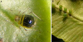
From 17 specimens. When alive, the dorsum of the abdomen is noticeably raised, resembling a large hump, and shiny black; and antennae, legs, siphunculi, ventral part of the body, and membranous parts of the dorsum are brown, greenish brown, or green. Nymphs completely green. When mounted, body is 1.470–1.920 mm including cauda.
(A) Gibbomyzus pteridophytorum live apterous viviparous female. (B) Gibbomyzus pteridophytorum live nymphs that will develop into apterous viviparae.

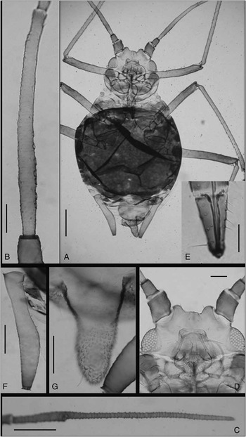
Gibbomyzus pteridophytorum apterous viviparous female: (A) habitus (scale bar, 0.20 mm); (B) II and III antennal segments (scale bar: 0.20 mm); (C) VI antennal segment (scale bar, 0.20 mm); (D) head (scale bar, 0. 05 mm); (E) ultimate rostral segment (scale bar, 0.10 mm); (F) siphunculus (scale bar, 0.20 mm); (G) cauda (scale bar, 0.10 mm).

Head. Pale-brown or brown. Frontolateral tubercles divergent (or sometimes apparently parallel), rounded and ∼0.5 times taller than the diameter of the antennal alveolus; frontomedial tubercle wide, low and rounded, making the frontal sinus undulate. Dorsum with clearly defined striae, particularly on the frontolateral tubercles, without scales or spinules; ventral face with few striae and spinulae, more numerous on frontolateral tubercles. One or two small spinal papillae sometimes present. Ventral margin of antennal socket carrying a striate protuberance. Dorsal setae with rounded apex, 17–25 μm and 0.7–1.3 times the basal diameter of antennal segment III (henceforth D); ventral setae pointed and slightly longer.
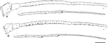
Antennae 1.420–2.050 mm and 0.96–1.17 times body length, as dark as or darker than head. Antennal segments I and II dorsally with spinules and ventrally with scales; carrying setae longer than those on flagellum. Antennal segment III 0.37–0.56 mm and 1.3–1.8 times IV, with scales, blunt setae 5–10 μm and 0.3–0.5 times D, and on its proximal half 3–15 secondary sensoria, circular with double-lined and nonciliate margin and placed in a ventral line. Antennal segments IV and V 0.21–0.39 and 0.19–0.32 mm, respectively, and imbricated, as is VI. Processus terminalis of antennal segment VI 0.38–0.48 mm, 3.0–3.5 times base (0.11–0.15 mm), and 0.8–1.0 times antennal segment III.
Rostrum more or less as pigmented as head, reaching coxae of metalegs. Third rostral segment with strong spinulae. Ultimate rostral segment long triangular, 0.11–0.14 mm, 2.0–2.6 times its basal diameter, 0.8–1.1 times base of antennal segment VI and 1.2–1.4 times second metatarsomere; carrying 8–11 accessory setae, 25–30 μm and 0.8–1.2 times D.
Thorax. Each segment with a dark, complete and wide sclerotised dorsal band, resembling an arch, with striated ornamentation; sometimes the metathoracic band coalesces with abdominal patch. Prothorax with six dorsal setae (two pleural, two marginal, and two spinal) not in a transverse line; and one or two small marginal papillae frequently present. Spiracular sclerites large and rugose; spiracular apertures rounded and only slightly wider than those on the abdomen, which are reniform.
Legs more or less as pigmented as antennae, with  $$$\tfrac{3}{4} $$$
distal part of femora, apex of tibiae and tarsi darker than other parts. Metafemur and metatibia 0.46–0.72, 0.84–1.32 mm, respectively. First tarsomeres with three setae. Second metatarsomere 0.08–0.11 mm. Setae on femora and tibiae sparse, blunt, and short; the longest ones on dorsum of metafemur 11–15 μm and 0.4–0.6 times D, those on outside margin of metatibiae 20–30 μm and 0.7–1.3 times D.
Abdomen. Segments I to V or VI with an extensive dorsal patch including the spiracular (darker) and intersegmental sclerites (paler), with a narrow membranous ring around the insertion of the siphunculi, with few and sparse spinules and striae. Small rounded marginal papillae frequently present on segments III and IV, sometimes also on II and V. Dorsal setae blunt, seven to nine per segment, 5–8 μm, 0.2–0.3 times D. Ventral setae more numerous, pointed and 20–25 μm. Abdominal segments VI (sometimes), VII and VIII with sclerites or transverse bands, frequently paler than patch but carrying spinules, especially postsiphuncular. Abdominal segment VIII with three to four setae, 27–30 μm and 1.2–1.4 times D.
Siphunculi swollen, with broad base and well-defined both preapical incision and flange, 0.31–0.45 mm, 0.20–0.24 times body length; swollen part asymmetrical (the outer edge is almost straight), its larger diameter is 0.05–0.09 mm, 0.9–1.4 times siphuncular diameter at base (0.06–0.08 mm), and 1.2–1.9 times the diameter of peduncle at middle (0.04–0.07 mm); pigmented like legs, markedly paler than the dorsal patch, and with a diverse cuticular ornamentation: striae on peduncle, few, small sparse spinules on swollen part, and an incipient reticulation with two or three lines on the incision.
Genital plate darker than siphunculi, with striae and spinulae, two discal and 9–14 posterior setae, all with rounded apex. Anal plate paler than genital plate. Cauda 0.13–0.20 mm and 0.4–0.5 times siphunculus, lanceolate (1.1–1.4 times its basal width) more or less pigmented as the legs and carrying four to six fine, curved, and pointed setae.
Alate viviparous females (Figs. 3–4)
From three specimens. As apterae except as noted.
Gibbomyzus pteridophytorum antennae (scale bar, 0.10 mm): (A) apterous viviparous female; (B) alate viviparous female.

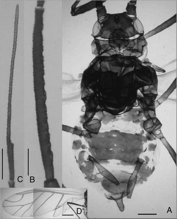
Gibbomyzus pteridophytorum alate viviparous female: (A) habitus (scale bar, 0.20 mm); (B) III antennal segment (scale bar, 0.10 mm); (C) VI antennal segment (scale bar, 0.10 mm); (D) wings (scale bar, 0.25 mm).

Head darker. Antennae darker and up to 1.19 times body length. Antennal segment III with 14–22 secondary sensoria, aligned and extending over entire length of segment. Antennal segments IV and V with 5–8 and 0–4 secondary sensoria, respectively. Legs more pigmented. Dorsal setae on metafemur up to 0.7 times D. Wing veins well pigmented; media of front wings with three branches. Abdomen with pleural–spinal plate on segments I and II, large rectangular pleural–spinal patch with a posterior–medial incision on segments III to VI, marginal plates from segments II to VII, and more or less wide setiferous sclerites on segments VII and VIII; intersegmental sclerites not always perceptible. Spinules present on abdominal I, II, patch, and on postsiphuncular sclerites. Dorsal setae on abdominal segments II–V up to 11 in number, 12 μm and 0.5 times D; and on segment VIII up to 22 μm, 1.0 times D.
Bionomics
The species is recorded from B. buchtienii and P. aquilinum (Linnaeus) Kuhn (Dennstaedtiaceae), but may also feed on other fern genera because the families Blechnaceae and Dennstaedtiaceae (Polypodiales) are in different, though closely related clades (Schneider et al. Reference Schneider, Smith and Pryer2009).
Geographical distribution
Collection data from southern Costa Rica and central Mexico indicate that the species inhabits the mountains of Mesoamerica, although it is not frequently collected and thus probably rare.
Taxonomic discussion
Morphological discussion
In the New World, 13 Macrosiphini genera with species characterised by the presence of swollen siphunculi and a strongly sinuous or deeply concave frons (U-shaped, V-shaped, or W-shaped), have been recorded (for the objective validity of the genera: Nieto Nafría and Favret (Reference Nieto Nafría and Favret2011); for the records from the New World south of the United States: Nieto Nafría and Mier Durante (unpublished data); and for the records from North America north of Mexico: Foottit and Richards (Reference Foottit and Richards1993), Blackman and Eastop (Reference Blackman and Eastop2006), and Foottit et al. (Reference Foottit, Halbert, Miller, Maw and Russell2006)). They are: Amphorophora Buckton, 1876; Carolinaia Wilson, 1921; Cryptomyzus Oestlund, 1923 (some species); Decorosiphon Börner, 1939; Delphiniobium Mordvilko, 1914; Illinoia Wilson, 1910; Hyperomyzus Börner, 1933; Microparsus Patch, 1909 (subgenus Picturaphis Blanchard, 1922); Neoamphorophora Mason, 1924; Rhopalomyzus Mordvilko, 1921 (subgenus Judenkoa Hille Ris Lambers, 1953); Rhopalosiphoninus Baker, 1920 (several species); Utamphorophora Knowlton, 1946; and Wahlgreniella Hille Ris Lambers, 1949.
Delphiniobium, Illinoia, and the species of Rhopalosiphoninus with parallel frontolateral tubercles, have isodiametrical subapical reticulations on siphunculi, and membranous abdominal dorsum; Gibbomyzus has sclerotised abdominal dorsum and has siphunculi not reticulated.
In Amphorophora, Carolinaia, Rhopalomyzus, and Wahlgreniella, the ventral margin of the antennal socket is nonprotuberant, but in Gibbomyzus it is protuberant. These genera additionally present the following differences in comparison with Gibbomyzus (see the description of G. peridophytorum). In Amphorophora, the frons is deeply V-shaped (if the medial tubercle exists it is very small) and the dorsum of the abdomen in both apterae and alatae is membranous. In Carolinaia (including the subgenus Glabromyzus Richards, 1960), the ultimate rostral segment carries only two (infrequently three or four) accessory setae, and the dorsal–abdominal surface is wrinkled. In the subgenus Jundekoa, the ultimate rostral segment is short, the first metatarsomere has only two setae, the abdominal–dorsal surface is membranous in apterous viviparae but with a patch in alatae, and the secondary sensoria are scattered. In Wahlgreniella, the dorsum of the abdomen is membranous in apterae and alatae, can carry a patch that is frequently incomplete or broken, the siphunculi are slender, and the margins of the secondary sensoria are ciliate.
In Cryptomyzus, Decorosiphon, Hyperomyzus, Neoamphorophora, Utamphorophora, and in the subgenus Picturaphis, the ventral margin of the antennal socket is protuberant. Each genus in this group can be separated from Gibbomyzus based on the following features: in Cryptomyzus (many of whose species do not have swollen siphunculi), the apterous viviparae have a membranous dorsal–abdominal surface and the antennal and dorsal setae are long and clavate; the alate viviparae have a dorsal–abdominal patch and the setae are shorter but the secondary sensoria are scattered. In Decorosiphon, the antennae and legs carry very long, erect, and pointed setae and the siphunculi are scabrous or squamous and have a very large flange. In Hyperomyzus, the head is smooth, the secondary sensoria are not aligned in the apterae and conspicuously scattered in the alatae; in several species of this genus the apterae show a patch on the thoracic and abdominal dorsum, but the alatae of this species have a largely membranous abdomen with cross-bands or isolated sclerites. In the subgenus Picturaphis, the frontolateral tubercles have a strong ornamentation, the dorsum of the abdomen is membranous and the wings exhibit a peculiar venation and pigmentation. In Neoamphorophora, the frontomedial tubercle is protruding, as tall as the lateral ones; the dorsum of the prothorax has two medial–anterior setae in addition to the habitual (in this group of genera), two medial–posterior and four lateral (two anterior and two posterior) setae, and the alatae have cross-bands and the secondary sensoria are scattered. In Utamphorophora, there are only two (infrequently three or four) accessory setae on the ultimate rostral segment, which is habitually short triangular, as in other genera inhabiting grasses, and neither apterae nor alatae have a dorsal–abdominal patch.
The global aphid fauna known to feed on ferns is composed of 60 species and subspecies (Blackman and Eastop Reference Blackman and Eastop2006), all of them belonging to Aphidinae. A small proportion belongs to the genera Aphis and Toxoptera (Aphidini) and the others are classified into 15 genera of Macrosiphini: Amphorophora Buckton, 1876; Aulacorthum Mordvilko, 1914; Idiopterus Davis, 1909; Macromyzella Ghosh, Basu, and Raychaudhuri, 1977; Macromyzus Takahashi, 1960; Macrosiphum Passerini, 1860; Mastopoda Oestlund, 1886; Micromyzella Eastop, 1955; Micromyzodium David, 1958; Micromyzus van der Goot, 1915; Myzus Passerini, 1860; Neomyzus van der Goot, 1915; Papulaphis Robinson, Reference Robinson1966; Shinjia Takahashi, 1938; and Taiwanomyzus Tao, 1963.
Only in Amphorophora, Taiwanomyzus and several species of Myzus, are the siphunculi swollen. See previous paragraph for the differences between Gibbomyzus and Amphorophora. The apterous females of Myzus have frontolateral tubercles convergent and a strong dorsal–cephalic ornamentation.
Taiwanomyzus has six species, five living on ferns. The apterous viviparous females of all of them have frontomedial tubercle reduced or absent (the bottom of the sinus is flat or weakly convex), and strong, although variably extended, cuticular ornamentation on the frontolateral tubercles; in G. pteridophytorum the frontomedial tubercle is conspicuous and there are no scales or spinules on the dorsum of the head. In addition: (1) the Japanese Taiwanomyzus babai Sorin and Arakawa, Reference Sorin and Arakawa2005; Taiwanomyzus chrysosplenii Miyazaki, Reference Miyazaki1971 (living on Chrysosplenium Linnaeus (Saxifragaceae)); and Taiwanomyzus filicis (Miyazaki, Reference Miyazaki1968) and the Indian Taiwanomyzus himalayensis (Chakrabarti and Banerjee Reference Chakrabarti and Banerjee1989), can be easily separated from G. pteridophytorum because the frontolateral tubercles are convergent; (2) the apterae of the European Taiwanomyzus alpicola (Hille Ris Lambers Reference Hille Ris Lambers1969), which have frontolateral tubercles parallel or divergent, have black siphunculi and one to two large secondary sensoria on antennal segment III (3–15 and smaller in G. pteridophytorum); and (3) the apterae of the Indian T. himalayensis (Chakrabarti and Banerjee Reference Chakrabarti and Banerjee1989), which have divergent frontolateral tubercles, have only four accessory setae on the ultimate rostral segment (8–11 in G. pteridophytorum).
Thus, the “key to aphids on Polypodium and other fern genera” by Blackman and Eastop (Reference Blackman and Eastop2006) can easily be modified to include the new species as follows. In this modification, we have used the terminology, abbreviations, and expressions used in these keys for uniformity.

Molecular results and discussion
A 710 base pair DNA fragment containing a portion of the mitochondrial COI gene was amplified through PCR; after removing primer sequences, useful sequences consisted of 658 nucleotides. Sequences for the sampled aphids were identical so that a single sequence was finally deposited in GenBank (accession number JX415255). The online identification engine available at the Barcode of Life Data Systems (BOLD) (Ratnasingham and Hebert Reference Ratnasingham and Hebert2007) using the COI species database, failed to find any record corresponding to any identified species that matched our sequence. After a BLASTN (nucleotide Basic Local Alignment Search Tool) search against the nonredundant nucleotide database at the National Center for Biotechnological Information (United States of America), sequences from different aphid genera of the tribe Macrosiphini, such as Utamphorophora, Hyperomyzus, and Micromyzus plus Myzus (M.) hemerocallis Takahashi, 1921, were most similar to our sequence (93–94% identical). However, as the COI sequence from the sample of G. pteridophytorum did not match any sequence available at the DNA barcode reference library, we must conclude that our sequence corresponds to a species not represented in the database, which currently hosts sequences from 295 species from 70 genera within the subfamily Aphidinae. Moreover, we were also unable to assign the new species to a particular previously described genus based on the COI sequence as neither strict tree-based nor best close match assignment criteria (Wilson et al. Reference Wilson, Rougerie, Shonfeld, Janzen, Hallwachs and Kitching2011) were fulfilled. Phylogenetic reconstructions based on this gene using a set of available sequences from different Macrosiphini representatives were not very informative about the phylogenetic affiliation of G pteridophytorum. Most relevant, it grouped, albeit with low support, with M. hemerocallis in a cluster that also included Utamphorophora humboldti (Essig, 1941) (results not shown). COI sequences from G. pteridophytorum and M. hemerocallis were separated by a relatively large genetic distance (6.2%), which would be in agreement with both species belonging to distinct but related genera. In this respect, it is relevant to note that average genetic distances among congeneric taxa for a collection of Aphididae COI sequences was 7.22% (range 0.46–11.3%) (Foottit et al. Reference Foottit, Maw, von Dohlen and Hebert2008). The divergence between Hyperomyzus lactucae (Linnaeus, 1758) and Nasonovia ribisnigri (Mosley, 1841) was 4.6%, while between Acyrthosiphon pisum (Harris, 1776) and Acyrthosiphon kondoi Shinji, 1938 it was 5.7% and a divergence as high as 7.9% was estimated between two species of Cavariella (Cavariella konoi Takahashi, 1939 and Cavariella theobaldi (Gillette and Bragg, 1918), which have siphunculi, respectively, swollen and subcylindrical). However, the use of COI sequences for phylogeny inference seems rather limited as the phylogenetic signal is somewhat weak as compared with other markers (Wilson Reference Wilson2010).
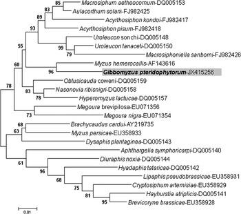
For the EF1α gene fragment, we obtained an identical sequence from the individuals analysed, 928 base pairs, which was deposited in GenBank with accession number JX415256. Using sequences available for EF1α at National Center for Biotechnology Information for different Macrosiphini species, a maximum likelihood tree was built that included the sequence obtained for our previously undescribed species (Fig. 5). Gibbomyzus pteridophytorum grouped with high support with a sequence from M. hemerocallis within a monophyletic clade that also included sequences representative of genera such as Hyperomyzus, Nasonovia Mordvilko, 1914, Acyrthosiphon Mordvilko, 1914, and Uroleucon Mordvilko, 1914, among others. With regard to the position of M. hemerocallis in this group and separated from other Myzus species, the opinion of Blackman and Eastop (Reference Blackman and Eastop2006: p. 1240) is interesting: “It is not a typical Myzus, and possibly would be more correctly placed in the genus Hyalomyzus”, although in this last genus the siphunculi are swollen and in M. hemerocallis they are cylindrical.
Maximum likelihood tree obtained for elongation factor-1 alpha (EF1α) sequences from different Macrosiphini species including Gibbomyzus pteridophytorum (highlighted). Sequences from aphid species other than G. pteridophytorum were obtained from the National Center for Biotechnological Information (United States of America) database and their accession numbers are indicated. A discrete Gamma distribution was used to model evolutionary rate differences among sites, five categories (+G, parameter = 0.2897). The rate variation model allowed for some sites to be evolutionarily invariable ([+I], 36.9534% sites). The tree is drawn to scale, with branch lengths measured in the number of substitutions per site. All positions containing gaps and missing data were eliminated leaving a total of 785 positions in the final data set. Percentage bootstrap support values obtained after 200 replicates are indicated above branches.

EF1α is widely used in phylogenetic reconstructions including insects (Simon et al. Reference Simon, Schierwater and Hadrys2010; Wilson Reference Wilson2010). Our phylogenetic analysis of EF1α sequences including representative species from diverse Macrosiphini genera strongly supported the grouping of G. pteridophytorum with the only sequence from M. hemerocallis available at GenBank. However, as already discussed for the COI sequence, both species were also relatively divergent for the EF1α gene (about 4.0%), which, as before, would support G. pteridophytorum belonging to a different, though related, genus. For comparison, the divergence between H. lactucae and N. ribisnigri was 2% and between Uroleucon sonchi (Linnaeus, 1767) and Uroleucon tanaceti (Linnaeus, 1758) 3%. However, the divergence between conspecific A. pisum and A. kondoi was 4.4%. In support of G. pteridophytorum and M. hemerocallis belonging to different genera rather than being congeneric species, it is necessary to note that the morphological differences between both species are very large.
Acknowledgements
This research was mostly supported by the Spanish Agency for International Development Cooperation (AECID) project D/010523/07. Partial support was also obtained from project CGL2011-27404 granted by the Spanish Government to D. Martínez-Torres. J. M. Nieto Nafría wishes to thank the Muséum national d'Historie naturelle of Paris for the grant he received in February 2011. The authors express their gratitude to Prof. Georges Remaudière (Muséum national d'Histoire naturelle, Paris, France), Prof. M. Pilar Mier Durante (Universidad de León, Leon, Spain), and Dr. Colin Favret (Université de Montréal, Montréal, Canada) for their support during the study and critical reading of the manuscripts.