Implications
This manuscript reviews effects of circulating progesterone (P4) on dairy cattle reproduction. Various methods to elevate P4 during growth of the preovulatory follicular wave have been shown to increase pregnancies/ artificial insemination (AI) and reduce double ovulation, providing methods to improve fertility and reduce twinning rate in lactating dairy cattle. Conversely, very low concentrations of P4 near AI are needed to optimize fertility. Finally, elevations of P4 after AI can impact embryonic development and also may elevate fertility. Thus, innovative strategies to optimize circulating P4 concentrations during selected reproductive periods enhance our management tools for improving reproductive efficiency of lactating dairy cows.
Discovery of Progesterone (P4)
The discovery of P4 begins with a clear description by Regnier deGraaf (1641–1673) of the corpus luteum (CL), calling them ‘globules’ and correctly surmising (for rabbits) that ‘the number of globules equals the number of offspring from a particular mating’ (deGraaf, 1672 in (Jocelyn and Setchell, Reference Jocelyn and Setchell1972)). A key discovery came in the laboratory of Gustav Born (1851–1900), an excellent histologist, who observed that the CL was a ductless gland and correctly advanced the idea that it was a gland of internal secretion that could be involved in pregnancy (see excellent reviews by Simmer (Reference Simmer1971); Magnus and Simmer (Reference Magnus and Simmer1972)). Two of his students later tested this hypothesis in their own laboratories. In Germany, Ludwig Fraenkel performed bilateral ovariectomy or electrocautery of all corpora lutea in mated rabbits and found that they did not maintain their pregnancies (Fraenkel and Cohn, Reference Fraenkel and Cohn1901). In Norway, Vilhelm Magnus also performed galvano-cautery of all CL as well as bilateral ovarian removal in mated rabbits and also reported that pregnancy was not maintained (Magnus, Reference Magnus1901). Both researchers reached the same conclusion that the CL was essential for maintenance of pregnancy. Additional experiments by Magnus showed that unilateral oophorectomy did not alter pregnancy. He also reported that bilateral oophorectomy produced atrophy of the uterus but that destruction of the CL did not produce atrophy of the uterus, in spite of lack of pregnancy in both treatments. He concluded that the ovarian stroma produced an endocrine substance that maintained the uterine structure but that the CL produced an endocrine secretion that maintained pregnancy, called ‘differentieringsstofe’ (differentiating stuff), the first name given to the hormone later known as P4 (Magnus, Reference Magnus1901). In addition, Magnus correctly predicted the differing functions of ovarian estrogens and gestagens. The initial reports were met with some skepticism, however, Fraenkel continued his research during the next decade on more than 160 rabbits eventually concluding that, ‘Thus by the power of large numbers my thesis is proven: The ovary, in particular the CL, regulates the implantation and initial development of the embryo’ (Fraenkel, Reference Fraenkel1910).
In order to isolate the luteal hormone, a bioassay was needed. Initial development was based on research that showed that the presence of the CL was required for the distinctive changes in the uterus that occurred during pregnancy in the rabbit (Bouin and Ancel, Reference Bouin and Ancel1910). George Corner and Willard Allen (Corner and Allen, Reference Corner and Allen1929) used a similar bioassay to evaluate the chemical nature of the luteal hormone. They found that an alcohol extract of the CL that was subsequently depleted of phospholipids could be injected into ovariectomized, mature rabbits and would result in distinctive ‘progestational proliferation’ when the uterus was removed after 5 days of treatment and evaluated grossly and histologically. Further, they demonstrated that this hormone was distinct from estrogen since follicular fluid did not produce these progestational changes. A second study demonstrated that daily administration of this CL extract could maintain pregnancy in rabbits that were ovariectomized at about 18 h after mating and the authors stated: ‘The evidence is now complete that… the corpus luteum is an organ of internal section which has for one of its functions the production of a special state of the uterine mucosa (progestational proliferation)… to nourish or protect the free blastocysts and to make possible their implantation’. By 1934, four different laboratories independently isolated the crystalline hormone that maintained pregnancy (Butenandt and Westphal, Reference Butenandt and Westphal1934; Fels et al., Reference Fels, Slotta and Ruschig1934; Hartmen and Wettstein, Reference Hartmen and Wettstein1934; Wintersteiner and Allen, Reference Wintersteiner and Allen1934) and reported the steroidal structure of the molecule (Allen, Reference Allen1974 ). Two names were being used for the hormone, progestin and luteosterone. Through a series of personal communications the name of P4 was chosen and accepted during an international conference on hormonal nomenclature (Allen et al., Reference Allen, Butenandt, Corner and Slotta1935).
Factors regulating circulating P4 concentrations
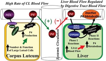
P4 is a steroid hormone primarily secreted by the CL and placenta. Production of P4 from cholesterol utilizes only two enzymes and is considered the simplest steroidogenic pathways to produce a biologically active steroid (Figure 1). All steroids are derived from the common precursor cholesterol. Cholesterol for steroidogenesis can come from multiple sources within a cell including: cellular membranes, cholesterol ester stores, circulating lipoproteins, or de novo cholesterol biosynthesis (Diaz et al., Reference Diaz, Anderson, Wu, Rabot, Tsai and Wiltbank2002). The major source of cholesterol for luteal cells is circulating lipoproteins and in particular high-density lipoprotein (HDL) in ruminants (Grummer and Carroll, Reference Grummer and Carroll1988; Wiltbank et al., Reference Wiltbank, Diskin, Flores and Niswender1990). Receptors for HDL and LDL have been identified in the CL. The HDL receptor increases dramatically after luteinization of granulosa cells in vitro and in vivo (Rajapaksha et al., Reference Rajapaksha, McBride, Robertson and Oshaughnessy1997; Li et al., Reference Li, Peegel and Menon1998). However, changes in luteal abundance of mRNA for LDL or HDL receptor did not explain the effects of various luteotrophic and luteolytic treatments on P4 production from ovine CL (Tandeski et al., Reference Tandeski, Juengel, Nett and Niswender1996).
Physiological model of the factors that regulate circulating P4 concentrations in the lactating dairy cow (center of model). Production of P4 (left side of model) is primarily a function of production in the corpus luteum (CL) through pathways that convert cholesterol from high density lipoprotein (HDL) into P4. The cholesterol transport protein, StAR, and the two steroidogenic enzymes, CYP11A1 and HSD3ß, are critical for this conversion. Although stimulators, such as LH, and inhibitors, such as PGF2α, can regulate P4 production by CL, it is emphasized in the model that the primary factor determining greater P4 production is likely to be CL volume and number of large luteal cells due the constitutive nature of P4 production in the bovine CL. On the right side of the model are pathways involved in P4 metabolism tending to decrease circulating P4. The Phase I pathways involve an aldo-ketoreductase (AKR) step, catalyzed by enzymes from the family of AKR enzymes, such as AKR1C and AKR1D, and a hydroxylation step catalyzed by a cytochrome P450 enzyme such as CYP3A4. The Phase II conjugation pathways are also important for the final production of hydrophilic P4 metabolites. Although stimulation and inhibition of the P4 metabolizing enzymes has been reported, this model emphasizes the critical role of the elevated liver blood flow in producing the extraordinary rates of P4 metabolism in high producing lactating dairy cows.

Cholesterol is primarily a hydrophobic molecule and this makes it difficult for cholesterol to freely diffuse through hydrophilic environments such as the cytoplasm. In addition, cholesterol has a hydroxyl group at the three position that produces a discrete hydrophilic region making it difficult for ‘flip-flop’ of cholesterol between membrane surfaces within the lipid bilayer of cellular membranes. Therefore, movement of cholesterol in the circulatory system (lipoproteins) or within the cell is dependent upon transport proteins. Provision of another hydroxyl-group at the other end of the cholesterol molecule alleviates the need for transport proteins. For example, when large or small luteal cells are treated with hydroxylated cholesterol, there is a dramatic increase in P4 production (25-OH>10-fold; 20-OH or 22-OH>100-fold) and factors that normally stimulate or inhibit P4 production by luteal cells no longer have effects on P4 production (Wiltbank et al., Reference Wiltbank, Belfiore and Niswender1993). Thus, regulation of P4 production in luteal cells is related more to transport of cholesterol within the cell than to changes in the activity of steroidogenic enzymes (Wiltbank et al., Reference Wiltbank, Belfiore and Niswender1993, Belfiore et al., Reference Belfiore, Hawkins, Wiltbank and Niswender1994). A number of proteins have been postulated to be involved with transport of cholesterol (sterol carrier protein-2, peripheral benzodiazepine receptor), however the key role of StAR has been the best characterized (Pescador et al., Reference Pescador, Soumano, Stocco, Price and Murphy1996; Stocco and Clark, Reference Stocco and Clark1996; Stocco et al., Reference Stocco, Wang, Jo and Manna2005). It is now clear that changes in StAR transcription, protein and activity account for increases in P4 production in response to trophic stimulation and acute decreases in P4 production after luteolytic treatments (Diaz et al., Reference Diaz, Anderson, Wu, Rabot, Tsai and Wiltbank2002).
The P450 cholesterol side chain cleavage enzyme (P450scc) is located on the inner mitochondrial membrane and catalyzes the conversion of cholesterol to pregnenolone. Three oxidation steps are catalyzed by this enzyme: hydroxylations at the 20 and 22 positions and then cleavage between these two carbons. The mRNA and protein for P450scc dramatically increase in CL after the LH surge and luteinization (Belfiore et al., Reference Belfiore, Hawkins, Wiltbank and Niswender1994; Niswender et al., Reference Niswender, Juengel, Silva, Rollyson and McIntush2000). Pregnenolone has two hydrophilic residues that increase the mobility through cellular membranes. Pregnenolone diffuses from the mitochondria to the smooth endoplasmic reticulum where it is converted to P4 by the enzyme 3-β hydroxysteroid dehydrogenase (3βHSD). This final reaction produces a double bond between the 4 and 5 carbon of the molecule and is the basis for the abbreviation of progesterone as P4. P4 then diffuses from the luteal cells to the bloodstream for transport to target tissues. The levels of 3βHSD mRNA and protein increase dramatically following the LH surge and subsequent ovulation to a maximum at days 8 to 11 after estrus in cattle (Couet et al., Reference Couet, Martel, Dupont, Luuthe, Sirard, Zhao, Pelletier and Labrie1990). After prostaglandin F2α (PGF2α) treatment, there was an abrupt and dramatic decrease in 3βHSD mRNA abundance (35% of control concentrations at 4 h; 15% of control at 12 h). However, there is no decrease in protein for P450scc or in 3βHSD enzyme activity even at 24 h after PGF2α treatment (Hawkins et al., Reference Hawkins, Belfiore, Kile and Niswender1993; Juengel et al., Reference Juengel, Meberg, McIntush, Smith and Niswender1998; Niswender et al., Reference Niswender, Juengel, Silva, Rollyson and McIntush2000; Atli et al., Reference Atli, Bender, Mehta, Bastos, Luo, Vezina and Wiltbank2012). Given that circulating P4 is decreased by 12 h after PGF2α treatment it seems unlikely that inhibition of P450scc and 3βHSD are a critical part of the acute inhibition of P4 production during luteolysis. Both small and large luteal cells have large amounts of P450scc and 3βHSD activity and neither enzyme appears to be the key step that acutely regulates luteal P4 production from these cells (Wiltbank et al., Reference Wiltbank, Belfiore and Niswender1993).
In ruminants, the role of LH in luteal function has been acknowledged for more than 3 decades (Niswender et al., Reference Niswender, Juengel, Mcguire, Belfiore and Wiltbank1994 and Reference Niswender, Juengel, Silva, Rollyson and McIntush2000) but there is still controversy on the precise role of LH in luteal function (Diaz et al., Reference Diaz, Anderson, Wu, Rabot, Tsai and Wiltbank2002; Wiltbank et al., Reference Wiltbank, Salih, Atli, Luo, Bormann, Ottobre, Vezina, Mehta, Diaz, Tsai and Sartori2012a; Haughian et al., Reference Haughian, Ginther, Diaz and Wiltbank2013). Indeed in our recent study we treated heifers with a potent gonadotropin releasing hormone (GnRH) receptor antagonist, acyline, for the first 6 days after the LH surge. Treatment with GnRH antagonist stopped follicle growth at the time of dominant follicle deviation, about 8 mm in diameter. In contrast, there was no change in development of the CL or in circulating P4 during the first 6 days of luteal development (Haughian et al., Reference Haughian, Ginther, Diaz and Wiltbank2013). It should be stated that all cows had the GnRH-induced LH surge in this study before initiation of acyline treatment. It seems clear that the LH surge is essential for ovulation and luteinization of the ovulated follicular cells into the luteal cells, however, the role for LH pulses, after the LH surge, is still controversial. Consistent with our results, other researchers have also found that treatment with a GnRH antagonist eliminated pulsatile LH secretion but had little or no effect on circulating P4 and CL function (Baird, Reference Baird1992; McNeilly et al., Reference McNeilly, Crow and Fraser1992). In contrast, when sheep are hypophysectomized on day 5 after estrus and CL recovered on day 12, circulating P4, luteal P4, and weight of CL are lower than expected for a day 12 CL but similar to a day 5 CL (Kaltenbach et al., Reference Kaltenbach, Graber, Niswender and Nalbandov1968; Denamur et al., Reference Denamur, Martinet and Short1973; Farin et al., Reference Farin, Nett and Niswender1990). However, acute treatments with LH have surprisingly small effects on circulating P4 (reviewed in Wiltbank (Reference Wiltbank1994)) and in contrast to primates, circulating P4 in ruminants is greatest at times of the cycle when LH pulses are least (Wiltbank et al., Reference Wiltbank, Salih, Atli, Luo, Bormann, Ottobre, Vezina, Mehta, Diaz, Tsai and Sartori2012a). Thus, the acute or even chronic effects of LH pulses on CL function in ruminants remain to be unequivocally defined in the scientific literature. In general, P4 production by the ruminant CL appears to be very high, due to high constitutive P4 production by the large luteal cells (Wiltbank, Reference Wiltbank1994; Diaz et al., Reference Diaz, Anderson, Wu, Rabot, Tsai and Wiltbank2002; Bogan and Niswender, Reference Bogan and Niswender2007; Wiltbank et al., Reference Wiltbank, Salih, Atli, Luo, Bormann, Ottobre, Vezina, Mehta, Diaz, Tsai and Sartori2012a).
The other key factor regulating circulating P4 is the rate of metabolism of P4, primarily by the liver (Figure 1). Primary regulation of P4 metabolism is due to changes in blood flow to the liver (Wiltbank et al., Reference Wiltbank, Lopez, Sartori, Sangsritavong and Gumen2006). For example, if liver blood flow increased from 1000 to 2000 l/h then circulating P4 concentrations will decrease to 50% even though P4 production and P4 metabolizing enzymes have not changed (Sangsritavong et al., Reference Sangsritavong, Combs, Sartori, Armentano and Wiltbank2002; Wiltbank et al., Reference Wiltbank, Lopez, Sartori, Sangsritavong and Gumen2006 and Reference Wiltbank, Souza, Giordano, Nascimento, Vasconcelos, Pereira, Fricke, Surjus, Zinsly, Carvalho, Bender and Sartori2012b). The role of feed intake in regulating liver blood flow, P4 metabolism, and circulating P4 was initially demonstrated in studies with pigs and sheep (Christenson et al., Reference Christenson, Ford and Redmer1985; Parr et al., Reference Parr, Davis, Miles and Squires1993; Prime and Symonds, Reference Prime and Symonds1993). For example, the studies in sheep showed convincingly that as feed intake increased, there was a clear increase in liver blood flow with a corresponding decrease in circulating P4 (Parr et al., Reference Parr, Davis, Miles and Squires1993). In addition, these studies demonstrated that the reduced P4 concentrations caused by overfeeding produced the reduction in fertility in overfed ewes (Parr et al., Reference Parr, Davis, Fairclough and Miles1987). Our results extended the results from previous studies to the lactating dairy cow and clearly demonstrated the relationship between dry matter intake, liver blood flow, and circulating concentrations of P4 (Sangsritavong, Reference Sangsritavong2002; Sangsritavong et al., Reference Sangsritavong, Combs, Sartori, Armentano and Wiltbank2002; Vasconcelos et al., Reference Vasconcelos, Sangsritavong, Tsai and Wiltbank2003; Wiltbank et al., Reference Wiltbank, Lopez, Sartori, Sangsritavong and Gumen2006).
In conclusion, circulating P4 concentrations represent a balance between production of P4 and metabolism of P4 (Figure 1). Production of P4 is regulated by development of the CL after the LH surge, number of granulosa cells that luteinize into large luteal cells, and constitutive production of P4 by the large luteal cells. Metabolism of P4 is primarily related to amounts of blood flow to the liver due to the abundance in the bovine liver of the enzymes that mediate metabolism of P4. Thus, practical regulation of circulating P4 will probably be most productive by focusing on increasing luteal tissue volume to increase P4 production and/or limiting liver blood flow and perhaps blocking P4 metabolizing enzymes.
Cellular mechanisms regulating P4 action
The receptors for P4 and the intracellular mechanisms of action have been extensively reviewed (Ellmann et al., Reference Ellmann, Sticht, Thiel, Beckmann, Strick and Strissel2009). The classical P4 receptors (PR) are members of the nuclear receptor superfamily. In other words, these receptors work through actions in the nucleus that regulate expression of specific genes. There are two types (isoforms) of the nuclear PR, termed PR-A and PR-B. These PR are products of the same gene with the key distinction that PR-B contains an additional 165 amino acids at the N-terminal end of the protein. Studies in mice in which PR-A or PR-B are selectively eliminated (knockout mice) show that these two receptors are functionally distinct and both are critical for successful reproduction (Arck et al., Reference Arck, Hansen, Jericevic, Piccinni and Szekeres-Bartho2007). The PR-A isoform alone is sufficient for establishment and maintenance of pregnancy; whereas, PR-B is not sufficient for establishment or maintenance of pregnancy but is essential for fertility, possibly through actions on tissues other than the uterus (Fernandez-Valdivia et al., Reference Fernandez-Valdivia, Mukherjee, Mulac-Jericevic, Conneely, DeMayo, Amato and Lydon2005). Surprisingly, there are also multiple types of plasma membrane P4 receptors that act through activation of intracellular signal transduction systems that are not related to the nuclear PR systems (Peluso, Reference Peluso2006).
The P4 concentration that reaches the receptors within each particular cell is the key determinant of the physiological actions of P4 in an animal. Almost all tissues have sufficient blood supply that the circulating P4 concentration is the primary determinant of cellular P4 concentrations. Therefore, factors regulating circulating P4 concentrations and intracellular P4 receptors primarily determine the magnitude of P4 responses throughout the body.
Studies on the effects of low v. high P4 during follicular growth before AI
Incidence of double ovulation
The incidence of increased twinning in lactating dairy cows is primarily due to a high percentage of cows with double ovulation as evidenced by over 93% of twins being non-identical (del Rio et al., Reference del Rio, Kirkpatrick and Fricke2006). There are numerous factors that regulate twinning rates, including age of dam, season, genetics, use of reproductive hormones or antibiotics, ovarian cysts and days open; however, there is increasing interest in the key role of peak milk production and circulating P4 concentrations on the incidence of double ovulation (Kinsel et al., Reference Kinsel, Marsh, Ruegg and Etherington1998; Wiltbank et al., Reference Wiltbank, Fricke, Sangsritavong, Sartori and Ginther2000 and Reference Wiltbank, Souza, Giordano, Nascimento, Vasconcelos, Pereira, Fricke, Surjus, Zinsly, Carvalho, Bender and Sartori2012b). For example, in a study with cows that were evaluated near natural estrus (Lopez et al., Reference Lopez, Caraviello, Satter, Fricke and Wiltbank2005b), almost 50% of cows that were above 50 kg/day of milk production had double ovulation, whereas <10% had double ovulation for cows producing below 40 kg/day. These effects were similar for multiparous or primiparous cows (Fricke and Wiltbank, Reference Fricke and Wiltbank1999; Lopez et al., Reference Lopez, Caraviello, Satter, Fricke and Wiltbank2005b).
Many results are consistent with reduced P4 concentrations being the key factor that links high milk production with increased double ovulation and twinning in lactating dairy cattle. For example, cattle that grow the ovulatory follicular wave in the absence of P4 have an increased percentage of cows with co-dominant follicles and double ovulation (Hayashi et al., Reference Hayashi, Matsui, Shimizu, Sudo, Sato, Shirasuna, Tetsuka, Kida, Schams and Miyamoto2008). Manipulative studies have now been performed that have demonstrated a decrease in the percentage of cows that double ovulate when circulating P4 is increased (Stevenson et al., Reference Stevenson, Portaluppi and Tenhouse2007a; Cunha et al., Reference Cunha, Guenther, Maroney, Giordano, Nascimento, Bas, Ayres and Wiltbank2008; Cerri et al., Reference Cerri, Chebel, Rivera, Narciso, Oliveira, Thatcher and Santos2011a). For example, a study from our laboratory found that cows with high P4 during follicular growth, had a much lower percentage of cows with double ovulation than cows with low P4 (7.0% v. 20.6%; P<0.01; Cunha et al., Reference Cunha, Guenther, Maroney, Giordano, Nascimento, Bas, Ayres and Wiltbank2008). Similarly, in cows synchronized with the Ovsynch protocol (Fricke and Wiltbank, Reference Fricke and Wiltbank1999), double ovulation was much greater in cows that were above average milk production (40.7 kg/day) than below (20.2% v. 6.9%; P<0.05). Alternatively, analysis of cows with co-dominant follicles during the first follicular wave demonstrated that these cows had greater milk production, lower circulating P4, and a distinct elevation in circulating FSH and LH concentrations during the 24 h before dominant follicle deviation (Lopez et al., Reference Lopez, Sartori and Wiltbank2005a). Thus, greater milk production may produce decreased circulating P4 concentrations due to the increased feed intake and corresponding increases in P4 metabolism. The depressed P4 concentrations may underlie a delay in the FSH nadir and increased LH pulses near dominant follicle deviation and thus selection of two or even three dominant follicles.
Fertility in dairy cows
The key role of greater P4 before AI on fertility of lactating dairy cows was recognized in early studies that compared fertile to non-fertile inseminations (Folman et al., Reference Folman, Rosenber, Herz and Davidson1973; Erb et al., Reference Erb, Garverick, Randel, Brown and Callahan1976; Meisterling and Dailey, Reference Meisterling and Dailey1987). In addition, concentrations of P4 12 days before first AI had a positive relationship with conception rate at first AI in lactating Holstein and Jersey dairy cattle (Fonseca et al., Reference Fonseca, Britt, Mcdaniel, Wilk and Rakes1983). Similarly, concentrations of P4 on the day of PGF2α-induced luteolysis had a positive linear correlation with subsequent fertility (Diskin et al., Reference Diskin, Murphy and Sreenan2006). More recent manipulative studies have shown some improvements (~5% to 7% increase in percentage pregnant) by using a controlled internal drug release (CIDR; releases P4) during the Ovsynch program before AI (Stevenson et al., Reference Stevenson, Pursley, Garverick, Fricke, Kesler, Ottobre and Wiltbank2006 and Reference Stevenson, Tenhouse, Krisher, Lamb, Larson, Dahlen, Pursley, Bello, Fricke, Wiltbank, Brusveen, Burkhart, Youngquist and Garverick2008; Chebel et al., Reference Chebel, Al-Hassan, Fricke, Santos, Lima, Martel, Stevenson, Garcia and Ax2010; Bilby et al., Reference Bilby, Bruno, Lager, Chebel, Moraes, Fricke, Lopes, Giordano, Santos, Lima, Stevenson and Pulley2013). Nevertheless, the results from these studies may not be due exclusively to the elevation in P4 produced by the CIDR, because improvements in synchronization may also accompany the use of a CIDR in the Ovsynch protocol. In the study mentioned above we tested the effects of elevated P4 on fertility to a timed AI during the Double Ovsynch program (Cunha et al., Reference Cunha, Guenther, Maroney, Giordano, Nascimento, Bas, Ayres and Wiltbank2008). Cows (n=564) were randomly assigned to have either high or low P4 during the Ovsynch protocol. The cows with low P4 had increased double ovulation rate, which would be expected to potentially increase fertility in these cows (ovulation of more follicles should result in greater chance for pregnancy). However, cows with lower P4 before AI had much lower fertility (37.1% pregnant at day 29 pregnancy diagnosis) compared with cows with high P4 (51.0%; P<0.001). This indicates that increasing P4 before timed AI can result in a substantial improvement in fertility, suggesting that the reason for the lower fertility in lactating dairy may be, at least partly, due to reduced P4 concentrations during the time period before AI. Obviously, the reduction in P4 in our study was done by hormonal manipulations in a commercial farm setting; however, physiologically, P4 concentrations may also be reduced in high-producing dairy cows due to high feed intake that produces increased gastro-intestinal/liver blood flow and increased P4 metabolism. A number of studies have targeted an elevation of circulating P4 in order to increase fertility.
Studies by Bisinotto et al. (Reference Bisinotto, Chebel and Santos2010a) demonstrated the importance of higher P4 during growth of the final follicular wave. Two studies were performed in which the effect of P4 during the Ovsynch protocol was evaluated. In the first study, cows were evaluated for P4 concentration at the time of the first GnRH of Ovsynch and 7 days before this first GnRH. Cows were classified as anovular (both samples low in P4), or as beginning Ovsynch with high P4 or low P4 (first sample had high P4, second had low P4). Cows that began Ovsynch with high P4 had greater pregnancies per AI (P/AI=43.0%) than cycling cows that had low P4 (31.3%) or that were anovular (29.7%) at the time of initiation of Ovsynch (Bisinotto et al., Reference Bisinotto, Chebel and Santos2010a). In a second manipulative study, cows received two PGF2α treatments and Ovsynch was initiated either 3 or 10 days after the second PGF2α. This design would produce cows that ovulated a dominant follicle from either the first (3 days) or second (10 days) follicular wave near the timed AI. Similar to the first study, cows ovulating a follicle of the second follicular wave (high P4 concentrations) had greater P/AI that cows ovulating the first follicular wave (low P4) (41.7% v. 30.4%) (Bisinotto et al., Reference Bisinotto, Chebel and Santos2010a). Other studies have also reported that low P4 at the beginning of the Ovsynch protocol results in decreased P/AI compared with cows with high P4 concentrations (Silva et al., Reference Silva, Sterry, Kolb, Wiltbank and Fricke2007; Denicol et al., Reference Denicol, Lopes, Mendonça, Rivera, Guagnini, Perez, Lima, Bruno, Santos and Chebel2012; Giordano et al., Reference Giordano, Wiltbank, Guenther, Ares, Lopes, Herlihy and Fricke2012a, Reference Giordano, Wiltbank, Guenther, Pawlisch, Bas, Cunha and Fricke2012b and Reference Giordano, Wiltbank, Fricke, Bas, Pawlisch, Guenther and Nascimento2013a). In addition, a recent study (Bisinotto et al., Reference Bisinotto, Ribeiro, Lima, Martinez, Greco, Barbosa, Bueno, Scagion, Thatcher and Santos2013) compared cows with a CL at the initiation of a synchronized ovulation program to cows either without a CL, that were untreated, or cows without a CL that were treated with two intravaginal P4 implants (two CIDRs). The cows treated with two CIDRs had similar fertility to cows with a CL and both of these groups were greater than untreated cows without a CL (Bisinotto et al., Reference Bisinotto, Ribeiro, Lima, Martinez, Greco, Barbosa, Bueno, Scagion, Thatcher and Santos2013). Thus, it seems clear that increasing P4 during growth of the preovulatory follicular wave increases fertility more than 10% to the subsequent timed AI.
Pregnancy loss in dairy cows
One further important observation related to low P4 before AI is that there is generally an increase in pregnancy loss following the subsequent AI. Pregnancy loss averages 6% to 20% in high producing dairy cows when pregnancy loss is evaluated between day 28 after AI (by ultrasound) and day 60 after AI. This time period is critical for implantation and development of the embryo. We observed a decrease (P=0.05) in pregnancy loss between day 29 and day 57 when cows had high P4 (6.8% loss) v. low P4 (14.3% loss) during growth of the follicular wave that produces the ovulatory follicle before AI (Cunha et al., Reference Cunha, Guenther, Maroney, Giordano, Nascimento, Bas, Ayres and Wiltbank2008). Thus, the high P4 group not only had increased number of pregnancies at the day 29 pregnancy diagnosis, these cows also had less risk of subsequent pregnancy loss after this time. This was not due to differences in circulating P4 after AI because P4 concentrations after AI were actually somewhat higher in the cows that had low P4 before AI (2.88 ng/ml) compared with the cows with high P4 before AI (2.49 ng/ml). Other studies have also reported increased pregnancy loss in anovular cows compared with cyclic cows (Santos et al., Reference Santos, Thatcher, Chebel, Cerri and Galvão2004 and Reference Santos, Rutigliano and Filho2009; Sterry et al., Reference Sterry, Welle and Fricke2006; Stevenson et al., Reference Stevenson, Pursley, Garverick, Fricke, Kesler, Ottobre and Wiltbank2006). Thus, there was a positive effect of elevated P4 before AI on subsequent maintenance of pregnancy even after 29 days following AI.
Effect of pre-AI P4 on embryo quality
An elegant study in superstimulated cows (Rivera et al., Reference Rivera, Mendonca, Lopes, Santos, Perez, Amstalden, Correa-Calderon and Chebel2011) showed that high P4 concentrations during superstimulation increased the subsequent quality of embryos flushed on day 7 after superovulation. Cows began superstimulation during the second follicular wave (high P4), during the first follicular wave (low P4), or during the first follicular wave with P4 supplementation using two CIDRs to increase P4 concentrations. Although, total structures that were collected (embryos/oocytes) was not different between groups, the percentage of structures that were transferable embryos were much less (P=0.01) for cows superstimulated during the first follicular wave (55.9%), than during the second follicular wave (88.5%) or during the first follicular wave with P4 supplementation (78.6%). This result is consistent with an effect of elevated P4 during follicle growth on embryo development before day 7. It is postulated that effects of P4 during growth of the follicles may allow production of a better oocyte for subsequent fertilization and embryo development (Rivera et al., Reference Rivera, Mendonca, Lopes, Santos, Perez, Amstalden, Correa-Calderon and Chebel2011). However, a recent study that flushed embryos from single ovulating cows that had follicles grow during low or high P4 did not find a difference in embryo quality on day 7 (58.3% v. 53.3%) (Cerri et al., Reference Cerri, Chebel, Rivera, Narciso, Oliveira, Thatcher and Santos2011a). A companion study (Cerri et al., Reference Cerri, Chebel, Rivera, Narciso, Oliveira, Amstalden, Baez-Sandoval, Oliveira, Thatcher and Santos2011b) indicated that although cows with low P4 had increased basal LH concentrations and altered follicular dynamics and follicular fluid composition that could alter oocyte quality, a particularly distinct difference in cows with low P4 was the premature development of pathways leading to uterine PGF2α secretion. Thus, altered uterine function could also have an important role in reducing fertility in cows that have low P4 concentrations before AI. Our laboratory (Wiltbank et al., unpublished) has recently completed a study evaluating day 7 embryo quality in single ovulating cows with follicle development occurring in low v. high P4. We found a greater percentage of cows with Grade 1 and two embryos in cows with high P4 than low P4 before AI (86.5% v. 61.5%; P=0.02). Thus, our recent results are consistent with the effects of low P4 on fertility being evident by day 7 after AI even in single-ovulating lactating cows, consistent with the results of Rivera et al. (Reference Rivera, Mendonca, Lopes, Santos, Perez, Amstalden, Correa-Calderon and Chebel2011) in superovulated cows.
Importance of low P4 near time of AI
Inadequate luteolysis can result in an elevation in circulating P4 near AI and a reduction in fertility. This has been clearly shown in numerous studies using timed AI programs (Souza et al., Reference Souza, Gumen, Silva, Cunha, Guenther, Peto, Caraviello and Wiltbank2007; Brusveen et al., Reference Brusveen, Cunha, Silva, Cunha, Sterry, Silva, Guenther and Wiltbank2008; Bisinotto et al., Reference Bisinotto, Ribeiro, Martins, Marsola, Greco, Favoreto, Risco, Thatcher and Santos2010b; Giordano et al., Reference Giordano, Wiltbank, Guenther, Pawlisch, Bas, Cunha and Fricke2012b and Reference Giordano, Wiltbank, Fricke, Bas, Pawlisch, Guenther and Nascimento2013b). In addition, studies on cows that receive AI after detection of estrus, have generally reported that minor elevations in P4 near AI are also detrimental to fertility (De Silva et al., Reference De Silva, Anderson, Gwazdauskas, McGilliard and Lineweaver1981; Waldmann et al., Reference Waldmann, Reksen, Landsverk, Kommisrud, Dahl, Refsdal and Ropstad2001; Ghanem et al., Reference Ghanem, Nakao, Nakatani, Akita and Suzuki2006), although some studies did not obtain this result (Erb et al., Reference Erb, Garverick, Randel, Brown and Callahan1976; Plym Forshell et al., Reference Plym Forshell, Andersson and Pehrson1991). During Ovsynch, the percentage of cows that do not have complete regression following the PGF2α treatment before timed AI has been reported to range from 5% to 30% (Moreira et al., Reference Moreira, de la Sota, Diaz and Thatcher2000; Gumen et al., Reference Gumen, Guenther and Wiltbank2003; Souza et al., Reference Souza, Gumen, Silva, Cunha, Guenther, Peto, Caraviello and Wiltbank2007; Brusveen et al., Reference Brusveen, Souza and Wiltbank2009; Martins et al., Reference Martins, Policelli, Neuder, Raphael and Pursley2011b; Giordano et al., Reference Giordano, Wiltbank, Fricke, Bas, Pawlisch, Guenther and Nascimento2013b). A recent extensive study of incomplete luteolysis evaluated multiple blood samples in cows at first AI (n=652) and second or greater AI (n=394) (Martins et al., Reference Martins, Policelli, Neuder, Raphael and Pursley2011b). They defined complete luteolysis and low P4 (<0.5 ng/ml) at 56, 72 and 96 h after PGF2α. At first AI, 80% of cows underwent complete luteolysis, whereas at second+ AI only 71% underwent complete luteolysis. Surprisingly, greater P4 concentrations at the time of PGF2α were associated with greater probability of luteolysis after PGF2α treatment and greater fertility (50% v. 28%).
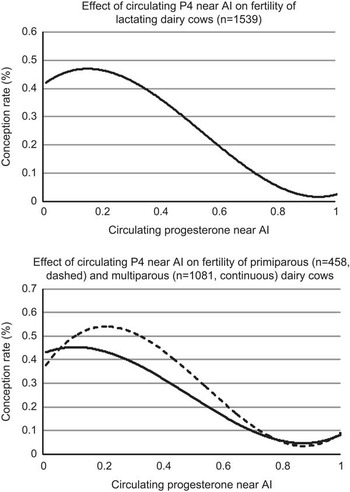
To study whether P4 near AI had an effect on fertility during timed AI protocols, our laboratory (Souza et al., Reference Souza, Gumen, Silva, Cunha, Guenther, Peto, Caraviello and Wiltbank2007; Brusveen et al., Reference Brusveen, Souza and Wiltbank2009) evaluated fertility in lactating cows synchronized with Ovsynch on three commercial dairy herds. We collected blood samples for P4 measurement near the time of the last GnRH of Ovsynch. The combined results from these two studies are shown in Figure 2. There was a dramatic decrease in P/AI as P4 increased above 0.4 ng/ml near the time of the second GnRH of Ovsynch (16 h before AI). Thus, slight elevations in circulating P4 near AI result in dramatic decreases in fertility to the AI. This decrease was observed even when only cows that ovulated to the second GnRH were evaluated for fertility. The decrease that we observed was similar to the decrease described in the pioneering study of De Silva et al. (Reference De Silva, Anderson, Gwazdauskas, McGilliard and Lineweaver1981) using visual detection of estrus in lactating cows and heifers.
Relationship between circulating P4 near AI and conception results in lactating dairy cows receiving Ovsynch (data from Souza et al. (Reference Souza, Gumen, Silva, Cunha, Guenther, Peto, Caraviello and Wiltbank2007) and Brusveen et al. (Reference Brusveen, Souza and Wiltbank2009) combined).

In the two studies analyzed for Figure 2 there was no effect of parity on incidence of luteal regression. However, in a different study from our laboratory (Giordano et al., Reference Giordano, Wiltbank, Guenther, Pawlisch, Bas, Cunha and Fricke2012b) lower rates of CL regression were observed for multiparous (83.9%) than primiparous (89.7%) cows. Other studies have also reported lower rates of luteal regression in multiparous than in primiparous lactating dairy cows (Martins et al., Reference Martins, Policelli, Neuder, Raphael and Pursley2011a).
Two methods have been used to increase regression of the CL during timed AI protocols, increasing the dose of PGF2α or increasing the number of PGF2α treatments. This is particularly important in cows treated with the 5-day Ovsynch protocol (Santos et al., Reference Santos, Narciso, Rivera, Thatcher and Chebel2010; Ribeiro et al., Reference Ribeiro, Bisinotto, Favoreto, Martins, Cerri, Silvestre, Greco, Thatcher and Santos2012; Nascimento et al., Reference Nascimento, Souza, Keskin, Sartori and Wiltbank2013a). Fertility was reduced if only a single PGF2α was given (Santos et al., Reference Santos, Narciso, Rivera, Thatcher and Chebel2010) or if two PGF2α treatments were given on the same d (5 days after GnRH) compared with giving one treatment on day 5 and a second on day 6 (Ribeiro et al., Reference Ribeiro, Bisinotto, Favoreto, Martins, Cerri, Silvestre, Greco, Thatcher and Santos2012). Using the 7-day Ovsynch protocol, an increased percentage of cows with complete CL regression (<0.4 ng/ml 56 h after PGF2α) was observed after two (days 7 and 8; 326/341=95.6%) compared with one (day 7; 301/356=84.6%) PGF2α treatment (Brusveen et al., Reference Brusveen, Souza and Wiltbank2009). In a recent study from our laboratory (Giordano et al., Reference Giordano, Wiltbank, Fricke, Bas, Pawlisch, Guenther and Nascimento2013b), increasing the dose of cloprostenol from 500 to 750 µg on day 7 of a 7-day Ovsynch protocol increased CL regression in multiparous (122/154=79.2% v. 135/154=87.7%; P=0.025) but not primiparous (131/146=89.7% v. 129/139=92.8%; P=0.181) cows. An indication of improved fertility (P=0.054) was observed at the 39 days pregnancy diagnosis with the 750 (247/544=45.4%) compared with the 500 (221/540=40.9%) µg dose of cloprostenol (Giordano et al., Reference Giordano, Wiltbank, Fricke, Bas, Pawlisch, Guenther and Nascimento2013b). Consistent with this idea, treatment with PGF2α one day earlier provided greater time for reduced P4 at the time of AI and improvements in fertility (Pereira et al., Reference Pereira, Sanches, Guida, Rodrigues, Aragon, Veras, Borges, Wiltbank and Vasconcelos2013). In this study, cows with P4 concentrations above 0.1 ng/ml near the time of AI had reduced fertility (Pereira et al., Reference Pereira, Sanches, Guida, Rodrigues, Aragon, Veras, Borges, Wiltbank and Vasconcelos2013). Thus, a small elevation in P4 near AI can reduce fertility. Treatment with an increased dose of PGF2α or increased number of PGF2α treatments can alleviate or reduce this problem. Based on the above results, it appears that two doses of PGF2α, 24 h apart, may be the most consistent method for causing CL regression and lowering P4 concentrations near AI. Nevertheless, in certain on-farm situations two treatments may be difficult, and treatment with a higher dose or leaving a longer time from PGF2α treatment until removal of the P4 vaginal implant is likely to enhance but possibly not optimize fertility.
There may be multiple physiological mechanisms that result in the reduced fertility observed when P4 is elevated near AI. First, P4 may alter sperm or oocyte transport by altering uterine or oviductal contractility and thus reduce fertilization (Hunter, Reference Hunter2005). Second, addition of P4 to in vitro fertilization (IVF) media reduced blastocyst rate (Silva and Knight, Reference Silva and Knight2000) suggesting that there may be direct effects of P4 during IVF on subsequent embryo development. This detrimental effect was reversed with a P4 receptor antagonist (mifepristone – RU486) indicating a specific role for P4 receptors in this action. Elevated P4 in vitro, also increased total α-inhibin production by the cumulus–oocyte complex, which may reduce embryo development after cleavage (Silva et al., Reference Silva, Groome and Knight1999). Further, the reduced endometrial thickness that accompanies slight elevations in P4 (Souza et al., Reference Souza, Silva, Cunha, Gumen, Ayres, Brusveen, Guenther and Wiltbank2011) may indicate other major effects of P4 on the blood flow and functionality of the uterus and these changes could underlie the reduced embryo development.
In summary, circulating P4 near AI, >0.1 ng/ml in a study using estradiol to induce ovulation (Pereira et al., Reference Pereira, Sanches, Guida, Rodrigues, Aragon, Veras, Borges, Wiltbank and Vasconcelos2013) or>0.3 to 0.5 ng/ml in studies using GnRH to induce ovulation (Souza et al., Reference Souza, Gumen, Silva, Cunha, Guenther, Peto, Caraviello and Wiltbank2007; Brusveen et al., Reference Brusveen, Souza and Wiltbank2009; Santos et al., Reference Santos, Narciso, Rivera, Thatcher and Chebel2010; Martins et al., Reference Martins, Policelli, Neuder, Raphael and Pursley2011b; Ribeiro et al., Reference Ribeiro, Bisinotto, Favoreto, Martins, Cerri, Silvestre, Greco, Thatcher and Santos2012; Giordano et al., Reference Giordano, Wiltbank, Guenther, Pawlisch, Bas, Cunha and Fricke2012b), is detrimental to fertility in dairy cattle, but the underlying physiological mechanisms that reduce fertility are not well understood. An additional PGF2α treatment in cows receiving Ovsynch and more precise detection of estrus may help to minimize the percentage of cows with elevated circulating P4 near AI.
Importance of high P4 after AI
The role of the ‘CL hormone’ after ovulation in maintaining pregnancy was described more than 100 years ago and was recognized 30 years before the chemical structure of P4 was identified (Fraenkel and Cohn, Reference Fraenkel and Cohn1901; Magnus, Reference Magnus1901). Recent studies continue to characterize the uterine effects of P4, inducing a uterine environment compatible with embryo growth, implantation, and maintenance of pregnancy (Arck et al., Reference Arck, Hansen, Jericevic, Piccinni and Szekeres-Bartho2007; Forde et al., Reference Forde, Carter, Fair, Crowe, Evans, Spencer, Bazer, McBride, Boland, O’Gaora, Lonergan and Roche2009; Bazer et al., Reference Bazer, Kim, Song, Ka, Tekwe and Wu2012; Mullen et al., Reference Mullen, Elia, Hilliard, Parr, Diskin, Evans and Crowe2012a and Reference Mullen, Forde, Parr, Diskin, Morris, Nally, Evans and Crowe2012b).
Although there is unequivocal evidence that there is an absolute requirement for P4 in pregnancy maintenance, results have been somewhat more equivocal about the relationship between levels of circulating P4 after AI and fertility in lactating dairy cows. A number of studies have reported lower P4 in non-pregnant than pregnant cows, whereas, other studies reported no relationship between post-AI P4 and fertility (Bulman and Lamming, Reference Bulman and Lamming1978; Larson et al., Reference Larson, Butler and Currie1997; Mann and Lamming, Reference Mann and Lamming1999; Gumen et al., Reference Gumen, Guenther and Wiltbank2003; Stronge et al., Reference Stronge, Sreenan, Diskin, Mee, Kenny and Morris2005; Lonergan et al., Reference Lonergan, Woods, Fair, Carter, Rizos, Ward, Quinn and Evans2007; Morris and Diskin, Reference Morris and Diskin2008). More extensive logistic regression modeling demonstrated a relationship in dairy cows between P/AI with circulating P4 on days 5, 6 and 7 after AI or with rate of P4 increase after AI (Stronge et al., Reference Stronge, Sreenan, Diskin, Mee, Kenny and Morris2005). They reported that 60% to 85% of dairy cows had sub-optimal circulating P4 for pregnancy, based on absolute P4 concentrations during the early luteal phase or rate of P4 increase (Stronge et al., Reference Stronge, Sreenan, Diskin, Mee, Kenny and Morris2005). For example, a milk P4 concentration of 7.4 ng/ml on day 5 was optimal for embryo survival, however 60% of cows had lower P4 concentrations than this optimum. A particularly intriguing recent study reported that one of the major hormonal differences found in cows with genetics for high fertility was 34% greater circulating P4 concentrations than cows with poor genetic merit for fertility (Cummins et al., Reference Cummins, Lonergan, Evans and Butler2012). Many recent studies have attempted to unravel the mechanisms involved in the complex relationship between circulating P4 concentrations and levels of fertility in lactating dairy cows.
Early embryos express different types and concentrations of P4 receptors (Clemente et al., Reference Clemente, de la Fuente, Fair, Al Naib, Gutierrez-Adan, Roche, Rizos and Lonergan2009) raising the possibility that P4 may be acting directly on the embryo to improve embryo development. An elegant series of experiments found no effect of P4 supplementation after fertilization, in vitro, on blastocyst yield, in the presence or absence of bovine oviductal epithelial cells (Clemente et al., Reference Clemente, de la Fuente, Fair, Al Naib, Gutierrez-Adan, Roche, Rizos and Lonergan2009). In a surprising twist, these researchers treated recipient cows with an intravaginal P4-releasing device (PRID) starting on day 3 after estrus with in vitro produced blastocysts transferred on day 7. Circulating P4 concentrations were elevated in the recipient cows from days 3 to 6 but were not elevated in treated cows after that time. Thus, the rise in P4 concentrations in treated cows occurred before the transfer of embryos. Nevertheless, embryos that were transferred into recipients that had received prior P4 exposure (days 3 to 6 increased P4) had longer embryos on day 14 and embryo area was much greater (Clemente et al., Reference Clemente, de la Fuente, Fair, Al Naib, Gutierrez-Adan, Roche, Rizos and Lonergan2009). The authors concluded that ‘P4-induced changes in the uterine environment are responsible for the advancement in conceptus elongation reported previously in cattle and that, interestingly, the embryo does not need to be present during the period of high P4 in order to exhibit advanced elongation’ (Clemente et al., Reference Clemente, de la Fuente, Fair, Al Naib, Gutierrez-Adan, Roche, Rizos and Lonergan2009). These results are consistent with the studies of (Larson et al., Reference Larson, Krisher and Lamb2011) that also failed to find a direct effect of P4 during either days 1 to 3 or days 4 to 7 of culture on percentage of embryos that developed to the morula or blastocyst stage; although small differences in glucose metabolism were observed. Further evidence for a lack of P4 effect in early embryo is found in the studies of Carter et al., Reference Carter, Forde, Duffy, Wade, Fair, Crowe, Evans, Kenny, Roche and Lonergan2008 and Reference Carter, Rings, Mamo, Holker, Kuzmany, Besenfelder, Havlicek, Mehta, Tesfaye, Schellander and Lonergan2010. In the first experiment, 210 crossbred beef heifers were used to analyze the effects of in vivo supplementation with P4 on embryo development. These researchers observed no difference in early embryo development by day 5 or 7 after AI, however dramatic effects of P4 supplementation on embryonic length could be observed on day 13 and 16 after AI (Carter et al., Reference Carter, Forde, Duffy, Wade, Fair, Crowe, Evans, Kenny, Roche and Lonergan2008). In an elegant study that continued this research focus (Carter et al., Reference Carter, Rings, Mamo, Holker, Kuzmany, Besenfelder, Havlicek, Mehta, Tesfaye, Schellander and Lonergan2010), in vitro produced embryos were transferred to the oviduct of beef heifers that received or did not receive a PRID on day 3 after estrus. There was no detectable effect of P4 on the proportion of embryos that developed to the blastocyst stage by day 7 when embryos were recovered or during subsequent culture of the embryos in vitro. However, there were subtle but intriguing differences in gene expression, detected by microarray, in the embryos recovered from recipients that received P4 supplementation (Carter et al., Reference Carter, Rings, Mamo, Holker, Kuzmany, Besenfelder, Havlicek, Mehta, Tesfaye, Schellander and Lonergan2010). Thus, it seems clear that increased P4 during days 3 to 7 induces changes in the uterus that increase embryo elongation by day 14. Whether a P4-induced increase in embryo development can improve fertility in lactating dairy cows continues to be an area of investigation, as discussed below.
Many studies have investigated the P4-induced changes in gene expression that occur in the endometrial tissue and these will not be extensively reviewed in this manuscript. However, it seems clear that there are dramatic differences in endometrial gene expression as the luteal phase progresses and that early supplementation with P4 can induce earlier expression of this P4 program (McNeill et al., Reference McNeill, Sreenan, Diskin, Cairns, Fitzpatrick, Smith and Morris2006; Forde et al., Reference Forde, Carter, Fair, Crowe, Evans, Spencer, Bazer, McBride, Boland, O’Gaora, Lonergan and Roche2009 and Reference Forde, Beltman, Duffy, Duffy, Mehta, O’Gaora, Roche, Lonergan and Crowe2011). The P4-induced changes in uterine gene expression can have dramatic consequences for the development of the embryos (Forde et al., Reference Forde, Beltman, Duffy, Duffy, Mehta, O’Gaora, Roche, Lonergan and Crowe2011).
There have been numerous studies that have evaluated the effects of P4 supplementation on fertility in cattle with the earliest experiments conducted in the 1950s (Herrick, Reference Herrick1953; Wiltbank et al., Reference Wiltbank, Hawk, Kidder, Black, Ulberg and Casida1956). Throughout the last 60 years (reviewed by Mann and Lamming, Reference Mann and Lamming1999), there have been numerous methods to increase P4 including: treatment with exogenous P4 (injectable P4; PRID; or CIDR) or by treatments attempting to ovulate a follicle and produce an accessory CL (human chorionic gonadotropin (hCG); or GnRH). These experiments have varied considerably in regard to type of animal (beef v. dairy, heifers v. cows), supplementation/administration day relative to AI, utilization of synchronization before AI, and number of animals in the trial (n). Of the 30 trials that we evaluated, the vast majority (25/30) showed a numeric improvement in fertility with P4 supplementation, although only six of these trials showed significance (P<0.05). Only two (Stevenson et al., Reference Stevenson, Portaluppi, Tenhouse, Lloyd, Eborn, Kacuba and DeJarnette2007b; Nascimento et al., Reference Nascimento, Bender, Souza, Ayres, Araujo, Guenther, Sartori and Wiltbank2013c) of these trials that found significance used >100 animals per comparison.
The most extensive trials to increase P4 have been done by inducing formation of an accessory CL with hCG or GnRH treatment. When hCG or GnRH is administered on day 5 after AI, there is generally formation of an accessory CL and increased P4 during the mid-luteal phase (Fricke et al., Reference Fricke, Reynolds and Redmer1993; Santos et al., Reference Santos, Thatcher, Pool and Overton2001). In our recent study we found that 93% of dairy cows treated with hCG on day 5 had ovulation (Nascimento et al., Reference Nascimento, Souza, Guenther, Costa, Sartori and Wiltbank2013b). Cows treated with hCG showed an increase in circulating P4 by 3 days after hCG treatment, day 8, until day 16 (Nascimento et al., Reference Nascimento, Souza, Guenther, Costa, Sartori and Wiltbank2013b). However, circulating P4 remained lower, even after hCG treatment, in lactating cows compared with normally ovulating heifers. Eight studies that evaluated the effect of hCG on P4 concentrations from days 4 to 7 after AI in lactating dairy cows reported significant increases in circulating P4 concentrations after treatments with different doses of hCG: 3300 IU (Schmitt et al., Reference Schmitt, Barros, Fields, Fields, Diaz, Kluge and Thatcher1996; Santos et al., Reference Santos, Thatcher, Pool and Overton2001), 2500 IU (Stevenson et al., Reference Stevenson, Portaluppi, Tenhouse, Lloyd, Eborn, Kacuba and DeJarnette2007b; Vasconcelos et al., Reference Vasconcelos, Sa Filho, Justolin, Morelli, Aragon, Veras and Soriano2011); 1500 IU (Walton et al., Reference Walton, Halbert, Robinson and Leslie1990; Hanlon et al., Reference Hanlon, Jarratt, Davidson, Millar and Douglas2005; Kendall et al., Reference Kendall, Flint and Mann2009) or 1000 IU (Rajamahendran and Sianangama, Reference Rajamahendran and Sianangama1992). Thus, treatment with hCG on day 5 after AI produces a consistent, fairly rapid (3 days delay) increase in circulating P4 in lactating dairy cows.
The effect of hCG treatment on subsequent fertility in lactating dairy cows has been the subject of many previous studies. Recently we did a meta-analysis of 10 previous trials that analyzed a total of 4397 lactating cows (Nascimento et al., Reference Nascimento, Bender, Souza, Ayres, Araujo, Guenther, Sartori and Wiltbank2013c). There was a modest (P=0.04) increase of 3% comparing hCG (37.0%; 808/2,184) to control cows (34.0%; 752/2,213). Based on these results, we designed a manipulative study that included data from 2979 lactating dairy cows on six commercial dairies (Nascimento et al., Reference Nascimento, Bender, Souza, Ayres, Araujo, Guenther, Sartori and Wiltbank2013c). Treatment with hCG 5 days after AI increased (P=0.01) fertility by 3.5% from 37.3% in controls (566/1519) to 40.8% in hCG-treated cows (596/1460). These results are consistent with some improvement due to the hCG treatment, however, it is unclear why the effect is not of a larger magnitude. It seems possible that formation of the CL and the increase in P4 following hCG or GnRH administration is not sufficiently early in the cycle to induce the uterine changes that are needed to optimize fertility. A particularly surprising result from this study was that all of the effect of hCG on fertility was due to a dramatic increase in primiparous cows (39.5%; 215/544 Control primiparous; 49.7%; 266/535 hCG-treated primiparous) with no change due to hCG treatment in multiparous cows (36.0%; 351/975 Control multiparous; 35.7%; 330/925 hCG-treated multiparous). Numerous other studies have reported that first parity cows generally have higher fertility than older cows particularly when cows are bred after protocols that provide excellent synchrony of ovulation for fixed time AI (Brusveen et al., Reference Brusveen, Cunha, Silva, Cunha, Sterry, Silva, Guenther and Wiltbank2008; Souza et al., 2008; Giordano et al., 2012b). However, our experiment does not provide a clear explanation for the differences between parities in the fertility effects of hCG. Further research is clearly needed to clarify whether the timing of the P4 increase or other factors can explain the relatively low effect of hCG on fertility and the unexpected parity influence on the hCG effect.
Conclusions
This manuscript has attempted to describe the underlying physiology that produces the changes in circulating P4 in lactating dairy cows and the potential reproductive challenges associated with sub-optimal P4 concentrations. Metabolism of P4 appears to be the primary cause of lowered P4 in lactating dairy cows, although increases in P4 production by the CL may be important in higher fertility genotypes of dairy cows. This manuscript reviewed the scientific literature on P4 and fertility with clear evidence for effects of P4 at all three-time periods that were analyzed. Before AI, there were very dramatic effects observed when the P4 concentrations before AI were compared with subsequent fertility or when manipulative studies were performed to increase P4 before AI. Improvements of more than 10% in P/AI were observed by increasing P4 concentrations. In addition, insufficient P4 at this time may, at least partially, underlie the high double ovulation rate as well as the lowered fertility that is characteristic of high-producing dairy cows. Near the time of AI, it is critical that P4 concentrations reach a nadir concentration. Even small increases in P4 near the time of AI were associated with dramatic reductions in fertility, either in cows bred to natural estrus or after timed AI protocols. Following AI, there are dramatic effects of increasing P4 on embryo elongation. In addition, there have been relationships found between P4 concentrations after AI and subsequent fertility when linear regression analyses were performed. However, manipulative studies have not provided consistent or dramatic effects of P4 supplementation on fertility in most studies that have evaluated large numbers of cows. Thus, although substantial research has investigated the role of P4 on fertility in lactating dairy cows for more than 6 decades, it seems clear that future research is needed to fully understand the physiology that underlies previous research observations and to unlock the practical improvements in fertility that are expected by consistent management of P4 concentrations in lactating dairy cows.
Acknowledgements
The work has been supported by grants from Wisconsin Experiment Station Hatch project WIS01240 to M.C.W., Pfizer Animal Health Global Academic Consortium, and FAPESP, Brazil.