Malignant melanoma is a cancer of specialised pigment cells of the skin called melanocytes. The incidence of malignant melanoma continues to rise more rapidly than the incidence of all other malignancies, except for lung cancer. In the USA, it is the fifth and seventh most common cancer in men and women, respectively (Ref. 1). If detected early, melanoma can be cured by surgical removal of the malignant tissue. Most melanomas are detected and excised early (i.e. stage I), and the 5-year survival rate after removal of nonulcerated, thin (<1.0 mm) melanomas is 97% (Ref. Reference Balch2). By contrast, the 5-year survival rate for more advanced melanoma is much worse, dropping to 39% for ulcerated primary tumours with a depth greater than 4 mm (Ref. Reference Mueller and Bosserhoff3).
The incidence of malignant melanoma has increased steadily in fair-skinned populations during the last few decades, being highest in Australia and New Zealand, followed by much of North America and northern Europe (Ref. Reference Garbe and Leiter4). According to the US National Cancer Institute's Surveillance, Epidemiology and End Results (SEER) database, it is estimated that 68 130 individuals will be diagnosed with melanoma worldwide in 2010 and that in the USA alone, 8700 people will die of melanoma (Ref. 1). The increased incidence of melanoma might be partly attributable to higher exposure to ultraviolet (UV) radiation and partly to increased awareness and higher reporting of the disease. Despite higher incidence, the mortality rates for malignant melanoma have remained constant.
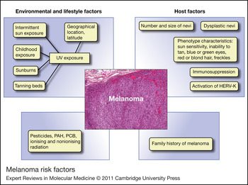
The risk factors for melanoma can be broadly classified as environmental or host related (see Fig. 1). Undoubtedly, exposure to UV radiation from sun exposure is the most important environmental risk factor for melanoma (Ref. Reference Markovic5). It is now well established that intermittent sun exposure, exposure during childhood, sunburns and tanning bed usage substantially increase the risk of malignant melanoma (Ref. Reference Markovic5). Exposure to some toxic chemicals, as well as cosmic ionising and nonionising radiation are also significant risk factors for melanoma (Ref. Reference Fortes and de Vries6). Melanoma risk increases with the size and total number of nevi, and number of dysplastic nevi. Specific phenotypic characteristics, such as eye colour (blue, green), hair colour (red, blonde), inability to tan, presence of freckles and sun sensitivity, have been shown to correlate with increased risk. Other important risk factors are a family history of melanoma and immunosuppression (Ref. Reference Markovic5). There is a growing body of evidence that human endogenous retroviruses (HERVs) might also have a role in the genesis of melanoma (Ref. Reference Haqq7).
Melanoma risk factors. Abbreviations: HERV-K, human endogenous retrovirus; PCB, polychlorinated biphenyl-PCB; PAH, polycyclic aromatic hydrocarbons.

Melanoma initiates in melanocytes or their precursors. Melanocytes derive from neural crest cells, which are progenitors of neurons, glial cells, secretory cells of the peripheral neuroendocrine system and connective tissue (Ref. Reference Le Douarin8). During embryogenesis, melanoblasts – the precursors of melanocytes – migrate from the neural crest to the skin, uvea, leptomeninges and mucous membranes – body sites where melanoma can occur (Ref. Reference Slominski9). After migration, they remain undifferentiated until stimulated, when they differentiate into pigment-producing cells. Melanocytes produce and transfer melanin to neighbouring keratinocytes, thus influencing hair and skin pigmentation (Ref. Reference Slominski9). Melanin is a paramagnetic biopolymer that protects us against UV damage by absorbing UV radiation and neutralising free radicals. The wavelength of light determines how it affects melanogenesis: UVA induces dark pigmentation mainly in basal keratinocytes; UVB induces melanin distribution throughout the epidermis; UVC, the most energetic and mutagenic, does not reach the earth's surface (Ref. Reference Slominski9).
A subject of continuing controversy is whether melanoma follows the canonical model of cancer stem cell renewal. Conceptually, it is believed that although cancer cells might have a phenotypically pleomorphic and undifferentiated bulk population, a select subset (0.1–0.0001%) of them – stem cells – are responsible for their perpetuation (Ref. Reference Quintana10). Supporting the fact that this notion is valid for melanoma, some experimental data (Ref. Reference Schatton11) have shown that only a small subpopulation of melanoma cells is tumourigenic. For example, only one in 1 million (0.0001%) human cancer cells xenotransplanted into NOD/SCID mice have tumourigenic potential (Ref. Reference Schatton11). This observation has been challenged by more recent experiments revealing that having a propitious host environment (highly immunocompromised NOD/SCID IL2Rγnull mice) yields a much larger percentage (25%) of tumourigenic melanoma cells (Ref. Reference Quintana10). Because there are currently no reliable cancer stem cell markers, neither of these studies could address issues such as initial stem cell density nor transplantation efficiency.
Clinical features of melanoma
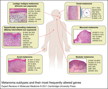
Melanoma is currently classified according to the anatomical site of the primary tumour and histopathological features of the skin lesion (see Fig. 2). The most common subtypes are the following: nodular melanoma (NM), which consists of raised nodules; acral lentiginous melanoma (ALM), which occurs on the palms of the hands, soles of the feet and in the nail bed; it accounts for about 50% of melanoma in non-Caucasians; mucosal melanoma (MM), which occurs in the upper respiratory tract and oral cavity; uveal melanoma (UM), which occurs in the eye; lentigo maligna melanoma (LMM), which occurs on sun-exposed skin in the elderly, associated with chronic sun exposure; and superficially spreading melanoma (SSM), which occurs on sun-exposed skin on the back in males and on the leg in females and is the most common form of melanoma (Ref. Reference Gray-Schopfer, Wellbrock and Marais12).
Melanoma subtypes and their most frequently altered genes. Uveal melanoma (UM), lentigo maligna melanoma (LMM), nodular melanoma (NM), acral melanoma (AM), mucosal melanoma (MM) and superficially spreading melanoma (SSM).

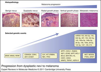
Patients with melanoma present with a variety of signs, including changes in size, shape or colour of a pigmented skin lesion or the development of a new skin growth that changes over a few weeks or more. Characteristic signs of malignant transformation of a pigmented lesion are bleeding, asymmetry, border irregularity, nonuniform pigmentation, elevation from flat and size greater than 6 mm. The Clark model describing molecular events correlated with initiation and progression of most melanomas can be summarised as follows (Ref. Reference Miller and Mihm13): (1) benign nevi with an increased number of nested melanocytes; (2) dysplastic nevi with irregular borders, multiple colours, increased diameter, and random and discontinuous cytological atypia; (3) radial growth phase melanoma, with malignant cells proliferating within the epidermis; (4) vertical growth phase melanoma, with cells transgressing the lamina propria; and (5) metastatic melanoma, with spread of neoplastic cells to other areas of the skin or other organs (Ref. Reference Clark14). Figure 3 shows the histopathological features and selected genetic changes associated with progression from dysplastic nevi to melanoma. The conventions for staging melanoma are not described here, but have been reported previously (Ref. Reference Balch2).
Progression from dysplastic nevi to melanoma. Some benign nevi transform into dysplastic nevi, and may follow radial and vertical growth phases, culminating in metastatic melanoma. Genetic events include amplification, deletion, upregulation or downregulation of certain oncogenes or tumour-suppressor genes, including cyclin-dependent kinase inhibitor 2A (CDKN2A), cyclin-dependent kinase 4a (CDK4), serine/threonine protein kinase B (PKB, also known as AKT), cyclin D1 (CCND1), KIT, ERBB4, retinoblastoma protein (pRb), TP53, osteopontin (SPP1), melanoma growth-stimulating activity (CXCL1) and RAB32, WIF1, ECM2, microphthalmia-associated transcription factor (MITF), BCL2, BCL-XL, E-cadherin (CDH1), N-cadherin (CDH2), matrix metalloproteinase (MMP2), transient receptor potential melastatin 1 (TRPM1) and human endogenous retrovirus K (HERV-K). V-raf murine sarcoma viral oncogene homologue B1 (BRAF), neuroblastoma RAS viral (v-ras) oncogene homologue (NRAS) and PERK are involved in early stages of progression from benign to dysplastic nevus. Hepatocyte growth factor receptor (MET), neural precursor cell expressed, developmentally down-regulated (NEDD9), phosphatase and tensin homologue (PTEN) and apoptotic peptidase activating factor (APAF1) are involved mainly in the progression of metastasis. Upregulated and downregulated miRNAs identified in melanoma stage progression are also shown.

Current molecular therapeutic approaches
Metastatic melanoma has poor prognosis and no effective therapies (Ref. 1). Its treatment includes surgery, immunotherapy, chemotherapy and radiation therapy. There are only three agents known to us that have been approved by the US Food and Drug Administration (FDA) for the treatment of metastatic melanoma: dacarbazine (DTIC; approved more than three decades ago) (Ref. Reference Serrone15), immunotherapy with high-dose IL-2 (approved in 1998) (Ref. Reference Garbe16) and ipilimumab (approved in 2011) (Ref. Reference Sondak17). DTIC as a single agent has shown clinical response in only 5–10% of cases (Ref. Reference Serrone15). Similarly, treatment with high-dose IL-2 produces a long-term complete response in approximately 5% of patients (Ref. Reference Agarwala18). Unfortunately, IL-2 immunotherapy does not improve overall survival and might induce significant adverse events (Ref. Reference Parkinson19). A recent report of a Phase III multicentre randomised clinical trial assessing the effect of ipilimumab administration with or without a glycoprotein 100 (gp100; a receptor subunit of the IL-6 family) peptide vaccine well illustrates its efficacy (Ref. Reference Hodi20). The median overall survival for patients receiving ipilimumab alone was greater (10.1 months) than that with either the vaccine alone (6.4 months) or the ipilimumab and vaccine combination (10.0 months) (Ref. Reference Hodi20). More than 28% of patients in this study showed a partial or complete response, or stable disease with ipilimumab monotherapy. The 1-year and 2-year survival rates were reported to be 45.6% and 23.5%, respectively (Ref. Reference Hodi20). Unfortunately, as with IL-2 immunotherapy, some patients have an adverse reaction to ipilimumab that can be life-threatening (Ref. Reference Hodi20).
Interferon alpha (IFN-α) is the most commonly used drug in patients with intermediate-risk or high-risk melanoma (Ref. Reference Kirkwood21). IFN-α(2a) and IFN-α(2b) have been tested as therapeutic agents in the clinical setting for melanoma, and only human recombinant IFN-α(2b) is approved for adjuvant treatment of this cancer (Ref. Reference Mocellin22). Randomised controlled clinical trials yielded conflicting results on the efficacy of IFN-α as an adjuvant treatment for melanoma (Ref. Reference Mocellin22). However, a recent meta-analysis, including 14 randomised controlled trials, suggested that adjuvant treatment with IFN-α has a statistically significant positive effect on disease-free survival (risk reduction = 18%) and overall survival (risk reduction = 11%) (Ref. Reference Mocellin22).
Molecular pathways altered in melanoma
Recent progress in understanding the molecular mechanisms of melanoma progression has led to the development of new therapeutic targets and strategies, some of which are currently being tested in clinical studies. As of July 2011, the US clinical trials website (http://clinicaltrials.gov/) listed 1102 registered clinical trials involving new therapies for melanoma (Ref. 23), many of which exploit melanoma-specific mutations or pathways.
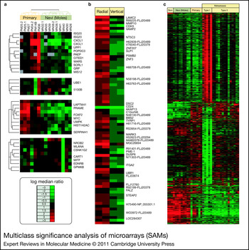
Melanoma, as well as many other cancers, develops through a multistep process involving sequential alterations of numerous proto-oncogenes, tumour suppressors and miRNA genes. The altered genes have key roles in the regulation of cell cycle progression, differentiation and apoptosis (Ref. Reference Croce24). Furthermore, transcriptional profiling of nevi, primary melanomas and melanoma metastases reveals specific patterns of gene expression at different stages of melanoma progression (Ref. Reference Haqq7). Although Figure 3 shows the most accepted genetic alterations in the dysplastic nevi–melanoma sequence, more than 1000 genes are either upregulated or downregulated during melanoma carcinogenesis (Ref. Reference Haqq7); furthermore, an excess of 2000 genes are differentially expressed in normal skin, nevi and primary or metastatic melanoma (Fig. 4) (Ref. Reference Haqq7).
Multiclass significance analysis of microarrays (SAMs). SAM applied to skin, nevi, and primary and metastatic melanoma subtypes predicts 2602 genes able to distinguish among these tissue types. (a) Differences between gene expression in primary melanoma and nevi. (b) Radial and vertical growth. (c) Skin, nevi, and primary and metastatic melanoma subtypes. Red indicates gene overexpression and green indicates gene expression lost (Ref. Reference Haqq7). Images reprinted with permission from Ref. Reference Haqq7, courtesy of Proceedings of the National Academy of Sciences of the United States of America.

Growing evidence links melanoma development and progression to germline mutations in genes encoding CDKN2A, CDK4 and MC1R (Refs Reference MacKie, Hauschild and Eggermont25, Reference Thomas26, Reference Kamb27, Reference de Snoo and Hayward28), as well as somatic mutations in proto-oncogenes (B-RAF, N-RAS and KIT) and tumour suppressor genes CDKN2A, p53 and PTEN (Refs Reference de Snoo and Hayward28, Reference Ivry, Ogle and Shim29, Reference Bataille30, Reference Whiteman31). The most frequently altered genes in the most common melanoma subtypes are shown in Figure 2.
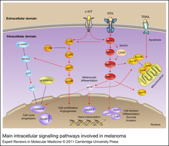
These genes act through several molecular pathways (Fig. 5), which, although they might also be affected in other human malignancies, exhibit specific alterations for melanoma. Perhaps the most studied signalling cascade in melanoma is the MAPK–ERK (mitogen-activated protein kinase–extracellular-related kinase) signal transduction pathway or the central proliferation pathway. This pathway regulates cell division, differentiation, survival and tumour invasion (Ref. Reference Miller and Mihm13). It is also referred to as the ERK or RAS–RAF–MEK–MAPK pathway. Members of this pathway are among the most frequently mutated in melanoma, with B-RAF (50–60%) and N-RAS (15%) currently undergoing intense research (Ref. Reference Herlyn32). Other pathways that might be deregulated in melanoma include KIT (2–45%); PI3K (type I phosphatidylinositol-3-kinase), involved in cell growth, apoptosis, protein synthesis and metabolism; retinoblastoma, involved in cell cycle progression; and the p53 pathway, involved in initiating DNA repair, and inhibition of cell cycle progression, senescence and apoptosis (Ref. Reference Fecher33).
Main intracellular signalling pathways involved in melanoma.

B-RAF
Human B-RAF, also known as V-raf murine sarcoma viral oncogene homologue B1, belongs to a family of three human RAF genes: A-RAF1, B-RAF and RAF-1 (also known as c-RAF) (Ref. Reference de Snoo and Hayward28). RAF proteins are serine/threonine kinases that link extracellular mitogenic stimuli to transcription of cell cycle progression or progrowth genes (Ref. Reference de Snoo and Hayward28).
Since its discovery in 2002, as a gene mutated in melanoma (Ref. Reference Davies34), B-RAF has proved to have a great impact on the quest for novel melanoma therapies (Ref. Reference Wellbrock and Hurlstone35). Somatic mutations in B-RAF are the most common genetic alterations in melanoma, occurring in up to 66% of malignant melanomas (Refs Reference Davies34, Reference Pollock and Meltzer36, Reference Brose37). B-RAF mutations are thought to be induced by UV radiation or reactive oxygen species that are generated as a byproduct of melanogenesis or phaeomelanin absorption of UVA radiation (Ref. Reference Thomas26). However, UV radiation alone might not be sufficient for B-RAF mutagenesis, because the presence of the B-RAFV600E mutation has been reported in a subset of acquired nevi from glabrous skin and genitalia (81.4%), and in congenital nevi (69%), implying that other factors are required as well (Ref. Reference Ichii-Nakato38).
There are at least 30 documented melanoma mutations of B-RAF that activate the MAPK pathway. The most common is V600E (previously mistakenly reported as position 599), which accounts for 89% of all B-RAF mutations (Refs Reference Wellbrock and Hurlstone35, Reference Davies and Samuels39). A significant novel observation is that melanomas with CDK4 amplification do not show concomitant N-RAS or B-RAF mutations (Ref. Reference Curtin40), indicating overlapping oncogenic functions in the MAPK and CDK4 pathways. B-RAF mutations are more frequent in skin without chronic sun-induced damage and without N-RAS mutations (Ref. Reference Curtin40).
A summary of the frequency of B-RAF mutations by melanoma subtypes is shown in Figure 2. B-RAF mutations are present in up to 82% of benign nevi (Ref. Reference Pollock41). Surprisingly, it has been reported that mutated B-RAF induces cell cycle arrest and senescence in nevi. The mechanism by which B-RAF induces senescence in melanocytes remains controversial and is not entirely understood (Refs Reference Michaloglou42, Reference Cotter43, Reference Houben44, Reference Wajapeyee45, Reference Kuilman46, Reference Scurr47). In nevi, it has been reported that B-RAF induces cell senescence by means other than telomerase attrition. It appears that B-RAF might directly induce p16INK4a and acidic β-galactosidase [SA-β-Gal (Ref. Reference Michaloglou42)]. A different group has shown that ectopic expression of B-RAFV600E induces melanocyte senescence by increasing the levels of MAPK activity and by upregulating the expression of cyclin-dependent kinase (CDK) inhibitors (Refs Reference Houben44, Reference Wajapeyee45). RNA interference data have suggested pro-inflammatory cytokines, particularly IL-6 and IL-8, as key regulators of melanocyte senescence (Ref. Reference Kuilman46). However, the role of insulin growth factor-like binding protein 7 remains controversial (Refs Reference Cotter43, Reference Scurr47). Hence, B-RAF mutations should be followed or accompanied by additional molecular events, such as loss of tumour suppressor genes or upregulation of oncogenes, for melanoma transformation (Refs Reference Dankort48, Reference Zhao49).
In general, patient treatment with selective B-RAF inhibitors as single-agent targeted therapy is usually followed by relapse as a result of acquired drug resistance. Intense research has recently focused on understanding the mechanism by which melanoma develops resistance to BRAF inhibition. Several other genes appear to be involved in the acquired resistance to B-RAF inhibitors. Among other genes, COT (Ref. Reference Johannessen50), CRAF (Ref. Reference Montagut51), RTK, NRAS (Ref. Reference Nazarian52), PERK (Ref. Reference Paraiso53), PTEN (Ref. Reference Paraiso54) and CCND1 (Ref. Reference Smalley55) become either upregulated or downregulated, whereas B-RAF inhibitors become ineffective. Some authors have suggested that drug resistance to RAF inhibitors could be overruled by the use of combined RAF and MEK inhibitors (Ref. Reference Johannessen50), or cotargeting MEK and PI3K pathways (Ref. Reference Villanueva56). An excellent review on the subject has been published (Ref. Reference Solit and Rosen57).
Sorafenib, originally developed as an inhibitor of RAF-1, is 10 times less potent as an inhibitor of B-RAF than as an inhibitor of RAF-1 (Ref. Reference Flaherty and McArthur58). Recent studies demonstrated that sorafenib is indeed a multikinase inhibitor that prevents tumour cell proliferation and angiogenesis by inhibiting the RAF–MEK–ERK pathway. Sorafenib also affects vascular endothelial growth factor (VEGF), KIT and other growth factor receptor tyrosine kinases in vitro and in vivo (Refs Reference Flaherty59, Reference Treisman and Garlie60). Sorafenib has already been approved by the FDA for the treatment of renal cell and hepatocellular carcinoma (Ref. Reference Treisman and Garlie60). Preclinical studies demonstrated that sorafenib inhibits the growth and survival of B-RAF mutant human melanoma cell lines, and slows the growth of melanoma xenografts in animal models (Ref. Reference Karasarides61). However, a Phase II study of sorafenib as a single agent showed little or no efficacy in the majority of advanced melanoma patients, even though 19% of 37 patients experienced stable disease (Ref. Reference Eisen62).
In vitro and in vivo studies show that sorafenib enhances the efficacy of regional chemotherapy (Refs Reference Murray63, Reference Augustine64). It was reported that sorafenib in combination with melphalan or temozolomide is more effective in slowing down tumour growth in an animal model of melanoma than either treatment alone (Ref. Reference Augustine64). Also, sorafenib plus etoposide, paclitaxel or temozolomide significantly inhibits the growth of melanoma cell lines in vitro (Ref. Reference Murray63). Similarly, a Phase I dose-escalation trial (Ref. Reference Flaherty59) reported that sorafenib in combination with carboplatin and paclitaxel (CP) had a better clinical outcome than CP alone. The combination of sorafenib and CP resulted in one complete response and nine partial responses among 24 patients with advanced melanoma. However, a subsequent randomised Phase III trial showed no difference in the clinical outcome for patients treated with CP and patients treated with sorafenib plus CP (Ref. Reference Hauschild65).
PLX4032, also known as RG7204 or RO5185426, is a newly developed and highly selective B-RAF inhibitor (Refs Reference Garber66, Reference Livingstone67, Reference Bollag68, Reference Flaherty, Puzanov and Sosman69). PLX4032 has higher affinity for mutated B-RAFV600E (IC50 = 31 nM) than for wild-type B-RAF (IC50 = 100 nM) (Ref. Reference Livingstone67). Preliminary reports characterised the antiproliferative effects of PLX4032 on melanoma cell lines with B-RAF mutations (Ref. Reference Yang70). PLX4032 exhibited encouraging results in a xenograft model of melanoma harbouring B-RAFV600E. In this model, PLX4032 induced partial or complete tumour regression and increased survival in a dose-dependent manner (Ref. Reference Yang70).
PLX4032 was the first selective B-RAF inhibitor clinically tested (Ref. Reference Flaherty71). Preliminary results of a Phase I trial showed a 79% overall response rate in patients with melanoma and other cancers (Ref. Reference Flaherty, Puzanov and Sosman69). Of 16 patients with melanoma expressing B-RAFV600E, 14 showed significant tumour regression (Ref. Reference Flaherty, Puzanov and Sosman69). The six-month survival of these patients also improved; however, progression-free survival is still being monitored for these patients.
Another Phase I clinical trial has shown that PLX4032, administered at 960 mg PO twice daily, has a remarkably high response rate (81%) in metastatic melanoma patients harbouring B-RAFV600E mutations (Ref. Reference Flaherty71). The trial also reported that 24 of 32 (75%) patients had partial response and 6.25% (2 of 32) had complete response. The median progression-free survival for all patients was estimated to be more than 7 months (Ref. Reference Flaherty71). Similar results were found in a Phase II trial that reported an 82% overall response rate to PLX4032. Thirty per cent of the patients experienced stable disease, and the median progression-free survival rate was reported to be 6.2 months (Ref. Reference Vultur, Villanueva and Herlyn72). This astounding response rate is unfortunately followed by relapse in most patients, caused by acquired drug resistance.
In vitro studies show that melanoma cells might acquire resistance to PLX4032 and other BRAF inhibitors, possibly by activating either the PI3K–Akt or RAF–MEK–ERK pathways (Refs Reference Villanueva56, Reference Livingstone67). One of the mechanisms of resistance to B-RAFV600E inhibitors is the generation of BRAF–C-RAF complexes, C-RAF–C-RAF homodimers or A-RAF kinase switching that will activate the MAPK pathway (Ref. Reference Villanueva56). Also, increased PI3K signalling promotes the survival of cells resistant to BRAF inhibitors (Ref. Reference Villanueva56). In addition, when PLX4032 is administered to N-RAS-mutated melanoma cell lines, resistance to the drug is accompanied by hyperactivation of MEK–ERK1/2 signalling (Ref. Reference Kaplan73). Parallel sequencing of 138 cancer genes from a melanoma tumour in a patient who, after an initial dramatic response, developed resistance to PLX4032 detected an activating mutation in MEK1 that was not present before treatment (Ref. Reference Wagle74). It was concluded that this particular MEK1 mutation conferred resistance to RAF and MEK inhibitors in vitro (Ref. Reference Wagle74). Finally, another proposed mechanism is N-RAS upregulation through RTK or NRAS upregulation (Ref. Reference Nazarian52); and by COT activation of ERK through MEK-dependent mechanisms that do not require RAF signalling (Ref. Reference Johannessen50).
The most commonly reported adverse effects to PLX4032 included rash, fatigue, photosensitivity, nausea, alopecia and arthralgia (Refs Reference Livingstone67, Reference Bollag68, Reference Flaherty, Puzanov and Sosman69). However, significant adverse effects were observed in up to 23% of patients. Well-differentiated cutaneous squamous cell carcinoma, kerato-acanthoma type, occurs in a significant number of patients (10 of 32 patients in an extension cohort) (Ref. Reference Flaherty71). It has been postulated that the development of squamous cell carcinoma is possibly due to PLX4032-mediated activation of ERK in keratinocytes (Ref. Reference Hatzivassiliou75).
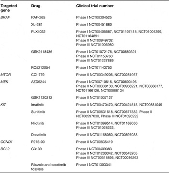
The efficacy of PLX4032 as a melanoma therapeutic continues to be studied in ongoing clinical trials (Table 1). Emerging broad-spectrum kinase inhibitors with greater potency and improved selectivity for B-RAF are RAF-265 and XL-281, which are currently being tested in clinical trials (Table 1) (Ref. Reference Flaherty and McArthur58). They are potent RAF1 inhibitors, with relevance for targeting N-RAS mutant melanomas as well. The success rate of selective and potent inhibitors of B-RAF, such as PLX4032, indicates that targeting an early mutation is a feasible way of treating melanoma.
Selected drugs investigated in clinical trials for patients with melanoma caused by specific gene mutations (http://clinicaltrials.gov/) (Ref. 23)

MEK
MEK is a member of the MAP kinase pathway that is infrequently mutated in cancer cells. MEK is an important oncotherapeutic target because it is the only known direct substrate of B-RAF, and because inhibiting MEK might inhibit the growth of melanomas with mutated B-RAF and RAS (Ref. Reference Flaherty and McArthur58). PD0325901 (Ref. Reference Ciuffreda76) and AZD6244 (also known as selumetinib, ARRY-142886) (Refs Reference Banerji77, Reference Denton and Gustafson78) are selective second-generation MEK inhibitors. In vitro studies show that PD0325901 strongly inhibits the growth of melanoma cell lines independent of their B-RAF mutation status by promoting apoptosis and decreasing the production of proangiogenic factors (Ref. Reference Ciuffreda76). However, Phase I and Phase II studies were discontinued in 2007 because of ocular and neurological toxicity (NCT00147550) (Ref. 23). Experiments in nude mice inoculated with the human melanoma cell line A375 demonstrated that AZD6244 effectively inhibits cell proliferation by inhibiting ERK phosphorylation (Ref. Reference Denton and Gustafson78). AZD6244 inhibited the in vitro proliferation of melanoma cell lines with mutant B-RAFV600E and N-RAS, but exhibited no significant effect on cells with wild-type B-RAF/N-RAS (Refs Reference Davies79, Reference Solit80). AZD6244 showed clinical efficacy in Phase I and Phase II clinical trials in patients with advanced melanoma and other advanced cancers (Refs Reference Banerji77, Reference Adjei81). Ongoing clinical trials involving AZD6244 are listed in Table 1.
KIT
KIT (also called c-KIT or CD117) is a receptor tyrosine kinase expressed on the surface of some cells. It is an upstream activator of the MAP kinase pathway and has a central role in melanocyte development and survival (Ref. Reference Berger and Garraway82). Mutation and/or amplification of KIT are present in ALM, LMM, MM and UM (Refs Reference Davies and Samuels39, Reference Berger and Garraway82, Reference Curtin83, Reference Beadling84, Reference Hofmann85, Reference Woodman and Davies86, Reference Garrido and Bastian87) (Fig. 2). KIT mutations in either the juxtamembrane domain or the kinase domain result in gain of function. KIT mutations are usually mutually exclusive with B-RAF and N-RAS mutations (Refs Reference Beadling84, Reference Garrido and Bastian87).
Cancers with KIT mutations have been successfully treated with KIT kinase inhibitors (Ref. Reference Garrido and Bastian87). As in gastrointestinal stromal tumours (GIST), most KIT mutations in melanoma occur in exon 11 (Refs Reference Berger and Garraway82, Reference Curtin83, Reference Flaherty, Hodi and Bastian88). Patients with GIST harbouring KIT mutations have been successfully treated with the KIT inhibitor imatinib mesylate or GLEEVEC. Partial responses have been reported in up to 84% of treated patients (Ref. Reference Berger and Garraway82).
Initial in vitro preclinical studies indicated that imatinib has potent antiproliferative and proapoptotic effects on melanoma cells by inhibiting MAPK and PI3K–AKT pathways (Ref. Reference Jiang89). Case report studies showed promising results in imatinib-treated melanoma patients with reported KIT mutations (Refs Reference Woodman and Davies86, Reference Jiang89, Reference Terheyden90, Reference Hodi91, Reference Lutzky, Bauer and Bastian92). Unfortunately, early Phase II clinical trial studies reported poor efficacy for treating metastatic melanoma (Refs Reference Wyman93, Reference Ugurel94). One possible explanation for these findings is that these studies were performed before the discovery of KIT mutations in melanoma. Early studies assessing the effect of imatinib did not set the presence of KIT mutations as a criterion for entry.
A newer Phase II clinical trial of imatinib indicated that one patient with metastatic ALM and the highest KIT expression of all enrolled patients showed significant improvement after 6 weeks of therapy and had partial response lasting 12.8 months (Ref. Reference Kim95). After 2 weeks of treatment, patients showed a marked increase in apoptosis of tumour cells (Ref. Reference Kim95). In general, imatinib was relatively well tolerated, with fatigue and oedema the most commonly reported adverse reactions. However, grade 3 or 4 toxicity occurred in >10% of patients (Ref. Reference Kim95). A clinical study of three patients exhibiting KIT mutations treated with imatinib and one with sorafenib showed that all four patients responded to treatment, but three developed brain metastasis. This is probably due to the inability of imatinib to cross the blood–brain barrier (Ref. Reference Handolias96). Other KIT inhibitors, such as dasatinib, appear to penetrate the blood–brain barrier better than imatinib (Ref. Reference Porkka97).
Multiple ongoing clinical trials target melanoma patients with KIT mutations (Table 1). Interim results from a Phase II study of imatinib in stage III or IV patients showed that two out of 12 patients had complete response, two patients had partial response, and six patients had stable disease with imatinib (Ref. Reference Carvajal98). The two patients with complete response had both amplification and mutation of KIT, whereas the two patients who showed no response had KIT mutations that confer resistance to imatinib in GIST (Ref. Reference Carvajal98). In conclusion, KIT inhibitor therapy is reasonably effective for selected patients with melanoma harbouring KIT mutations.
Bcl-2 family
The disruption of key apoptotic regulatory genes makes malignant melanoma resistant to chemotherapy (Ref. Reference Hocker, Singh and Tsao99). The Bcl-2 family of genes comprises both antiapoptotic (such as Bcl-2, Bcl-xL, Bcl-w, Mcl-1 and A1) and proapoptotic proteins [multidomain proteins (Bak and Bax) and BH3-only proteins (Bid, Bim, Bad, NOXA and PUMA)] (Ref. Reference Hocker, Singh and Tsao99). The role of Bcl-2 and Bax in melanoma progression remains controversial (Ref. Reference Bush and Li100). Although some studies report that Bcl-2 expression is upregulated in advanced melanoma, other studies report the opposite effect (Ref. Reference Tron101). Similarly, Bax expression is reported to be both downregulated (Ref. Reference Bush and Li100) and upregulated during melanoma progression (Ref. Reference Tang102). By contrast, antiapoptotic family members Mcl-1 and Bcl-xl are consistently reported to be overexpressed in melanoma cells (Refs Reference Bush and Li100, Reference Tang102, Reference Zhuang103), most probably at an early stage in melanoma development (Ref. Reference Tang102). Keuling et al. demonstrated a progression-related upregulation of Mcl-1 and that inhibition of Mcl-1 using RNAi sensitised melanoma cells to the Bcl-2-selective inhibitor ABT-737 (Ref. Reference Keuling104).
The in vivo role of Bcl-2 in melanoma was shown to differ significantly from its role in vitro (Ref. Reference Benimetskaya105). In this study, downregulation of Bcl-2 protein and mRNA expression in 518A2 melanoma cells was achieved by using the novel Bcl-2 inhibitor G3139 (Ref. Reference Benimetskaya105). G3139 (oblimersen) is an antisense oligonucleotide targeted to the initiation region of the Bcl-2 mRNA (Ref. Reference Bedikian106). The results of the study indicated that in vitro downregulation of Bcl-2 expression does not affect tumour cell growth rate or sensitivity to standard cytotoxic chemotherapeutic agents (Ref. Reference Benimetskaya105). In addition, G3139-induced apoptosis was independent of Bcl-2. However, in vivo results on immunodeficient mice with xenografted melanoma cells having silenced Bcl-2 demonstrated that the tumour either failed to grow or grew to a low volume with subsequent regression (Ref. Reference Benimetskaya105). By contrast, mice with xenografted melanoma cells with normal levels of Bcl-2 exhibited large, necrotic tumours (Ref. Reference Benimetskaya105).
G3139 combined with DTIC has been tested on 771 patients with advanced melanoma enrolled in a Phase III clinical trial (Ref. Reference Bedikian106). The study reported that the drug combination produced better clinical outcomes than DTIC alone. The clinical trial reported a better median overall survival when the drugs were combined (9 months) compared with DTIC only (7.8 months). In the same manner, better outcomes were observed by the dual therapy on progression-free survival: median (2.6 versus 1.6 months), overall response (13.5% versus 7.5%), complete response (2.8% versus 0.8%) and durable response (7.3% versus 3.6%) (Ref. Reference Bedikian106).
HERV-K
Approximately 8% of the human genome is composed of sequences derived from HERVs (Ref. Reference Singh107). Most HERV sequences are defective as a result of mutations, deletions or termination signals in their coding regions (Ref. Reference Serafino108). However, HERV-K has been implicated in the pathogenesis of breast (Ref. Reference Golan109), ovarian (Ref. Reference Wang-Johanning110) and haematological cancer (Ref. Reference Depil111). HERV-K might also have a role in both the malignant transformation of melanocytes (Refs Reference Haqq7, Reference Singh107, Reference Serafino108, Reference Hahn112) and the ability of melanoma cells to evade immune surveillance (Ref. Reference Oricchio113). Metastatic melanoma cell lines express HERV-K-type antigens, whereas primary melanocytes do not (Ref. Reference Haqq7). Although HERVs are poorly expressed in normal somatic tissues, HERV-derived RNA transcripts can be induced in response to UV radiation (Refs Reference Hohenadl114, Reference Reiche, Pauli and Ellerbrok115), chemicals (Ref. Reference Khan, Muller and Sears116) and other endogenous factors (Ref. Reference Ono, Kawakami and Ushikubo117). For example, it was shown that UV induces activation of HERV-K in normal human epidermal melanocytes 24 h after exposure to 10 mJ/cm2 UVC (Ref. Reference Reiche, Pauli and Ellerbrok115).
Recently, detectable levels of anti-HERV-K antibodies were found in 51 of 312 (16%) melanoma patients. By contrast, no retroviral antigens were found in any healthy controls (Ref. Reference Hahn112). Higher levels of anti-HERV-K antibodies were found in patients with melanoma at sun-protected sites (ALM, MM or UM) compared with patients affected by LMM, NM or SSM (Ref. Reference Hahn112). The data suggested that the presence of anti-HERV-K antibodies in patient serum at early stages of disease correlates with lower overall survival (Ref. Reference Hahn112).
In vitro studies show that cellular stress promotes the transformation of human melanoma cells from adherent to nonadherent, less to more proliferative, and less to more aggressive and malignant (Ref. Reference Serafino108). This transformation occurs simultaneously with the activation of HERV-K and the production of a large amount of viral-like particles (Ref. Reference Serafino108). The transition from adherent to nonadherent might be prevented by the downregulation of HERV-K expression by RNA interference (Ref. Reference Serafino108).
The above findings indicate a possible role of HERV-K in melanoma development and progression. Furthermore, understanding the molecular events leading to increased HERV expression and determining why ALM, MM and UM paradoxically express higher levels of HERVs might represent a fertile area of research for new melanoma therapeutics.
MicroRNA-based melanoma therapeutics
MicroRNAs (miRNAs) are small noncoding RNA molecules 21–23 nucleotides long that modulate gene expression at the post-transcriptional or translational level (Ref. Reference Wang and Lee118). miRNAs have been found in all multicellular organisms. Although they represent only 1% of the mammalian genome, there are approximately 650 human miRNAs, each of which can regulate the expression of hundreds of genes and can directly or indirectly modulate the expression of thousands of proteins (www.microRNA.org) (Refs 119, Reference Selbach120). Approximately 30% of protein-coding genes in the human genome are thought to be regulated by miRNAs (Ref. Reference Chen and Rajewsky121).
About half of all miRNA genes are located in genomic regions that are frequently amplified or deleted in cancer cells (Ref. Reference Calin122), and miRNA deregulation has been specifically associated with brain, thyroid, breast, lung, liver, gastrointestinal, gynaecological, prostate and other cancers (reviewed in Ref. Reference Wang and Lee118). Some miRNAs are deregulated in several cancers whereas others are deregulated only in specific tumour types (Ref. Reference Wang and Lee118).
There are several reasons and approaches for pursuing miRNAs as therapeutic targets: (1) miRNAs can be efficiently downregulated or upregulated using miRNA antagonising molecules (antagomiRs/antimiRs) or mimics; (2) miRNAs simultaneously regulate many target genes that might function in several oncogenic pathways; (3) miRNAs can be used as noninvasive analysis tools for detecting cancers, because of their presence in the blood of cancer patients in addition to cancer tissue (Refs Reference Mueller and Bosserhoff3, Reference Leidinger123).
miRNA-focused study of melanoma is an important emerging research area (Ref. Reference Sigalotti124). In this regard, miRNA-193b and miR-211 are of particular interest, because they are downregulated in melanoma relative to nevi. When miR-193b was overexpressed in melanoma cells in vitro, the expression of approximately 300 mRNAs decreased. Once this list was crossed with potential in silico targets from Targetscan, the list was reduced to 18, including cyclin D1 and Mcl-1 (Ref. Reference Chen125). A signature of 18 miRNAs was identified, one of which was miRNA-193b, whose overexpression is significantly correlated with longer survival (Ref. Reference Segura126). miR-211 is also markedly downregulated in melanoma relative to benign nevi. When reintroduced into melanoma cell lines, a significant reduction in melanoma invasiveness was noted. miR-211 might decrease invasiveness by directly regulating the expression of TGFBR2 and NFAT5 (Ref. Reference Levy127).
One therapeutic approach would be to reintroduce a deficient miRNA in a cancer, in order to reverse the oncogenic process. Indeed, miRNA replacement has recently been shown to be effective in a mouse lung cancer model. miR-34a was introduced using a lipid-based delivery system, resulting in reduced tumour growth (Ref. Reference Wiggins128). Thus, in melanoma, one potential therapeutic-based approach might be for miRNA replacement of miRNAs such as miR-193b and miRNA-211.
Targeting multiple pathways
The relative inefficiency of small-molecule inhibitors, such as rapamycin, sorafenib, U0126 or PD98059 (Refs Reference Lasithiotakis129, Reference Smalley and Herlyn130), as well as the poor outcomes of targeting single oncogenic proteins in treating patients with advanced melanoma, led to the testing of synergetic inhibition of multiple pathways (Refs Reference Lorigan, Eisen and Hauschild131, Reference Smalley and Flaherty132, Reference Smalley133). Simultaneously targeting the PI3K–Akt and MAPK pathways significantly inhibits melanoma development (Refs Reference Smalley and Flaherty132, Reference Tran134, Reference Krasilnikov135, Reference Lopez-Bergami, Fitchman and Ronai136). Simultaneous siRNA (small interfering RNA) targeting of Akt and B-RAF using liposomes inhibited in vitro or xenograft melanoma tumour growth (Ref. Reference Tran134). Synergistically inhibiting Akt with nanoliposomes containing ceramide and RAF kinase with sorafenib has been shown to decrease melanoma proliferation (Ref. Reference Tran137). Inhibiting MAPK (with sorafenib or MEK inhibitors) and mTORC1 (with rapamycin) is more effective in reducing melanoma cell growth compared with either agent alone (Refs Reference Dankort48, Reference Lasithiotakis129). Topical application of LY-294002 and U0126, inhibitors of PI3K/Akt and RAF/MAPK, respectively, inhibits the growth of TPras transgenic melanomas in mice compared with either agent alone (Ref. Reference Bedogni138). Targeting simultaneously the PI3K pathway with sorafenib, and the MEK pathway with rapamycin, was more effective than use of the single agents (Ref. Reference Lasithiotakis129). Concurrent inhibition of MEK and CDK4 kinases by PD98059 and 219476 significantly increased apoptosis compared with single agents (Ref. Reference Li139).
Another promising type of combination therapy being developed for melanoma seeks to inhibit an oncogenic target while increasing the expression of a tumour suppressor (Ref. Reference Zhao49). For example, B-RAFV600E can be inhibited using siRNA, whereas expression of tumour suppressor CDKN2A is increased (Ref. Reference Zhao49). Unfortunately, combination therapy has yielded only partial but not complete regression of melanoma tumours, indicating that other targets or combinations of targets should be identified.
siRNA-based therapy
RNA interference (RNAi) is a biological process of post-transcriptional gene silencing in animals and plants. In 2001 it was demonstrated that siRNAs mediate RNAi in mammalian cells without generating an interferon response, thus opening the landscape for the use of RNAi in humans (Ref. Reference Elbashir140). siRNAs are ~21 base-pair double-stranded RNA molecules that are processed within the cell by the RNA-induced silencing complex (Refs Reference Elbashir140, Reference Tran, Watts and Robertson141, Reference Kawakami and Hashida142).
The administration of naked siRNA is not compatible with clinical applications because siRNAs have poor cellular internalisation, are unstable and are degraded rapidly by circulating enzymes (Refs Reference Tran, Watts and Robertson141, Reference Kawakami and Hashida142). As a result of these shortcomings, there are only a few clinical trials utilising this technology (Ref. Reference Chen and Zhaori143). The most important, and perhaps the rate-liming, factor for widespread use of in vivo RNAi is the paucity of effective siRNA carriers (Ref. Reference Fire144). Currently, the most commonly used delivery agents for siRNA include polyethylenimine (PEI) modified with stearic acid (StA), cationic liposomes, polyelectrolyte complex micelles, water-soluble lipopolymers, polycationic dendrimers and aptamers (see below) (Refs Reference Kawakami and Hashida142, Reference Alshamsan145). Nevertheless, siRNAs are promising therapeutic agents because of their high target selectivity and predicted low toxicity. In vitro and in vivo experiments indicate that siRNA targeting of various genes, such as MYC (Ref. Reference Chen146), AKT, BRAF V600E (Ref. Reference Tran134), CD147 (Ref. Reference Su, Chen and Kanekura147), BIRC7 (livin) (Ref. Reference Wang148) and STAT3 (Ref. Reference Alshamsan145) can inhibit melanoma tumour growth and decrease proliferation and invasion.
It has been demonstrated that siRNA can be packed effectively into complexes suitable for cell internalisation by using a lipid-substituted PEI (PEI-StA) (Ref. Reference Alshamsan145). PEI-StA complexes were used to deliver siRNA efficiently to B16 melanoma cells, resulting in potent silencing of the gene encoding signal transducer and activator of transcription 3 (STAT3) (Ref. Reference Alshamsan145). STAT3 upregulates VEGF and antiapoptotic proteins, such as Bcl-2, Bcl-xL and Mcl-1. Melanoma cell viability was significantly reduced to 16% after the third daily dose with STAT3-siRNA–PEI–StA complexes, and in vivo experiments demonstrated regression of tumours, higher levels of IL-6, and lower levels of VEGF and STAT3 (Ref. Reference Alshamsan145).
The first-in-human (recruiting) Phase I clinical trial has been initiated to assess the safety of CALAA-01, an siRNA, using a targeted nanoparticle delivery system for treating solid tumours, including melanoma (NCT00689065) (Refs 23, Reference Davis149). Another recruiting Phase I clinical trial is assessing the safety and efficacy of an antimelanoma vaccine generated using siRNA techniques (NCT00672542) (Ref. 23).
Novel delivery modalities
A major impediment to successful melanoma therapy is the lack of an effective system for delivering traditional and novel antimelanoma agents. Because of this, researchers have turned to nanotechnology and stem cell research, research areas that have recently developed promising delivery modalities as described below.
Nanoparticles
Nanotechnology has advanced tremendously during the last two decades. A variety of delivery systems now available for tumour therapeutics use nonorganic polymers (Refs Reference Zhang150, Reference Hughes151), silica (Ref. Reference Zhao152), silicon, gold, iron (Refs Reference Zhang150, Reference Timko, Dvir and Kohane153, Reference Kim, Ghosh and Rotello154, Reference Cobley155) and carbon nanoparticles (Ref. Reference Timko, Dvir and Kohane153). The most commonly used nanoparticle delivery agents are liposomes (Refs Reference Hughes151, Reference Timko, Dvir and Kohane153, Reference Chen156, Reference Villares157), lipid-like materials (Ref. Reference Love158), quantum dots (Ref. Reference Tran, Watts and Robertson141) and viral particles (Ref. Reference Zhang150). Nanoparticle delivery systems can be loaded with several pharmacological agents or molecular agents. The advantages of this approach include protection of particle contents from degradation and interaction with the biological environment, enhanced drug uptake by tumours and reduced toxicity (Refs Reference Tran, Watts and Robertson141, Reference Peer159). Excellent reviews on the general topic of nanoparticle-based drug delivery (Refs Reference Kawakami and Hashida142, Reference Hughes151, Reference Peer159, Reference Xu and Anchordoquy160, Reference Bawa161) and more specifically on liposome-based delivery applications for melanoma (Ref. Reference Tran, Watts and Robertson141) have been published.
Nanoparticles made up of materials that react to changes in their environment, and nanoparticles controlled by a remote device are novel modalities that are being explored in cancer therapy. Light, pH, ultrasound and magnetic-field-sensitive nanoparticles are of therapeutic importance as a result of their targeting abilities (Refs Reference Timko, Dvir and Kohane153, Reference Cobley155, Reference Gao, Chan and Farokhzad162). Metallic and ceramic nanoparticles, such as silica, titanium oxide and aluminium oxide, can be fabricated to assume a porous structure, suitable for the delivery of antineoplastic agents (Ref. Reference Zhang150). Cellular uptake will be dependent on their size, functionalisation and crystalline structure, among other characteristics (Ref. Reference Buzea, Pacheco and Robbie163).
Efficient targeting of neoplastic cells can be achieved by carefully designing delivery systems conjugated to antibodies, aptamers or other cancer-specific ligands (peptides, vitamins) (Refs Reference Tran, Watts and Robertson141, Reference Zhang150, Reference Peer159). It is expected that to maximise specificity, cancer cells should overexpress a surface marker relative to normal cells (Ref. Reference Peer159). Using antibodies as targeting agents leads to the binding of immune cells to the antibody and the initiation of a signalling cascade that kills the tumour cells (Ref. Reference Peer159). Aptamers are short single-stranded DNA or RNA oligonucleotides selected to bind to a wide variety of targets, such as intracellular proteins, transmembrane proteins and, most importantly, tumour receptors (Refs Reference Barbas and White164, Reference White, Sullenger and Rusconi165).
Iron oxide, coated with phospholipids, surfactants or other compounds that improve their stability, can be used in thermotherapy (Ref. Reference Zhang150). A rodent model of melanoma has proved that thermotherapy, based on the magnetic-field-induced excitation of iron oxide nanoparticles, effectively prolonged the survival of the treated group (a 4.5-fold increase) when compared with controls (Ref. Reference Jordan166). Gold nanoparticles are of interest because of their optical properties, suitable for high infrared phototherapy (Ref. Reference Zhang150).
Liposomes can be used to encapsulate chemotherapeutic agents, siRNA, miRNA or radioactive particles, and are being explored as tools for delivery of antimelanoma agents, as discussed above (Refs Reference Tran, Watts and Robertson141, Reference Fink167, Reference Hwang168).
Polymer nanoparticles chemically conjugated to PD98059 have been used to target the MAPK signal transduction cascade in melanoma cells and melanoma-bearing mice (Ref. Reference Basu169). These experiments showed that this system effectively inhibits in vitro proliferation and induces apoptosis of melanoma cells (Ref. Reference Basu169). The nanoparticle–PD98059 complex also enhanced the antitumour activity of cisplatin chemotherapy (Ref. Reference Basu169). In one recent report, miRNA and siRNA were codelivered using a liposome–polycation–hyaluronic-acid nanoparticle-based formulation modified with a single-chain antibody fragment. This novel system proved to have an enhanced therapeutic effect (Ref. Reference Chen156).
Hundreds of clinical trials are already using encapsulated drugs on patients with various diseases, including melanoma (Ref. 23). A clinical Phase II study evaluated the efficacy of second-line treatment with liposomal doxorubicin in 30 patients with metastatic melanoma, previously unsuccessfully treated with chemotherapy (Ref. Reference Fink167). Liposomal doxorubin was well tolerated; the median survival was 214 days, with seven patients surviving for more than 300 days and five patients for more than 400 days (Ref. Reference Fink167).
Stem cells
Preclinical studies suggest that genetically modified neural stem cells (NSCs) can be targeted to solid tumours with high selectivity (Refs Reference Aboody, Najbauer and Danks170, Reference Aboody171, Reference Aboody172). Some researchers have used NSCs that express prodrug-activating enzymes, interleukins, interferon (IFN)-β and apoptosis-promoting factors (Ref. Reference Aboody, Najbauer and Danks170). Systemically administered progenitor cells migrate to and infiltrate solid tumours. Thus, this antineoplastic delivery system is quite promising (Ref. Reference Aboody, Najbauer and Danks170). Stem cells selectively migrate not only to solid tumours, but also to metastases at sites far away from the primary site of the tumour (Ref. Reference Porada and Almeida-Porada173). The tropism of NSCs has been used to deliver therapeutic genes selectively to tumours of various origin (brain tumours, metastatic melanoma to brain, metastatic breast carcinoma to lung and neuroblastoma) (Ref. Reference Aboody, Najbauer and Danks170). The exact mechanism that drives stem cell migration to tumours is still unknown; however, it is assumed that tumour cells produce chemotactic factors (Ref. Reference Brown174).
The extraordinary tropism of NSCs has been show to effectively target melanoma metastases in the brain (Ref. Reference Aboody171). In mice, NSCs injected into the peripheral circulation migrate to tumours such as glioma, neuroblastoma, prostate cancer and melanoma (Ref. Reference Brown174). When injected into the brain, NSCs migrate through normal brain tissue to specifically target tumour cells (Ref. Reference Aboody172). NSCs can be genetically engineered to express cytosine deaminase (CD) enzyme which is able to convert the nontoxic compound 5-fluorocytosine into a chemotherapeutic drug 5-fluorouracil (5-FU) (Ref. Reference Aboody172). In animal models, intracranial implantation of genetically engineered NSCs expressing CD resulted in a 71% reduction in tumour mass (Refs Reference Aboody171, Reference Aboody172).
A recent study evaluated the effect of NSCs secreting carboxylesterase, which converts CPT-11 into SN-38, a potent topoisomerase-1 inhibitor, on the growth of melanoma cells (Ref. Reference Gutova175). Exposure of melanoma cells to conditioned medium from the enzyme-secreting NSCs and CPT-11 enhanced cell killing 100-fold compared with the use of CPT-11 alone (Ref. Reference Gutova175). High concentrations of IFN-β inhibit malignant cell growth in vitro. Mesenchymal stem cells (MSCs) have been used to target the delivery of IFN-β to tumour cells in a mouse xenograft model with pulmonary metastases derived from breast carcinoma and melanoma cells (Ref. Reference Studeny176). In vitro coculture of MSCs and breast carcinoma or melanoma cells inhibited tumuor cell growth (Ref. Reference Studeny176). Intravenous injection of MSCs into mice with pulmonary metastases suppressed the growth of metastases and prolonged mouse survival compared with control or with mice injected with recombinant IFN-β (Ref. Reference Studeny176).
A recent study indicated that human metastatic melanoma cells can be reprogrammed in the neural-crest-rich microenvironment of the chick embryo, adopting a neural-crest-cell-like phenotype (Ref. Reference Kulesa177). This suggests that it will be important in future studies to determine the molecular events that allow melanoma cells to modulate or be modulated by constituents of the immediate microenvironment.
Emerging melanoma therapies that deserve to be mentioned, but are not covered in this review, include melanoma immunotherapy, with a focus on vaccines, cytotoxic T-lymphocyte antigen-4 antibodies and adoptive cell transfer (Refs Reference Weber178, Reference Homsi, Grimm and Hwu179).
Conclusions
Malignant melanoma arises after a complex series of molecular or genetic events that inactivate tumour suppressor genes and activate proto-oncogenes in melanocytes. Although it is recognised that melanoma cells grow aggressively even in the early stages of tumour development, the molecular mechanisms that promote early metastasis of melanoma, and the mechanisms that allow melanoma cells to escape immune surveillance and develop resistance to chemotherapy, are poorly understood. Nevertheless, distinct molecular signatures have been identified that correlate with the most frequent clinicopathological subtypes of malignant melanoma. DNA sequencing and gene expression profiling have identified many oncogenes that are important for melanoma progression. As our understanding of melanoma initiation and progression continues to grow and deepen, and more therapeutic targets are identified and validated, it might be possible to introduce individualised evidence-based or mutation-analysis-based therapies for melanoma. Ultimately, it is our belief that the most fruitful approaches will involve simultaneous targeting of several oncogenic targets or pathways, and tumour suppressor genes, using novel nanoparticle- or stem cell-mediated delivery systems.
Acknowledgements and funding
We acknowledge funding provided by the Canadian Institutes for Health Research (CIHR) in the form of a research grant to V.T. We would also like to thank the reviewers for their input and helpful advice.