Introduction
Cardiac catheterisations are among the radiologic diagnostic procedures with the highest patient radiation dose. About 7% of all cardiac angiography procedures are performed for children 0–15 years of age. Reference Chida, Ohno and Kakizaki1 A number of specific challenges unique to the setting of paediatric cardiac catheterisation, such as higher heart rates, smaller cardiovascular structures, smaller body size, and wider variety of unusual anatomic variants with the potential need for relatively lengthy and complex studies, result in relatively high radiation doses. In addition, the improved survival of patients with complex anatomy implies that many such children with chronic cardiac disease require frequent catheterizations within the first few years of life. These factors, coupled with the increased radiosensitivity of children and a longer life span ahead of them during which radiation-related sequelae can develop, can create potentially ominous consequences. These diagnostic and therapeutic procedures contribute greatly to improved outcomes in children with heart diseases, at the cost of exposure to high doses of ionising radiation. Reference Beauséjour Ladouceur, Lawler and Gurvitz2 They can cause deterministic effects like skin ulceration and hair loss, which are directly proportional to the amount of radiation dose exposure, whereas stochastic effects such as cancer can occur even in low doses. Tissue reactions result from radiation-induced cell death or damage and are only very rarely seen in children because individual procedural doses typically do not exceed threshold levels. Stochastic effects are induced mutations and occur more commonly in rapidly dividing cells, such as breast, bone marrow, stomach, colon, and lung tissues. 3 The International Commission on Radiological Protection publication 60 states that the possibility of inducing cancer is 2–3 times higher for children than adults. Rassow et al. state that the radiation risk is 4–8 times higher for infants than for adults. Reference Rassow, Schmaltz, Hentrich and Streffer4
Attempts have been made to quantify the increased lifetime risk of cancer-associated mortality from paediatric cardiac catheterisation using models from national and international expert organisations (from 0.003 to 0.016 %/ mSv depending on gender and age of exposure). Although >2,000 mGy was used as the threshold dose (as this is typically cited as the earliest detectable effect of radiation on the skin, e.g., transient erythema), for causing deterministic effects, cancer risk is possible even with the smallest exposure to ionising radiation. 5,Reference Ahmed6 Accordingly, ionising radiation doses should be kept as low as reasonably achievable principle during cardiac catheterisation. There is little published data from this part of the world describing radiation dose exposure during common congenital cardiac interventions in children. Our centre is a major referral centre in eastern India, performing >500 catheterisations and interventions for structural heart. Therefore, we have set out to analyse the various parameters used to detect radiation dose in our centre and compare them with other international studies, to establish local dose reference levels and pave a path for future studies aimed at optimising these parameters.
Materials and methods
NSH Ethics Committee approval was obtained (anonymized) and informed consent was taken. Data were collected on 356 paediatric patients under 18 years of age, undergoing diagnostic and interventional catheterisation procedures in NSH cardiac catheterisation laboratory over an 11-months period. Patients who underwent more than one intervention/ diagnostic procedure were not included in the study. All examinations were carried out by the same staff using INNOVA 2100, manufactured by Wipro GE Healthcare.
The system consisted of an under-couch tube with an over-couch image intensifier in the posteroanterior position. In line with local protocols, the grid was routinely removed for all patients. The radiation exposure measurements used were dose area product, fluoroscopy time, cumulative air kerma, and absorbed radiation dose. Dose-area product, air kerma, and fluoroscopy time were calculated by the machine, while absorbed radiation dose was measured using a Bhabha Atomic Research Centre dosimeter (the semiconductor device designed by Bhabha Atomic Research Centre, Department of Atomic Energy, Government of India) as shown in Figure 1 fixed on the patient’s chest on the anterior chest wall.
( a ) BARC dosimeter, the semiconductor device designed by Bhabha Atomic Research Centre, Department of Atomic Energy, Government of India (schematic diagram). ( b ) BARC dosimeter, the semiconductor device designed by Bhabha Atomic Research Centre, Department of Atomic Energy, Government of India.

All dose-area product metres were calibrated by the assigned hospital physicists before any exposures were recorded. In addition, regular quality assurance and maintenance tests were performed throughout the study. The dose-area product reading recorded was a single value for the whole procedure (this included all exposures during digital cine acquisition and fluoroscopy from both X-ray tubes).
Data were recorded by the radiographer involved in performing the examination. The data recording sheet was piloted among the radiographic staff and subsequently re-designed to ensure there was no ambiguity of questions and to ensure accurate data recording. The radiographer recorded the procedure type as (a) diagnostic catheterisation, (b) therapeutic intervention. Other data recorded included the grade of operator (cardiologist), patient gender, patient age, weight, height, and complexity score using the Cathetarisation Risk score for Pediatrics (CRISP) score. Standard descriptive and inferential statistics were calculated using Statistical Package for the Social Sciences version 26.0.
All continuous data were expressed as mean ± SD when data were normally distributed or median (1st quartile, 3rd quartile) when data were not normally distributed. Differences in measures of radiation exposure across procedural and interventional types were tested using the normality of continuous data by Kolmogorov–Smirnov test. Spearman’s correlation coefficient was calculated as data violated normality. Factors associated with increased radiation exposure were identified using linear regression with natural log-transformed values of radiation measures. All p-values < 0.05 were considered statistically significant.
Results
Ours was a single-centre study comprised of 356 patients over a span of 11 months. We divided them according to diagnostic cardiac catheterisations and intervention groups, with intervention group further divided into subgroups. Out of the 356 patients (195 were males and 161 females), 117 patients were in the diagnostic group and rest 239 in the intervention group. The mean age of the total study population was 4.8 years (0–18 years) with a mean weight of 14.49 kg. (1.9–50 kg). Diagnostic catheterisation procedures included pre-Glenn, pre-Fontan cath for all single ventricle physiologies, pre-op Tetralogy of Fallot cath, and oximetry cath, which were all recorded individually but combined under a single group for the purpose of data analysis. Amongst the intervention group, maximum number of cases were recorded for Patent Ductus Arteriosus (PDA) device closure (n = 50). The median dose-area product for the whole cohort was 300 mcGy.m2(17–12680). The diagnostic group had median dose-area product of 619 mcGy.m2. The intervention group had a median dose-area product value of 217 mcGy.m2. The median fluoroscopy time was 10.5 minutes ( 1.16–126.54) for the entire population. The median fluoroscopy time was 11.2 minutes and 10.2 minutes for the diagnostic and the intervention cohort, respectively.
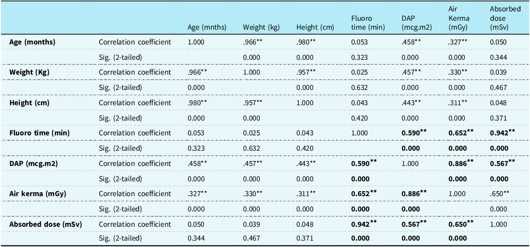
There were no significant differences in the parameters when compared between the two categories—diagnostics and interventional procedures. Therefore, the data for the whole cohort were taken together to further analyse by patient age, weight, fluoroscopy time, dose-area product, air kerma, and absorbed radiation dose. There was very strong correlation of fluoroscopy time with the absorbed radiation dose (Rho = 0.942, p < 0.05) of the entire cohort (Figure 2). Fluoroscopy time showed a fair correlation with air kerma (Rho = 0.65, p < 0.05) and dose-area product (Rho = 0.59). Absorbed radiation dose showed fair correlation with dose-area product (Rho = 0.56, p < 0.05) (Figure 3) and air kerma (Figure 4). There was a modest correlation of dose-area product with weight (Rho = 0.457). Fluoroscopy time correlated poorly with weight (Rho = 0.33), whereas absorbed dose did not have any correlation with weight.
Showing correlation of absorbed dose with fluoro time.

Showing correlation of absorbed dose with DAP.

Showing correlation of absorbed dose with air kerma.

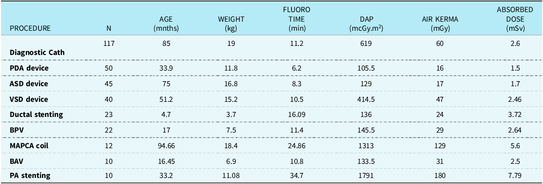
The differences in radiation exposure parameters measured across procedure types were significant (p = 0.0001), as shown in Table 1. The highest median dose-area product was recorded for posteroanterior stenting, 1791 mcGy.m2, followed by Major Aorto Pulmonary Collaterals (MAPCA) coil embolisation with dose-area product of 1313 mcGy.m2. Least median dose-area product was for balloon atrial septostomy (47 mcGy.m2), followed by Coarctation Of Aorta balloon plasty (127 mcGy.m2). The median fluoroscopy time was highest for posteroanterior stenting (34 minutes) and least for PDA device closure (6 minutes). The median value of air kerma did not increase linearly with the complexity of the procedure. It was maximum for pulmonary vein dilatation, branch posteroanterior stenting, and pulmonary valve perforation i.e. 134 mGy, 180 mGy, and 226 mGy, respectively.
Showing the interpersonal correlation of the radiation characteristics in the form of fluoro time, DAP value, air kerma, total absorbed dose, and their correlation with the demographic variables i.e. age, weight, and height for all the procedures

Age, weight, and height showed no correlation with either the fluoro time or the absorbed dose. (Rho<0.1and had no statistical significance (p > 0.05)).
There was very strong correlation of Fluoro time with the absorbed radiation dose (Rho = 0.942, p < 0.05). Fluoro time showed a fair correlation with air kerma (Rho = 0.65, p < 0.05) and DAP (Rho = 0.59, p < 0.05). Absorbed radiation dose showed a fair correlation with DAP ( Rh0 = 0.56, p < 0.05) and air kerma (Rho = 0.65, p < 0.05).
The overall median absorbed dose measured directly using a Bhabha Atomic Research Centre dosimeter was 2.4 mSv. The diagnostic procedures had a median absorbed dose of 2.6 mSv, and the intervention group had a median absorbed dose of 2.3 mSv. The highest median absorbed radiation doses were seen for pulmonary vein dilatation, pulmonary valve perforation, and branch posteroanterior stenting, measuring 18 mSv, 12.9 mSv, and 7.7 mSv, respectively. Only one procedure recorded an absorbed dose of >20mSv ( 27.9 mSv) in a 24-month-old patient, where Left Pulmonary Artery (LPA) stenting was done for post-operative LPA stenosis after total correction for Tetralogy of Fallot. These were the rare procedures done. The median absorbed dose for Atrial septal defect (ASD), PDA, and coarct balloon dilatation were 1.7 mSv, 1.5 mSv, and 1.9 mSv, respectively, which were amongst the least incurred in our study.
Discussion
This is the first study from India to report radiation exposure data for common congenital cardiac interventions in children. Ours is a tertiary centre with a well-equipped adult and a dedicated paediatric cath lab. Around 550 interventions per year were done in the preceding two years.
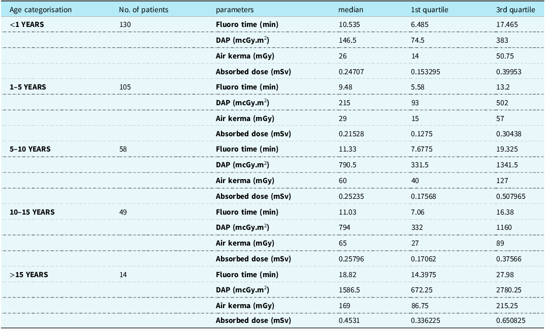
Based on our study, we have seen no significant difference between diagnostic and interventional catheterisation procedures. As rightfully pointed out by Ghelani et al. and also established in our present study, fluoroscopy time alone cannot be the only metric to monitor radiation dose. Reference Ghelani7 Therefore, we propose median radiation dose values as local dose reference levels or baseline for both diagnostic and interventional paediatric cardiac catheterisation processes combined. They were further stratified according to age to account for the wide range of patient sizes. The age stratification was done following previous studies of Rassow et al. and few others. Reference Rassow, Schmaltz, Hentrich and Streffer4,Reference Ghelani7,Reference McFadden, Hughes and D’Helft8 Various groups have reported dose reference levels using different methods of calculation. Closest to our study was the study performed by McFadden et al., where 354 paediatric patients <16 years of age reporting a median dose-area product of 288.5 cGycm2 (10–7961 cGycm2) and median fluoroscopy time of 8.3 min (1.0–75.1 min), respectively, and the mean dose-area product doses according to age stratification as local dose reference levels. Reference McFadden, Hughes and D’Helft8 Our results are also compared with those of previous studies by Rowley et al., who calculated a mean dose-area product of 489 cGycm2. Reference Rowley9 Other studies done by Onnasch et al. and more recently by Manica et al. proposed dose reference levels calculated by the patient body weight (dose-area product/Body Weight (BW)). Reference Onnasch, Schroder, Fischer and Kramer10,Reference João Luiz, Oliveira Duarte and Ribeiro11 Whereas studies done by Bacher et al. and Rassow et al. have calculated effective doses using the Monte Carlo simulation and maximum skin entrance dose using thermoluminescence dosimeters. Reference Rassow, Schmaltz, Hentrich and Streffer4,Reference Bacher, Bogaert, Lapere, De Wolf and Thierens12 El Sayed et al. calculated effective dose from dose-area product/BW using an average conversion factor. Reference El Sayed, Roushdy, El Farghaly and El Sherbini13 Even though the aim of all these studies is the same, i.e. to establish a benchmark which can be used for further optimisation and ultimately decrease patient exposure dose, the standards reported are not the same and therefore a direct comparison cannot be made.
Absorbed dose
The overall median absorbed dose measured directly using a Bhabha Atomic Research Centre dosimeter was 2.4 mSv. The diagnostic procedures had a median absorbed dose of 2.6 mSv, whereas the intervention group had a median absorbed dose of 2.3 mSv, respectively.
This is neither the effective dose nor the peak skin dose; however, this acts as an external/secondary reference or monitoring device independent of the equipment used for the procedure. These dosimeters give real-time values, unlike thermoluminescence dosimeters, which can only be read after the procedure. They are easy to use and easy to carry. Unlike thermoluminescence dosimeters in earlier studies Reference Bacher, Bogaert, Lapere, De Wolf and Thierens12 which were placed at the point of entry of the radiation, we have placed them on the anterior chest wall. Since the equipment used here had an under-couch tube with an over-couch image intensifier in the posteroanterior position, trying to put the dosimeter at the point of entry would have proven both cumbersome and counterproductive in our case. As shown in Table 1, the absorbed radiation measurements obtained from the Bhabha Atomic Research Centre dosimeter had a strong to fair correlation with the other radiation parameters measured through the equipment. Therefore, we believe that the absorbed radiation, while not a direct measure of the tissue effective dose, may help to monitor radiation exposure in real time.
Correlation
According to Walsh, catheterisation procedures follow a common method. Reference Walsh14 The majority of procedures start with percutaneous femoral access, followed by angiograms of the relevant vessels/area. Balloons are guided to and dilated at the area of a stenosis. Devices, for example, stents or occluders, can be guided and deployed at the site of pathology. It is anticipated that similar examinations would incur similar fluoroscopy times and dose-area product readings.
But, in this current study, we recorded a wide range of dose-area product readings across the range of examinations due to the increased complexity and longer fluoroscopy time required in complex cases. We classified the procedures into the CRISP stratification system and found that there were significant differences in all measures of radiation exposure across procedure types (p = 0.0001). Reference Nykanen, Forbes and Du15 The more complex procedures in the current study had a significant impact on the associated dose (p = 0.003). Reference McFadden, Hughes and D’Helft8
For diagnostic procedures, absorbed dose had strong correlation with fluoroscopy time (Rho = 0.979), fair correlation withair kerma (Rho = 0.62), and average correlation with dose-area product (Rho = 0.596). All were statistically significant. (p < 0.05). Patient’s age, weight, total fluoroscopy time, and the proportion of the overall exposure derived from digital acquisition were all found to be significant independent predictors of radiation exposure. Even when fluoroscopy times are comparable, air kerma and dose-area product levels can be quite different. This reinforces the notion that fluoroscopy time alone is not sufficient to estimate radiation exposure.
A recent study by Finke et al. compared radiation parameters from their team with a previously published successful radiation reduction programme. They showed that through the application of simple radiation exposure reduction steps and taking advantage of the advanced radiation reduction technologies, they could achieve a significant radiation reduction as measured by dose-area product, dose-area product/kg, and Air Kerma. Reference Finke, Mainzer and Yitzhak16 It is important to note that reduction in radiation exposure can be accomplished by using the lowest possible fluoroscopy pulse rate, decreasing cine acquisitions, and using storing fluoroscopy images or other modalities such as ultrasound and magnetic resonance. This can further be enabled by the technological advancements of the equipment. Awareness of the team and their commitment towards reducing radiation exposure also contribute greatly.
Limitations
One limitation of this study is the lack of effective radiation dose data. While effective dose is calculated based on phantom modelling using the Monte Carlo method or restricted number of conversion factors without taking into account the real exposure settings and geometry. However, it is of prime importance as it helps to assess the potential risk for stochastic effects such as cancer and leukaemia. Further investigations of organ-specific dosimetry are necessary, and the risks associated with cardiac catheterisation and interventional procedures to each organ should be assessed. There is a complete lack of standardised parameters for paediatric catheterisation procedures, and ours is the first single-centre data reported from India. As already discussed above, various studies have shown variation in the reported parameters based on different centres, instruments, or skills of the personnel. The reference levels reported should best be used as benchmarks for technique optimisation rather than as guidelines. CT imaging radiation doses may also be looked into. Diagnostic procedures were not subdivided into various groups but may be done in future studies.
Conclusion
Direct comparison with previous studies cannot be made as many different variables exist, such as procedure complexity, which is affected by underlying pathology as well as the type and quality of previously obtained imaging. Varying patient age and size, equipment specifications, and performer skills add to this challenge. Patients’ age, weight, total fluoroscopy time, and the proportion of the overall exposure derived from digital acquisition were all found to be significant independent predictors of radiation exposure. While we instinctively monitor how “long” the exposure was during the procedure but while monitoring exposure or establishing a baseline, it is important that all parameters are taken into account. We recommend the reported local dose reference levels to be used as benchmarks for further technique optimisation and reduction of radiation exposure rather than as guidelines. While safety measures have been set by the Atomic Energy Regulatory Board, no reference doses or benchmarks have been established in the Indian cohort for paediatric patients undergoing cardiac catheterisation. Therefore, we look forward to conducting multi-centre studies from India for establishing a more standardised radiation reduction programme.
Showing the radiation characteristics in the form of fluoro time (median), DAP value (median), air kerma (median), total absorbed dose (median), for various procedures are shown in Table 2 and demographic profile for the study group

The median radiation doses were stratified according to age in groups of 5 years each as shown in Table 3

Acknowledgements
None.
Financial support
This research received no specific grant from any funding agency, commercial, or not-for-profit sectors.
Ethical standard
The authors assert that all procedures contributing to this work comply with the ethical standards of the relevant national guidelines on ductal stenting and with the Helsinki Declaration of 1975, as revised in 2008, and have been approved by the institutional ethics committees NSH, Howrah.