Introduction
Felids (Felidae) are a biodiverse group of animals that are mostly highly endangered. All Felidae species, excluding the domestic cat, are threatened with extinction in their natural habitat.
All over the world, endangered species conservation programmes are being supported with the help of assisted reproductive techniques (ART). Domestic cats (Felis catus) are frequently used for research as a model for other felid species threatened with extinction. Many assisted reproduction methods are tested using the domestic cat model (Felis catus) as the most available source of research material. The biotechnological methods of assisted reproduction in felids have been developed based on research into fertilization mechanisms and development of embryos in vitro. Cat embryos can be obtained in vitro by several methods: in vitro fertilization (IVF, ICSI) (Goodrowe et al., Reference Goodrowe, Wall, O'Brien, Schmidt and Wildt1988; Johnston et al., Reference Johnston, O'Brien and Wildt1989; Jewgenow et al., Reference Jewgenow, Blottner, Lengwinat and Meyer1997; Pope et al., Reference Pope, Johnson, McRae, Keller and Dresser1998; Ringleb et al., Reference Ringleb, Rohleder and Jewgenow2004), cloning (Shin et al., Reference Shin, Kraemer, Pryor, Liu, Rugila, Howe, Buck, Murphy, Lyons and Westhusin2002; Gómez et al., Reference Gómez, Pope, Giraldo, Lyons, Harris, King, Cole, Godke and Dresser2004) or artificial activation of oocytes (Grabiec et al., Reference Grabiec, Max and Tischner2007; Rascado et al., Reference Rascado, Martins, Minto, de Sá Lorena and Landim-Alvarenga2010). In all of these methods, similar to IVF, activation of the oocyte is essential for triggering cleavage to form embryos. For this, the crucial mechanism that is common for most species is a calcium signal that simulates early symptoms of oocyte activation such as exocytosis of cortical granules and unblocking of meiosis. Later stages of the activation process include second polar body extrusion, pronucleus formation, DNA synthesis initiation and triggering embryonic cleavage (Cuellar, Reference Cuellar1977). During in vivo fertilization, the spermatozoon provides oocyte activating factor. However, artificial activation in vitro can be initiated without spermatozoa by mechanical, physical or chemical stimulation of oocytes (Cuellar, Reference Cuellar1977).
In felids, oocytes may be activated within the ovary (natural activation) as a result of changes in LH levels during copulation (Pushett et al., Reference Pushett, Gunn and Trounson1997). However, during in vitro maturation (IVM), oocytes may be spontaneously activated when simply exposed to in vitro culture conditions (Pincus & Enzmann, Reference Pincus and Enzmann1935; Keefer & Schuetz, Reference Keefer and Schuetz1982; Lechniak et al., Reference Lechniak, Cieślak and Sosnowski1998).
The methods used for artificial activation of oocytes by mechanical, physical or chemical stimulation are not complicated, but the results depend on the choice of appropriate stimuli or a combination of stimuli for activation (Grabiec et al., Reference Grabiec, Max and Tischner2007; Rascado et al., Reference Rascado, Martins, Minto, de Sá Lorena and Landim-Alvarenga2010). Parthenogenetic embryos of mammals obtained as a result of artificial activation do not survive longer than half of the pregnancy period as a result of parental genomic imprinting, which contradicts the theory of both sexes providing genetic material of equal value (Barlow, Reference Barlow1995). It is assumed that differences in parental genomes arise during gametogenesis as specific DNA modifications in certain parts of the female and male parental chromosomes. This imprinting is thus a barrier to parthenogenetic reproduction of mammals (Barlow, Reference Barlow1995).
Throughout the last few years, the main focus of research was to improve in vitro techniques for the ICSI method and for cloning of domestic cats in order to apply the results to wild cats (Moro et al., Reference Moro, Sestelo and Salamone2014; Pope, Reference Pope2014; Fernandez-Gonzalez et al., Reference Fernandez-Gonzalez, Hribal, Stagegaard, Zahmel and Jewgenow2015).
During the ICSI procedure, a spermatozoon is injected directly into the oocyte cytoplasm, which creates the possibility of uncontrollable parthenogenetic cleavages due to the mechanical stimulus (Pope et al., Reference Pope, Johnson, McRae, Keller and Dresser1998). Therefore, it seems necessary to calculate the risks involved in artificial oocyte activation caused by sperm injection during ICSI.
Somatic cloning is one of the most dynamically developing techniques of assisted reproduction, bringing new hope to endangered species conservation activities (Shin et al., Reference Shin, Kraemer, Pryor, Liu, Rugila, Howe, Buck, Murphy, Lyons and Westhusin2002; Gómez et al., Reference Gómez, Pope, Giraldo, Lyons, Harris, King, Cole, Godke and Dresser2004; Herrick et al., Reference Herrick, Bond, Magarey, Bateman, Krisher, Dunford and Swanson2007). To achieve successful cloning artificial oocyte activation is necessary. Therefore, optimization and development of the most efficient activation method should increase cloning efficiency.
The last decade has seen some reports of oocyte activation in domestic cats but they mostly referred to searching for effective artificial chemical methods for activation of reconstructed oocytes during the cloning procedure (Karja et al., Reference Karja, Otoi, Murakami, Wongsrikeao, Budiyanto, Fahrudin and Nagai2005; Grabiec et al., Reference Grabiec, Max and Tischner2007; Wang et al., Reference Wang, Swanson, Herrick, Lee and Machaty2009; Rascado et al. Reference Rascado, Martins, Minto, de Sá Lorena and Landim-Alvarenga2010). However, there have been no comprehensive, comparative studies on the parthenogenetic development of feline embryos after activation of oocytes and during consecutive stages of development.
With respect to the development of ART in domestic cats and the application of these techniques to conservation programmes for endangered wild cats, it appears crucial to define the factors that influence oocyte activation and normal development of embryos produced in vitro.
The aim of the study was to compare the development of cat oocytes and embryos after parthenogenetic stimulation using different methods.
Materials and Methods
Experiments were conducted according to guidelines approved by the Local Ethics Committees for Animal Experimentation in Wroclaw and Cracow, Poland.
Source of ovaries and oocyte collection
Ovaries were collected after ovariohysterectomy from 30 healthy adult domestic cats at a local veterinary clinics and a shelter for homeless animals in Cracow. The fertility history of these cats was not known. Ovaries were kept in Dulbecco's phosphate-buffered saline (DPBS) supplemented with 100 µg/ml streptomycin and 100 IU/ml penicillin at 4°C in a thermal box. Oocytes were obtained within 1–3 h of ovariohysterectomy.
Cumulus–oocyte complexes (COCs) were obtained by scarification in a washing medium (TCM199 with Earle's salts, HEPES-buffered and supplemented with 3 mg/ml BSA, 0.1 mg/ml cysteine, 0.25 mg/ml sodium pyruvate, 0.6 mg/ml sodium lactate, 0.15 mg/ml l-glutamine, and 0.055 mg/ml gentamycin).
2.2. Experimental groups
Three types of oocyte activation were analysed. The first one – spontaneous activation: natural (Group I) and activation during IVM (Group II). The second one – artificial activation: induced by chemical substances (Group III) and ‘sham injection’ by mechanical stimulation (Group V). These two types of activation produced parthenogenetic embryos. The third type of activation by spermatozoa during ICSI (Group IV) was analysed to demonstrate the development of embryos after fertilization.
In each group of our research we used random oocytes from many queens of different ages and different breeds to eliminate the individual predisposition.
2.3. Natural activation (Group I)
After collecting oocytes, the percentage of parthenogenetic embryos was recorded relative to the total number of oocytes. The parthenogenetic embryos reported in this group were considered as a group of oocytes that underwent natural activation within the ovaries before collection. Then, these parthenogenetic embryos were cultured in vitro to continue their development.
In vitro maturation (IVM) of oocytes and spontaneous activation (Group II)
Only oocytes with uniform and dark cytoplasm, surrounded by compact layers of cumulus cells, were selected for IVM. Selected COCs were placed into 400 µl of maturation medium, described above as washing medium, supplemented with 0.02 IU follicle stimulation hormone (FSH)/ml and 0.02 IU luteinizing hormone (LH)/ml under mineral oil and cultured for 24 h at 38.5°C and 5% CO2 in air. Afterwards, IVM cumulus cells were removed mechanically by pipetting.
The percentage of oocytes activated spontaneously during IVM was calculated relative to the total number of oocytes maturing in vitro. Parthenogenetic embryos were moved to in vitro culture medium, while oocytes with only a first polar body were used for further procedures.
Chemical activation (Group III)
Oocytes with the first polar body were incubated for 5 min in 5 mM ionomycin in HEPES-buffered TCM199 with Hanks salts. After incubation, the oocytes were washed in TCM199 supplemented with 10% fetal bovine serum (FBS) and transferred to 2 mM 6-DMAP/ml TCM199 for 3 h of incubation. Then presumptive embryos were transferred to in vitro culture medium.
Intracytoplasmic sperm injection (Group IV)
All manipulations were performed using micromanipulators (Narishige, Japan) according to the protocol described by Hribal et al. (Reference Hribal, Braun, Ringleb and Jewgenow2012). Frozen/thawed semen was used for ICSI. Immediately before injection, 1 μl of sperm suspension was placed in 3 μl of Sp-TALP containing 10% PVP. Sperm injection was carried out in a 50 μl drop of HEPES-buffered TCM199 containing 10% FBS. A spermatozoon was injected into the oocyte cytoplasm using an injection pipette (inner diameter 5 µm) and holding pipette (inner diameter 30 µm). After micromanipulation oocytes were cultured in vitro.
Mechanical activation- ‘sham ICSI’ (Group V)
Mechanical activation was carried out by oocyte injection following the same method as for ICSI as described above but with a small volume of the ICSI medium without spermatozoon. Oocytes after injection were moved to in vitro culture medium.
Embryo culture
Presumptive parthenogenetic zygotes and embryos from each group were cultured in vitro in medium Ham's F10 (Modified Ham's F10), supplemented with 5% FCS, 1 mM sodium pyruvate, 1 mM l-glutamine, 0.1 mg/ml streptomycin and 100 IU/ml penicillin under mineral oil at 38.5°C in a 5% CO2 atmosphere for 7 days. Every second day, part of the culture medium was exchanged for fresh medium.
Evaluation of embryo development
Embryos from each group were examined under an inverted microscope every 24 h for up to 7 days to assess morphological changes occurring in the oocytes/embryos including cleavage or degeneration. Embryos showing arrested cleavage were fixed in 2% formalin for 15 min and subsequently the chromatin was stained with Hoechst 33342 (2 μg/ml solution, 10 min incubation) in order to assess the embryo development stage.
The success rate of parthenogenetic activation was defined by cleavage rate (the percentage of oocytes that became activated, determined by the presence of at least two blastomeres); morula rate (the percentage of all cleaved embryos that developed to the morula stage, determined by the presence of at least 16 blastomeres); and blastocyst rate (the percentage of all cleaved oocytes that developed into blastocysts, determined by the presence of a well developed blastocoel cavity and a distinct embryoblast).
Statistical analysis
Differences between groups in the mean percentage of each developmental stage were assessed by chi-squared test. For all analyses, P < 0.05 was considered significant.
Results
Natural activation (Group I)
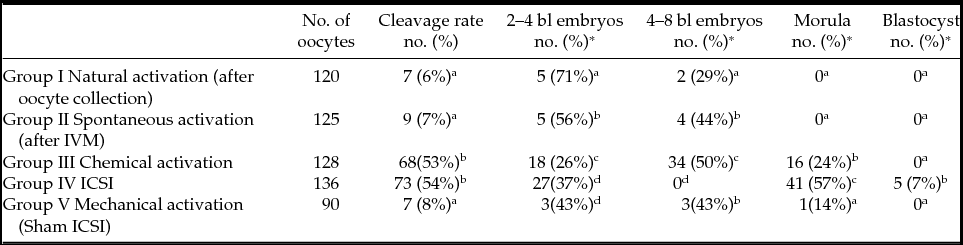
In order to determine the rate of natural activation, 120 oocytes were analysed directly after collection from the ovary. Of these, 7/120 (6%) parthenogenetic embryos were observed, mainly at the 2–4-blastomere stages (Table 1). These embryos were cultured in vitro after which only single cell divisions were observed. After Hoechst staining, a nucleus was confirmed in 7/120 embryos, which equalled 6% of all oocytes collected, the remaining presumptive embryos were considered as degenerate oocytes with fragmentation of the cytoplasm.
Development of parthenogenetic embryos at each stage using various in vitro embryo production procedures

* Percentages shown are % of cleaved oocytes.
a,b,c,d Within a column, means without a common superscript differ (P < 0.05).
Spontaneous activation (Group II)
In this group, 125 in vitro maturing oocytes were analysed. After 24 h of IVM, parthenogenetic activation was observed in 9/125 oocytes (7%). These presumptive parthenogenetic embryos were transferred to in vitro culture and more cell divisions were noted compared with the natural parthenogenetic embryos in Group I. Some of the embryos morphologically resembled a morula. However, after staining, fewer nuclei were confirmed relative to the total numbers of visible blastomeres.
Chemical activation (Group III)
Here, 128 oocytes in metaphase II were parthenogenetically activated using of ionomycin and 6-DMAP and 68/128 (53%) cleaved embryos were obtained. Following artificial chemical activation, morula stage embryos 16/68 (24%) were obtained after in vitro culture (IVC), however, none reached the blastocyst stage.
Intracytoplasmic sperm injection (Group IV)
The cleaved rate after ICSI (54% cleaved embryos) was not significantly different from the results in Group III using chemical activation (53% cleaved embryos), but many more cells at the morula stage were observed (57% vs. 24%). Only after ICSI were blastocysts obtained (5/73.7%) as a result of in vitro culture. Moreover, embryos after ICSI were of the best morphological quality with only a minor level of fragmentation among them.
Mechanical activation - ‘sham ICSI’ (Group V)
In total, 90 oocytes in metaphase II were injected with an injection pipette without spermatozoon (analogously to ICSI procedure) in order to trigger mechanical activation and provide a control for the ‘true’ ICSI. This procedure produced 7/90 (8%) cleaved embryos with one (14%) embryo reaching the morula stage. After staining, a high level of cytoplasmic fragmentation was observed. Mechanical activation proved to be significantly less efficient than chemical activation.
Discussion
Assisted reproduction techniques (ART) were found to be useful tools in medicine and veterinary practice for supporting production of pregnancy. In humans, these methods appear in some cases to be the only opportunity to have offspring and, in the case of animal species, in immediate danger of extinction they may serve to augment conservation programmes. Wild felidae species are among the most endangered populations of animals all over the world. To introduce ART in wild cats, it is essential to carry out research using domestic cats as a model. This model is easily accessible due to thousands of gonadectomies performed in many countries every day in programmes for pet population control. Unfortunately, methods for in vitro fertilization (IVF, ICSI) in domestic cats are still far from being optimized and many discrepancies are reported in the literature concerning the effectiveness of these procedures (Pope et al., Reference Pope, Johnson, McRae, Keller and Dresser1998; Comizzoli et al., Reference Comizzoli, Wildt and Pukazhenthi2003, Reference Comizzoli, Wildt and Pukazhenthi2006; Merlo et al., Reference Merlo, Iacono, Regazzini and Zambelli2008; Thuwanut et al., Reference Thuwanut, Arya, Comizzoli and Chatdarong2015). In most studies, the fertilization rate that denotes the percentage of oocytes fertilized by spermatozoa during IVF or ICSI procedures ranges from 50 to 80%, depending on the precise method used for fertilization, culture conditions and whether spermatozoa used for the study were (fresh or cryopreserved) (Johnston et al., Reference Johnston, Donoghue, O'Brien and Wildt1991; Pope et al., Reference Pope, Keller and Dresser1993, Reference Pope, Schmid and Dresser1999; Waurich et al., Reference Waurich, Ringleb, Braun and Jewgenow2010). The best results with IVF or ICSI are reached using fresh semen and in vivo matured oocytes (Pope et al., Reference Pope, Johnson, McRae, Keller and Dresser1998; Herrick et al., Reference Herrick, Bond, Magarey, Bateman, Krisher, Dunford and Swanson2007; Vick et al., Reference Vick, Bateman, Lambo and Swanson2012). When cryopreserved spermatozoa and in vitro matured oocytes are used, the effectiveness of fertilization significantly decreases (Penfold et al., Reference Penfold, Jost, Evenson and Wildt2003; Waurich et al., Reference Waurich, Ringleb, Braun and Jewgenow2010; Zambelli et al., Reference Zambelli, Raccagni, Cunto, Andreani and Isani2010). In our study 53% embryos development was obtained using in vitro matured oocytes and cryopreserved spermatozoa and the ICSI procedure. Moreover, most cat embryos arrested at the morula stage, with only a few morulae forming blastocysts. With current ICSI techniques, a maximum of 20–40% of embryos reach the blastocyst stage, which is not a satisfactory result results with other species (Pope et al., Reference Pope, Johnson, McRae, Keller and Dresser1998; Comizzoli et al., Reference Comizzoli, Wildt and Pukazhenthi2003, Reference Comizzoli, Wildt and Pukazhenthi2006; Merlo et al., Reference Merlo, Iacono, Regazzini and Zambelli2008; Thuwanut et al., Reference Thuwanut, Arya, Comizzoli and Chatdarong2015).
The crucial determinant for increasing the efficiency of cat embryo in vitro production methods is a thorough recognition of all the factors that activate the oocyte at each stage of the procedures. During the research presented here, immediately after collection from the ovary natural activation was observed in 6% of oocytes, consistent with another reported study (7% cleaved embryos) (Pushett et al., Reference Pushett, Gunn and Trounson1997). The time and temperature of ovary storage directly prior to collection of oocytes may influence the proportion of activated oocytes within the ovary. In our study, the ovaries were stored in the same way as reported by other authors at 4°C (Goodrowe et al., Reference Goodrowe, Wall, O'Brien, Schmidt and Wildt1988; Johnston et al., Reference Johnston, O'Brien and Wildt1989; Jewgenow et al., Reference Jewgenow, Blottner, Lengwinat and Meyer1997; Pope et al., Reference Pope, Johnson, McRae, Keller and Dresser1998) and the storage time did not exceed 1 h.
Domestic cat oocytes are very sensitive to in vitro conditions and may be spontaneously activated during IVM (Murakami et al., Reference Murakami, Otoi, Karja, Ooka and Suzuki2002; Grabiec et al., Reference Grabiec, Max and Tischner2007; Nagano et al., Reference Nagano, Uchikura, Takahashi and Hishinuma2008; Rascado et al., Reference Rascado, Martins, Minto, de Sá Lorena and Landim-Alvarenga2010; Waurich et al., Reference Waurich, Ringleb, Braun and Jewgenow2010). In our study, 7% parthenogenetic embryos was obtained after in vitro maturation of oocytes, while others reported 2% (Grabiec et al., Reference Grabiec, Max and Tischner2007), 5% (Waurich et al., Reference Waurich, Ringleb, Braun and Jewgenow2010) parthenogenetic embryos. In contrast, high rates (23–31%) of spontaneous parthenogenetic cleavage of cat oocytes after 24–30 h of IVM were reported by others (Murakami et al., Reference Murakami, Otoi, Karja, Ooka and Suzuki2002; Nagano et al., Reference Nagano, Uchikura, Takahashi and Hishinuma2008). Such a large discrepancy in results is most likely related to the different types of media used for in vitro maturation of oocytes.
Spontaneous oocyte activation in vitro is also reported in other species: in rabbits 21% of oocytes undergo spontaneous activation, in humans 12%, and in cattle 9% (Keefer & Schuetz, Reference Keefer and Schuetz1982; Lechniak et al., Reference Lechniak, Cieślak and Sosnowski1998; Combelles et al., Reference Combelles, Cekleniak, Racowsky and Albertini2002).
Additionally, numerous studies have shown that the number of parthenogenetic embryos increases in relation to increased time allowed for IVM (Keefer & Schuetz, Reference Keefer and Schuetz1982; Combelles et al., Reference Combelles, Cekleniak, Racowsky and Albertini2002; Bogliolo et al., Reference Bogliolo, Leoni, Ledda, Zedda, Bonelli, Madau, Santucciu, Naitana and Pau2004). Cattle embryos were cytogenetically analysed after spontaneous activation in vitro and 62% were reported to have the regular diploid number of chromosomes, while 25% were haploid, 4.2% triploid and 8.3% tetraploid (Lechniak et al., Reference Lechniak, Cieślak and Sosnowski1998).
For some species (sheep, mice, cattle), a single mechanical stimulus from injection is sufficient to trigger oocyte division (Gómez et al., Reference Gómez, Catt, Evans and Maxwell1998; Chung et al., Reference Chung, Keefer and Downey2000; Suttner et al., Reference Suttner, Zakhartchenko, Stojkovic, Muller, Alberio, Medjugorac, Brem, Wolf and Stojkovic2000). This is an undesirable phenomenon occurring during the ICSI procedure because there is a risk of triggering parthenogenetic development during injection of the oocyte with a pipette. Therefore, it is highly advisable during ICSI to prepare a control group (‘sham ICSI’) in which oocytes are injected with an empty pipette. In our research 8% of parthenogenetic embryos were obtained after injection without spermatozoa. Waurich et al. (Reference Waurich, Ringleb, Braun and Jewgenow2010) did not report formation of any embryos after ‘sham ICSI’, Penfold et al. (Reference Penfold, Jost, Evenson and Wildt2003) noted 7%, whereas Pope et al. (Reference Pope, Johnson, McRae, Keller and Dresser1998) reported more than 30% (Pope et al., Reference Pope, Johnson, McRae, Keller and Dresser1998; Penfold et al., Reference Penfold, Jost, Evenson and Wildt2003; Waurich et al., Reference Waurich, Ringleb, Braun and Jewgenow2010).
The discrepancies among these results may be related to the choice of medium or in vitro culture conditions at all stages of the ICSI procedure. Cleavage of embryos after sham intracytoplasmic sperm injection (ICSI) demonstrates the influence of puncturing oocytes on the activation of embryonic development. However, only a combination of mechanical puncture and interactions between spermatozoa and oocytes during ICSI supports development to the blastocyst stage.
The presence of parthenogenetic embryos among the total number of embryos after ICSI may lead to overestimation to the results with this technique.
Along with the development of cloning techniques, research into optimized methods of artificial activation of oocytes by chemical or electrical stimulus has gained importance. In the case of endangered felids, the availability of oocytes is very limited and so oocytes of domestic cats have been used as suitable recipients of nuclei derived from somatic cells of wild felids in nuclear transfer procedures (Gómez et al., Reference Gómez, Pope, Giraldo, Lyons, Harris, King, Cole, Godke and Dresser2004).
Several methods are able to induce parthenogenetic activation of oocytes, but a common, universal method or activation agent suitable for all species has not been developed because the process is highly species specific. Therefore, the activation step needs to be optimized for each species. Consequently these requires diverse methods for each species, with different type of activating stimulus or combination of stimuli, their intensity and duration of exposure. In some species, such as sheep or mice, there are methods that enable the development of parthenogenetic foetuses (Loi et al., Reference Loi, Branca, Dattena, Gallus, Ledda, Naitana and Cappai1996). In the case of cats, however, protocols for chemical parthenogenetic activation are not well documented.
In our research, we used a combination of ionomycin (an activator of intracellular calcium reserve) and 6-DMAP [an M-Phase promoting factor (MPF) kinase inhibitor]. We obtained 68 (53%) dividing embryos with 16 (24%) developing to the morula stage. Other authors tested a variety of chemical activators (ethanol, cytochalasin, thimerosal, strontium ions, ionomycin, calcium ionophore, cycloheximide and 6-DMAP) (Bogliolo et al., Reference Bogliolo, Leoni, Ledda, Zedda, Bonelli, Madau, Santucciu, Naitana and Pau2004; Gómez et al., Reference Gómez, Pope, Giraldo, Lyons, Harris, King, Cole, Godke and Dresser2004; Karja et al., Reference Karja, Otoi, Murakami, Wongsrikeao, Budiyanto, Fahrudin and Nagai2005; Grabiec et al., Reference Grabiec, Max and Tischner2007). With cat oocytes, Grabiec et al. (Reference Grabiec, Max and Tischner2007) obtained 18% cleavage of oocytes using a combination of ethanol and cycloheximide, Kitiyanant et al. (Reference Kitiyanant, Saikhun and Pavasuthipaisit2003) reported a 70% rate of development to the 2-cell stage and 40% morulae using 6-DMAP and a calcium ionophore, while Rascado et al. (Reference Rascado, Martins, Minto, de Sá Lorena and Landim-Alvarenga2010) obtained 47% cleaved oocytes and 22% morulae using calcium ionophore and roscovitine (Kitiyanant et al., Reference Kitiyanant, Saikhun and Pavasuthipaisit2003; Gómez et al., Reference Gómez, Pope, Giraldo, Lyons, Harris, King, Cole, Godke and Dresser2004; Grabiec et al., Reference Grabiec, Max and Tischner2007). Despite these promising results, there has been no success in obtaining parthenogenetic cat blastocysts. Problems with obtaining parthenogenetic cat embryos at advanced stages of development may be related to the phenomenon of developmental block observed in cats at the 5–8-cell stage (Hoffert et al., Reference Hoffert, Anderson, Wildt and Roth1997).
At this stage, most cat embryos arrest developmentally during in vitro culture to inefficient activation of their genome resulting from incomplete nuclear programming.
Intensification of research into activation mechanisms is crucial in the case of cloning procedures. There is a risk that embryos formed as a result of cloning arrest division when the artificial stimulation is too weak or undergo cytoplasmic fragmentation when it is too strong.
In conclusion, according to results presented here, parthenogenetic embryos might emerge et every step of in vitro embryo production (IVP) procedures. Therefore, it is advisable to maintain negative controls in parallel to the ICSI techniques, to avoid confounding results. Instead of focusing on the lack of competence for parthenogenetic embryos to attain advanced development they could well be used as a research model to study of the mechanisms involved in fertilization (Barlow, Reference Barlow1995; Kharche & Birade, Reference Kharche and Birade2013).
In vitro production of parthenogenetic embryos is an inexpensive and fast method. Thus, it enables experiments focused on the optimization of in vitro embryo culture conditions. This is especially important in the case of species with limitations on in vitro fertilization. Moreover, as parthenogenetic embryos almost always cease development because of improper genome activation, they provide a means to study the mechanism of this critically important process. Parthenogenetic embryos may be used to isolate a line of stem cells (Vrana et al,, Reference Vrana, Hipp, Gross, McCoo and Riddle2003).
Artificial activation also needs further study because it is an essential part of the cloning procedure. It appears that there is still a lack of comprehensive studies to validate the presence of parthenogenetic embryos at each stage of in vitro procedures and their influence on the final outcome of these procedures.
Acknowledgements
We acknowledge the financial support from the National Centre for Research and Development grant NCBiR PBS3/B8/16/2015.