Implications
The results of this study indicate that high-grain (HG) diets affect the rumen bacterial community structure and rumen metabolism, which in turn caused systemic effects, from the rumen to the liver and then to the peripheral circulation. The results in this study may provide a basis for further research on the metabolic process in the rumen–liver–peripheral circulation and the prevention of negative effects caused by HG feeding, which may reduce the economic losses caused by HG feeding.
Introduction
Feeding excessive cereals to ruminants is a common strategy to achieve high yields and improve cost efficiency. However, this feeding pattern also leads to a high incidence of metabolic disorders such as subacute ruminal acidosis, acute ruminal acidosis, fatty liver and laminitis (Plaizier et al., Reference Plaizier, Krause, Gozho and McBride2008; Dong et al., Reference Dong, Wang, Jia, Ni, Zhang, Zhuang, Shen and Zhao2013). The long-term use of HG diets has negative effects on ruminant health, production performance and economic benefits (Enemark, Reference Enemark2009; Kleen and Cannizzo, Reference Kleen and Cannizzo2012) and hence has received increasing attention in recent years.
As the main fermentation site of feed and an important organ for digestion and nutrition, the rumen has been extensively studied in terms of changes to its microbial composition and metabolites. The reshaped rumen microbial structure induced by HG diets leads to the accumulation of volatile fatty acids (VFAs), followed by a reduction in rumen pH, which is the generally accepted microbial mechanism of occurrence of subacute ruminal acidosis (Enemark, Reference Enemark2009). Two studies using the metabolomics approach demonstrated that rumen metabolism changes during adaptation to HG diets, and amino acids (e.g. lysine, leucine, valine), VFAs (butyrate and propionate), and toxic and inflammatory compounds (putrescine, methylamines, ethanolamine and lipopolysaccharide) are the main factors whose concentrations are changed (Ametaj et al., Reference Ametaj, Zebeli, Saleem, Psychogios, Lewis, Dunn, Xia and Wishart2010; Saleem et al., Reference Saleem, Ametaj, Bouatra, Mandal, Zebeli, Dunn and Wishart2012). However, studies simultaneously investigating both the rumen microbiota and metabolites as well as their intrinsic association in ruminants fed HG diets were limited. This simultaneous measurement would provide a more comprehensive understanding of rumen functions in response to HG diets.
The liver, a central tissue for metabolism, is involved in digestion, absorption, excretion, detoxification, material transportation, energy metabolism and immunity. Nutrients absorbed from the digestive tract in ruminants pass through the liver into the circulatory system (Jiang et al., Reference Jiang, Ni, Zhang, Zhang and Shen2014). Nutrients arrive at different tissue via peripheral circulation, for example, amino acids transported to the mammary glands can be utilized for protein synthesis (Remond et al., Reference Remond, Bernard and Poncet2000). Therefore, the metabolic process of transporting nutrients from the rumen to the liver and then to the peripheral circulation is closely related to ruminant performance. To our knowledge, limited research has been conducted on how HG diets induce changes in the metabolic profile of the entire axis (rumen–liver–peripheral circulation).
In this study, we hypothesized that HG diets may reshape the rumen microbiota and alter systemic metabolism in the rumen, liver and serum (peripheral blood) of goats. The objective of our study was to explore the effects of HG diets on rumen microbiota and the metabolic profiles in the rumen, liver and serum of goats by using the gas chromatography (GC)/MS-based metabolomics approach. Our findings may provide more information on how HG diets affect host health and production performance from a metabolic viewpoint.
Material and methods
Animals, diets and sampling
Data for rumen VFAs and lipopolysaccharides had been reported previously (Table S1, Liu et al., Reference Liu, Xu, Liu, Zhu and Mao2013). In the present study, rumen-related basic parameters were integrated with metabolomics data for an in-depth analysis. As described previously (Liu et al., Reference Liu, Xu, Liu, Zhu and Mao2013), 10 rumen-cannulated Boer crossbred male goats were randomly divided into two different groups according to a completely randomized design, one group received a diet containing 0% grain (hay), while the other group received a diet containing 65% grain (HG). The detailed ingredients and nutrient compositions of these two diets are shown in Table S2. To allow better adaptation to the diet, the proportion of grain in the HG diets was incrementally increased over 5 days. Each goat was fed two equal portions (750 g/dry matter (DM) per day) at 0830 and 1630 h every day for 7 weeks. There was no significant difference in DM intake or BW gain between these groups during the experimental period, and has been reported previously (Liu et al., Reference Liu, Xu, Zhu and Mao2014).
On day 50, 4 to 5 h after the morning feeding, jugular vein blood was collected, the goats were slaughtered and the ruminal digesta and liver tissues were immediately collected. The blood was centrifuged at 1500 rpm (4°C, 10 min), and the serum (the supernatants) was transferred and kept at −80°C until metabolomics determination. The rumen digesta of goat were mixed and homogenized, and the homogenized digesta were collected for DNA extraction. The harvested rumen fluid samples were kept at −80°C until the metabolome analysis. Similarly, the liver tissues were stored in liquid nitrogen until metabolome analysis.
Microbial DNA isolation, pyrosequencing and sequence analysis
After thawing, 1-ml sample was used for DNA isolation. The bacterial cells were first mechanically broken using a mini-bead beater (Biospec Products, Bartlesville, OK, USA), and DNA was extracted using phenol-chloroform reagent. A NanoDrop spectrophotometer (Nyxor Biotech, Paris, France) was used to measure the concentration and purity of the extracted DNA. The amplification of 16S ribosomal RNA V1 to V3 regions was conducted using the universal primers (8F: 5′-AGAGTTTGATCCTGGCTCAG-3′, 533R: 5′-TTACCGCGGCTGCTGGCAC-3′). The experimental program of the PCR consisted of an initial denaturation at 95°C (2 min); 25 cycles of 95°C (30 s), 55°C (30 s) and 72°C (30 s); 72°C (5 min); finally kept at 10°C until halted by user. The amplicons were sequenced by Roche 454 FLX pyrosequencer (Roche, Mannheim, Germany).
Following sequencing, raw sequences were trimmed, filtered and classified using MOTHUR (v 1.29.0). To ensure that any subsequent analysis was highly accurate, sequences shorter than 200 bp, or with a homopolymer longer than 8 bp, or with an average quality score lower than 35 or with ambiguous bases were discarded. The sequences finally harvested were aligned using the SILVA database, and the taxonomic analysis of representative operational taxonomic unit (OTU) sequences at 97% similarity was performed using the Ribosomal Database Project Bayesian Classifier. The bacterial richness (Chao 1) and diversity (the Simpson and Shannon index) index were calculated. The differences between two groups were measured using the principal coordinate analysis and the analysis of similarities (ANOSIM). Linear discriminant analysis effect size was conducted to determine the differentially abundant genera in rumen bacterial community.
In the present study, we used Phylogenetic Investigation of Communities by Reconstruction of Unobserved States (PICRUSt) in combination with Kyoto Encyclopedia of Genes and Genomes data base to predict the potential functions of rumen bacteria. The differences in predicted functions between two groups were determined using a principal component analysis and ANOSIM.
Gas chromatography/MS analysis and data processing
Serum, rumen fluid and liver samples were pre-treated, extracted and derivatizated according to the report of Mao et al Reference Mao, Huo and Zhu. (2016). In brief, 50 µl of liquid samples (or 30 mg liver samples) were added to 200 µl methanol (for liver samples, 900 µl) containing [13C2]-myristic acid (Sigma-Aldrich, St. Louis, MO, USA) as the internal standard. After mixtures were vortexed and sequentially treated as follows: first, the samples were kept at 4°C and maintained for 1 h, followed by being centrifuged at 20,000×g for 10 min. Subsequently, 100 µl of the supernatant was evaporated to dryness using a SpeedVac concentrator (Savant Instruments, Framingdale, NY, USA), followed by the process of methoxidation of trimethylsilylation. Finally, the solution was vortexed again and maintained for further analysis. Metabolites were identified using an Agilent 6890 GC system installed with a fused-silica capillary column (10 m×0.18 mm i.d.). Helium served as carrier gas, and its flow rate was set to 1.0 ml/min. After raw data were collected, the identification of metabolites is accomplished by comparing the retention index of chromatographic separation and mass spectrum with the reference standards and the data in the National Institute of Standards and Technology library 2.0 (Palisade Corp., Newfield, NY, USA). The acquired GC/MS data were further analyzed by the principal component analysis and partial least squares discriminant analysis using SIMCA-P+ 14.1 (Umetrics, Umea, Sweden). The metabolome view map of identified metabolites was conducted using MetaboAnalyst web server (http://www.metaboanalyst.ca).
Statistical analysis
Data related to metabolomics (rumen fluid, serum and liver) and the bacterial community (rumen) were statistically analyzed using SPSS 18.0 software (SPSS, Chicago, IL, USA). The effects of diet on metabolites and bacterial taxa were determined using the independent-samples t test. The statistical model used in the present study was as follows: Y = μ+s+e. where μ is the overall mean; s the effect of diet treatments; and e the residual error. Significant difference was declared when P < 0.05.
The metabolites in rumen fluid, liver and serum with variable importance projection (VIP) as harvested from the partial least squares discriminant analysis >1 and P < 0.05 were defined as significantly different metabolites. Correlations between rumen microbiota and metabolites or those among metabolites were assessed using Spearman’s correlation test using XLStat (Addinsoft, Paris, France), and metabolites with a |r|>0.7 and P < 0.05 were selected to construct the correlation network using the Gephi 0.8.2 beta software.
Results
The composition of rumen microbial communities
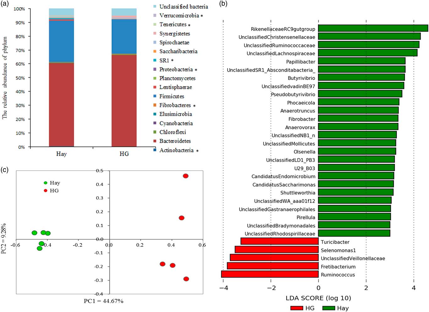
Results showed that the values of Chao 1 and Shannon index were decreased and the value of Simpson was increased (P <0.05) by the HG diets (Table 1). Within the detected 16 phyla, Bacteroidetes and Firmicutes were the dominant phyla, representing an average of 63.21% and 27.36%, respectively. The proportion of other phyla (Spirochaete, Synergistetes, SR1, etc.) was less than 1%. The changes at the phylum level after HG feeding are showed in Figure 1a. Feeding HG diets decreased (P < 0.05) the abundance of Actinobacteria, Fibrobacteres, Proteobacteria, SR1, Tenericutes and Verrucomicrobia.
Summary overview of the estimated operational taxonomic units (OTUs) through Chao 1 and Shannon index of ruminal bacterial community averaged across the goats between the Hay and the high-grain (HG) diets

Values are means ± SEM.
Effects of the high-grain (HG) diet on the composition of the rumen microbial communities in goats at different taxonomic levels. (a) The composition of the rumen microbial community at the phylum level; *P < 0.05. (b) Histogram of the linear discriminant analysis scores for genera differentially abundant between the hay and HG groups. Only the genera with a linear discriminant analysis significant threshold >2 are shown. (c) Principal coordinate (PC) analysis score plots between the hay and HG groups at the operational taxonomic unit level. LDA = Linear discriminant analysis.

At the genus level, the results of linear discriminant analysis effect size (Figure 1b) showed that 30 genera were identified as discriminative features among the samples taken from the hay and HG groups. Among them, five genera including Selenomonas, Turicibacter, Ruminococcus and unclassified Veillonellaceae were significantly enriched in the HG group. However, 25 genera (e.g. Rikenellaceae RC9 gut group, Pseudobutyrivibrio, Papillibacter) were significantly enriched in the hay group. In addition, the enriched taxa in the hay group also included 11 unclassified members, such as unclassified Christensenellaceae and unclassified Ruminococcaceae.
At the OTU level (Supplementary Figure S1), the shared OTU numbers (379 OTUs) accounted for 16.3% of the total OTUs, and the number of unique OTUs in the hay group was highest when compared with the HG group (1376 OTUs v. 565 OTUs, representing 59.3% and 24.4% of the total OTUs, respectively). The differences in rumen microbiota between the hay and HG groups were further analyzed using the principal coordinate analysis (Figure 1c), and the results showed that two sets of samples were separated clearly. In addition, this difference was confirmed by ANOSIM to have reached a statistically significance level (R = 1, P = 0.008).
Functional predictions of rumen microbiota
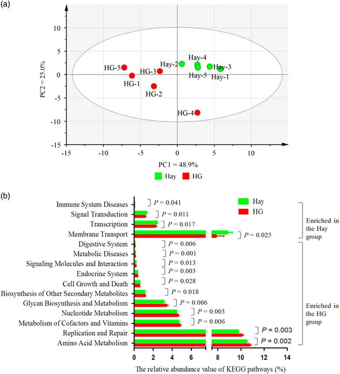
In the present study, the functional predictions were made using PICRUSt, and 39 metabolic pathways were identified. The principal component analysis indicated that samples from the hay and HG groups clustered differently (Figure 2a), and the results of ANOSIM showed that this difference was significant (R = 0.596, P = 0.016). Compared with the rumen microbiota from hay-fed goats, that from the HG-fed goats was predicted to have a lower (P <0.05) capability of influencing membrane transport, transcription, signal transduction and immune system diseases but a higher (P < 0.05) capability of influencing amino acid metabolism, replication and repair, metabolism of cofactors and vitamins, nucleotide metabolism, glycan biosynthesis and metabolism, biosynthesis of other secondary metabolites, cell growth and death, endocrine system, signaling molecules and interaction, metabolic diseases and digestive system (Figure 2b).
The metabolic functions of rumen microbiota in goats. (a) Principal component (PC) analysis of Kyoto Encyclopedia of Genes and Genomes (KEGG) pathways of rumen microbiota in hay and high-grain (HG) group. (b) Effects of the HG diet on the relative abundance of KEGG pathways of rumen microbiota in goats. Only the pathways whose relative abundance were >0.01% (in at least one group) and were significantly affected by dietary treatments were presented.

Correlations between disruption of the rumen microbiota and rumen metabolite profile
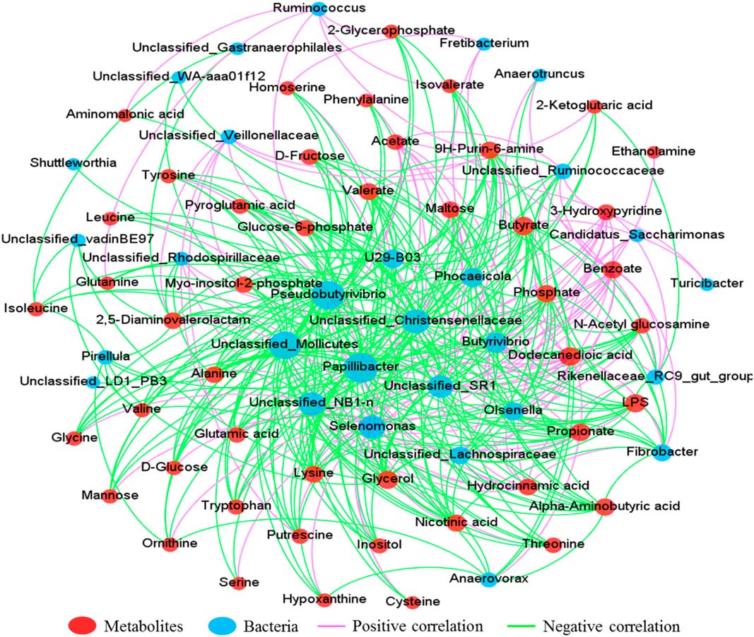
Correlation networks were examined using the data regarding disrupted genera obtained from the linear discriminant analysis effect size and significantly different metabolites (VIP >1 and P < 0.05) in the rumen of the goats fed hay and HG diets (Figure 3). The results indicated that the top 10 bacterial taxa that showed the strongest correlation linkages with metabolites were Papillibacter, Pseudobutyrivibrio, unclassified Mollicutes, unclassified Christensenellaceae, unclassified NB1-n, Selenomonas, unclassified SR1, Butyrivibrio, Phocaeicola and Olsenella. In addition, the top 10 metabolites that shared the strongest correlation linkages with bacterial taxa were dodecanedioic acid, butyrate, lipopolysaccharide, glycerol, propionate, benzoate, phosphate, α-aminobutyric acid, nicotinic acid and lysine. The correlation linkages between rumen bacteria and metabolites were extremely complex. For example, rumen lipopolysaccharides were positively correlated with Selenomonas and negatively correlated with Fibrobacter, Butyrivibrio, Papillibacter, Pseudobutyrivibrio, Phocaeicola, Rikenellaceae RC9 gut group, Olsenella, Anaerotruncus and seven unclassified bacterial taxa.
The correlation network between perturbed rumen microbiota and altered rumen metabolites in goats between the hay and the high-grain groups. The correlation coefficients that with a statistical P value <0.05 and the absolute value >0.7 were used to construct the network graph.

Common metabolites and metabolic pathways in the rumen fluid, liver and serum
In total, 84, 83 and 69 compounds were annotated in the rumen fluid, liver and serum by GC/MS, respectively, of which 34, 20 and 13 metabolites were unique to the analyzed component, respectively (Supplementary Figure S2). Thirty-nine common metabolites were identified in these three sites, while nine were common in rumen fluid and liver; two in the rumen fluid and serum; 15 in the liver and serum. The numbers of metabolites that were identified in any two sites are also illustrated in Supplementary Figure S2. Detailed information on all identified metabolites in these three sites is provided in Table S3.
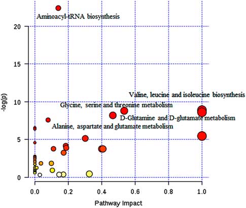
A correlation analysis of metabolites in these three sites was also conducted in the present study (Supplementary Figure S3). The number of correlation linkages between the rumen fluid and liver and between the liver and serum was 25 and 38, respectively. For example, the concentrations of cysteine, lysine, phenylalanine and pyroglutamic acid in liver were correlated with multiple rumen metabolites, and the concentrations of 3-hydroxybutyric acid, d-glucose, hypoxanthine and lactate in serum were correlated with multiple liver metabolites. The pathway analysis of common metabolites in these three sites (Figure 4) showed that 16 pathways were enriched (P < 0.05), while the top five pathways were aminoacyl-transfer RNA (tRNA) biosynthesis, valine, leucine and isoleucine biosynthesis, glycine, serine and threonine metabolism, d-glutamine and d-glutamate metabolism, and alanine, aspartate and glutamate metabolism. The metabolites in the relevant pathways were mainly involved in carbohydrate and amino acid metabolism.
Metabolome view map of the common metabolites identified in rumen fluid, liver and serum from goats (n = 10) fed the hay diet and the high-grain diet. The larger size indicates higher pathway enrichment, and the darker color indicates higher pathway impact values. tRNA = transfer RNA.

Differences in the metabolic profiles in the rumen fluid, liver and serum between the diets
The principal component analysis and the partial least squares discriminant analysis of metabolites in the rumen fluid, liver and serum revealed clearly different clusters in the two diet groups (Supplementary Figure S4). As shown in Table S4, of 47 significantly different metabolites (VIP >1 and P< 0.05) in the rumen fluid, 43 had higher concentrations in the HG group than the hay group, of which two (including putrescine and ethanolamine) were amines and 19 (including cysteine, glutamic acid, glutamine, etc.) were amino acids; only four (including acetate, benzoate, dodecanedioic acid and hydrocinnamic acid) had higher concentrations in the hay group. Among 10 significantly different metabolites in the liver including organic acids and amino acids, five (including cystine, glycine, glycerol, γ-aminobutyric acid (GABA) and pyroglutamic acid) were found at higher concentrations in the HG group, while the concentrations of another five (α-aminoisobutyric acid, lysine, valine, heptadecanoic acid and adenosine monophosphate) were higher in the hay group than in the HG group. Further, 10 among 17 significantly different serum metabolites were found at higher concentrations in the HG group than in the hay group.
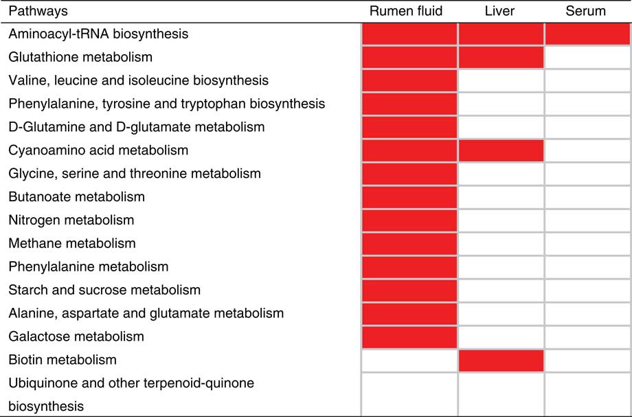
Since it is necessary to comprehensively evaluate how multiple pathways changed in response to HG diets and explore the key differential metabolites during the movement of metabolites from the rumen to the serum, an additional pathway analysis was conducted. The result showed that 14, 4 and 4 pathways were enriched (P <0.05) in the rumen fluid, liver and serum, respectively (Figure 5), of the HG diet-fed goats compared to the hay-fed goats. Notably, the only enriched metabolic pathway that simultaneously occurred in all three sites was that for aminoacyl-tRNA biosynthesis. The common metabolites of aminoacyl-tRNA biosynthesis in these three sites were glycine, lysine and valine, and these three amino acids were altered in all three sites (Supplementary Figure S5). In addition, the main altered metabolites in the rumen fluid of goats fed HG diets were amino acids; thus, the associations between amino acids involved in this common pathway and pathways of common metabolites in these three sites were manually linked in the present study (Supplementary Figure S6).
The enriched (P < 0.05) pathways of differential metabolites identified in the rumen fluid, liver and serum, respectively, of the high-grain-fed goats compared to the hay-fed goats. The red indicated that this pathway was enriched in this site of the high-grain-fed goats compared to the hay-fed goats. tRNA = transfer RNA.

Discussion
Consistent with many previous studies (Mao et al., Reference Mao, Zhang, Wang and Zhu2013; Plaizier et al., Reference Plaizier, Li, Tun and Khafipour2017), our study confirmed that HG diets have a potent ability to reshape the rumen microbial community, in terms of reducing community richness and diversity as well as altering the microbial composition at the levels of phylum, genus and OTU. More specifically, Firmicutes and Bacteroidetes were the dominant phyla, accounting for approximately 90% of the total bacterial population and neither was affected by dietary treatment, a finding consistent with the report of Hua et al. (Reference Hua, Tian, Tian, Cong, Luo, Geng, Tao, Ni and Zhao2017). In contrast, Mao et al. (Reference Mao, Zhang, Wang and Zhu2013) and Plaizier et al. (Reference Plaizier, Li, Tun and Khafipour2017) observed a decrease in Bacteroidetes, or also an increase in Firmicutes in the rumen after HG diets feeding. At the genus level, Selenomonas and Ruminococcus in the HG group, according to the results of the linear discriminant analysis effect size, while Butyrivibrio and Fibrobacter were enriched in the hay group. These changes were also reported by Petri et al. (Reference Petri, Forster, Yang, McKinnon and McAllister2012) and Fernando et al. (Reference Fernando, Purvis, Najar, Sukharnikov, Krehbiel, Nagaraja, Roe and Desilva2010) as being caused by the alterations in fermentable substrates in the rumen (increase in starches and reduction in fiber). It is worth noting that, besides these changes in classified bacteria, 12 unclassified taxa such as unclassified Christensenellaceae, unclassified Ruminococcaceae and unclassified Ruminococcaceae were also significantly affected by dietary treatments. The precise role of these unclassified bacteria is not clear, but they may be important for maintaining the micro-ecological stability and normal functions in the rumen. At the OTU level, the results of the Venn diagram and the principal coordinate analysis in the present study further indicated that HG diets altered the composition of the rumen microbiota. Because of the small number of experimental goats used in the present study, large variations in rumen microbiota were observed among individual animals, and this may have masked some differences caused by dietary treatment.
In response to the reduced community richness and diversity and the altered microbial composition, the potential functions of rumen microbiota in the HG diet-fed goats differed from those in the hay-fed goats, as indicated by the PICRUSt analysis. Our data showed that the rumen microbiota from HG-fed diets had an enhanced capacity to influence metabolism (amino acid metabolism, metabolism of cofactors and vitamins, nucleotide metabolism, etc.), replication and repair, and cell growth and death. These functional changes indicated that the rumen microbiota in the HG group was in a non-healthy state with relatively fluctuation, enhanced metabolism and faster turnover rates. This finding was consistent with the reduced abundance of rumen bacteria and an increase in some metabolites (such as lipopolysaccharides and amino acids) in HG-fed goats, compared to hay-fed goats.
It is well established that changes in the concentrations of various rumen metabolites can affect the rumen and host health (Ametaj et al., Reference Ametaj, Zebeli, Saleem, Psychogios, Lewis, Dunn, Xia and Wishart2010; Saleem et al., Reference Saleem, Ametaj, Bouatra, Mandal, Zebeli, Dunn and Wishart2012; Mao et al., Reference Mao, Huo and Zhu2016). In this study, a significant increase in the concentrations of putrescine and ethanolamine were observed in HG-fed goats. In ruminants, putrescine is produced by the decarboxylation of arginine/ornithine, and the activity of decarboxylase increases when the pH decreases to acidic level (Saleem et al., Reference Saleem, Ametaj, Bouatra, Mandal, Zebeli, Dunn and Wishart2012). Correspondingly, a significant correlation was observed between rumen putrescine and ornithine (P = 0.013, r = 0.818; data not shown), not between rumen putrescine and arginine (P = 0.905, r = 0.051; data not shown). Therefore, rumen putrescine in the current experimental conditions may only be produced from ornithine. The increased putrescine resulted in the generation of aldehyde and hydrogen peroxide (Yamashita et al., Reference Yamashita, Bowen, Burne and Kuramitsu1993), which are extremely toxic for cells (Ronchi et al., Reference Ronchi, Bernabucci, Lacetera and Nardone2000). Ethanolamine can be used by some pathogenic bacteria to accelerate their growth (Saleem et al., Reference Saleem, Ametaj, Bouatra, Mandal, Zebeli, Dunn and Wishart2012), so the higher ethanolamine in the rumen fluid may affect the microbiota composition, its role in the rumen needs further study. Metabolites (lipopolysaccharides, hypoxanthine, ornithine and alanine) are the degradation products of rumen bacteria, as many microbial species are not able to survive in a low pH and undergo widespread lysis (Saleem et al., Reference Saleem, Ametaj, Bouatra, Mandal, Zebeli, Dunn and Wishart2012). Our previous study showed that the low pH and high-lipopolysaccharide concentration in the rumen could initiate an inflammatory response and damage the rumen, whereby the lipopolysaccharide are translocated into the blood (Liu et al., Reference Liu, Xu, Liu, Zhu and Mao2013). Excess circulating lipopolysaccharide in turn activates a systemic inflammatory response, with elevated concentrations of acute phase proteins and pro-inflammatory cytokines in the blood (Dong et al., Reference Dong, Wang, Jia, Ni, Zhang, Zhuang, Shen and Zhao2013; Wang et al., Reference Wang, Jia, Yang, Liu, Yang, Li, Zhu, Wang and Han2017). Collectively, the increased concentrations of these metabolites in the rumen fluid after grain feeding have the potential to adversely affect rumen metabolism and the overall metabolic health of goats.
It is well accepted that rumen metabolites are associated with changes in bacterial flora. Therefore, a correlation network analysis was conducted in the present study to better explore these connections. Our data showed that these correlations were very complicated and inter-related. The ruminal bacterial taxa or metabolites that had the strongest correlation linkages may be the ones important for goats to adapt HG diets. For example, the genus Selenomonas is abundant in the rumen, and some species can utilize starch and sugars (Plaizier et al., Reference Plaizier, Li, Tun and Khafipour2017). Ruminal VFAs, especially butyrate, may stimulate the proliferation of rumen papillae to enhance the absorption of VFAs (Shen et al., Reference Shen, Seyfert, Löhrke, Schneider, Zitnan, Chudy, Kuhla, Hammon, Blum, Martens, Hagemeister and Voigt2004). Rumen lipopolysaccharide, as mentioned above, is considered important pro-inflammatory substances. Since these connections were derived from indirect evidence in the present study, more research is needed to further validate these relationships.
Recent research has demonstrated that cows were susceptible to oxidative stress after high-concentrate feeding (Guo et al., Reference Guo, Xu, Zou, Yang, Li and Cao2013), and the lipopolysaccharide derived from the digestive tract could provoke oxidative stress in the liver of ruminants fed HG diets (Abaker et al., Reference Abaker, Xu, Jin, Chang, Zhang and Shen2017). Our findings confirmed this conclusion from a metabolic viewpoint. In the present study, the HG diets increased cysteine, glycine, pyroglutamic acid and GABA concentrations but decreased lysine and valine concentrations in the liver. An optimal concentration of glutathione is required for the immune system to function well (Dröge and Breikreutz, Reference Dröge and Breitkreutz2000). Pyroglutamic acid (5-oxoproline) can serve as an intermediate in the glutathione biosynthesis pathway. A reduction in the hepatic glutathione concentration correlates with an increase in the concentrations of pyroglutamic acid (Geenen et al., Reference Geenen, Yates, Kenna, Bois, Wilson and Westerhoff2013); hence, the increased concentration of pyroglutamic acid in the livers of HG-fed goats suggests an inferior antioxidative capacity. Further, glutathione consists of glutamic acid, glycine and cysteine; thus, the increase in cystine (the precursor for cysteine) and glycine concentrations in the HG group indicated oxidative stress in the liver and that more glutathione was required for antioxidation. γ-Aminobutyric acid can play an important role in cell proliferation (Shilpa et al., Reference Shilpa, Pretty, Anitha and Paulose2013); when a hepatectomized rat liver was treated with GABA, cell signaling mechanisms during hepatic regeneration improved over those in a control group (Shilpa et al., Reference Shilpa, Pretty, Anitha and Paulose2013). In the present study, excess GABA may have been utilized to alleviate the liver injury through regeneration in the HG group. In terms of immunity, it has been suggested that the importance of branched-chain amino acids for immune cells is related to the synthesis of acute phase proteins, immunoglobulins, cytokines, etc. (Calder, Reference Calder2006). The lower valine concentration in the liver of HG-fed goats might have been an indication of an inferior immune capacity.
After metabolism in the liver, nutrients are transported via peripheral circulation to tissues or organs depending on physiological needs (Psychogios et al., Reference Psychogios, Hau, Peng, Guo, Mandal, Bouatra, Sinelnikov, Krishnamurthy, Eisner, Gautam, Young, Xia, Knox, Dong, Huang, Hollander, Pedersen, Smith, Bamforth, Greiner, McManus, Newman, Goodfriend and Wishart2011). The liver can redistribute nutrients, which mainly manifested as amino acids in the present study. Our results showed that HG diets elevated the concentrations of glycine, proline and tyrosine and decreased the concentrations of isoleucine, lysine, threonine and valine in the peripheral blood (serum). The changes of glycine, lysine and valine in serum were consistent with those in the liver, and more serum amino acids were altered, which may be highly indicative of liver damage after HG feeding in goats. Threonine could be used by hosts to produce acute phase proteins (Faure et al., Reference Faure, Choné, Mettraux, Godin, Béchereau, Vuichoud, Papet, Breuillé and Obled2007). Thus, the decreased threonine in serum may be caused by utilization for production of acute phase proteins, which is further linked with a decline in antibody production. Proline is important for protein synthesis and antioxidative reactions (Wu et al., Reference Wu, Bazer, Burghardt, Johnson, Kim, Knabe, Li, Li, McKnight, Satterfield and Spencer2011). The concentrations of serum proline increased noticeably in the HG-fed goats, which was consistent with observations in piglets after weaning (Sugiharto et al., Reference Sugiharto, Hedemann and Lauridsen2014); the upregulation of proline biosynthesis seemed to be a response to oxidative stress. Tyrosine is an essential component needed to produce thyroid hormones, melanine pigment, catecholamines epinephrine, norepinephrine and dopamine (Kim et al., Reference Kim, Mateo, Yin and Wu2007). The increase in serum tyrosine indicated that HG feeding may change physiological homeostasis of goats, although further experiments are needed to confirm this theory.
In the present study, 39 common metabolites were identified in the rumen fluid, liver and serum, mainly amino acids, sugars and organic acids. To further understand the association of these metabolites in the rumen fluid, liver and serum, a correlation analysis was conducted. The results indicated that these correlations in the rumen fluid and liver mainly involved fatty acids (stearic acid, oleic acid, palmitoleic acid, etc.) and amino acids (glutamine, tryptophan, lysine, etc.), and those in the liver and serum mainly involved amino acids (glycine, lysine, glutamine, etc.). Fatty acid metabolism (including degradation, de novo synthesis and biohydrogenation) and protein degradation mainly occurred in the rumen; although the main absorption site of these long-chain fatty acids and amino acids was the small intestine, the rumen fatty acids and amino acids profiles could reflect the absorption pattern in the small intestine, which may be the main cause of these correlations between the rumen and liver. The liver is an important hub of nutrient conversion, and the fate of amino acids that enter it were protein synthesis (such as albumin), oxidation for energy, gluconeogenesis and urea synthesis. The pathway analysis of the 39 common metabolites also showed that the top five were related to amino acid biosynthesis or metabolism. After liver metabolism, nutrients are redistributed and reach different tissues by peripheral circulation. Correspondingly, a high positive correlation was observed between sugars (such as fructose, glucose and glucose-6-phosphate), amino acids (such as glycine, lysine and ornithine), serum urea and liver nutrients.
The pathway analysis of significant differential metabolites in the rumen fluid, liver and serum showed that the only common pathway among these three sites was that for aminoacyl-tRNA biosynthesis, and the common metabolites in this pathway in the three sites were glycine, lysine and valine. Glycine is an important glycogenic amino acid and is a building block for glutathione, so an increase in its serum concentration may be needed to ensure proper immune functioning during HG diet feeding. A previous study reported that when dairy cows were fed high-concentrate diets, lysine and methionine were the first and second limiting amino acids, followed by branched amino acids (Li, Reference Li2005). Lysine and branched amino acids also show the greatest partitioning toward the mammary gland (Pacheco et al., Reference Pacheco, Tavendale, Reynolds, Barry, Lee and McNabb2003). The changes in lysine and valine exhibited similar trends (the only two amino acids had this trend) in our results: an increase in the rumen fluid, a reduction in the liver and a further reduction in the blood (HG group v. hay group). Therefore, we strongly suspected that the decline in the production performance of ruminants fed HG diets may be partly due to the limiting effects of lysine and valine. However, further research is required to verify this hypothesis.
In conclusion, pyrosequencing and GC/MS-based metabolomics were used to investigate the changes in rumen microbiota and metabolites in the rumen, liver and serum in response to HG diets in goats. The results demonstrated that HG diets altered rumen microbial composition and rumen metabolism, and these changes further (at least in part) lead to the metabolic alterations in the liver (mainly organic acids and amino acids) and peripheral circulation (mainly organic acids, amino acids and sugars). After HG feeding, the changes in the rumen–liver–serum were mainly related to amino acid metabolism, which may play a vital role in oxidative stress and the immune response induced by the HG diets. In addition, the decrease in lysine and valine concentrations in the liver and serum may be the key limiting factor affecting the overall performance of goats fed HG diets. These findings provided new insights into our understanding of the effects of HG feeding on host. Nonetheless, more research with large sample sizes is needed to validate the results of the present study.
Acknowledgements
This work received funding from the National Natural Science Foundation of China (No. 31572436), and Jiangsu Agriculture Science and Technology Innovation Fund (CX(18)2003).
Declaration of interest
No conflicts of interest are declared by all authors.
Ethics statement
The experiment proposals, the process of animal breeding and treatments were agreed by the Animal Care and Use Committee of Nanjing Agricultural University, in compliance with the requirements of the Regulations for the Administration of Affairs Concerning Experimental Animals (the State Science and Technology Commission of China, 1988).
Software and data repository resources
The pyrosequencing reads harvested in the present study were deposited at the Sequence Read Archive (SRA; http://www.ncbi.nlm.nih.gov/Traces/sra/) under accession no. SRP136963.
Supplementary materials
To view supplementary material for this article, please visit https://doi.org/10.1017/S1751731118003671