In countries with a low prevalence of tuberculosis (TB), healthcare workers (HCWs) represent a population at risk of tuberculosis. Routine screening for latent TB infection (LTBI) is therefore recommended. Until recently, only tuberculin skin testing (TST) could be used to assess the prevalence and incidence of LTBI. TST has high sensitivity but is limited by cross-reactivity with the bacille Calmette-Guerin (BCG), especially in countries that recommend BCG vaccination.
Recently, interferon-γ release assays (IGRAs) have been developed as alternatives to TST for diagnosing LTBI. These tests include the QuantiFERON TB Gold in Tube assay (QFT-G, Cellestis, Valencia, CA, USA) and T-SPOT TB (Oxford Immunotec Ltd, Abingdon, UK). The major advantages of IGRAs are: higher specificity than TST without being affected by the BCG vaccination, the absence of a boost response observed with TST, the absence of reader bias and requirement of only 1 HCW visit for testing.Reference Pai, Zwerling and Menzies 1
In countries with low TB incidence, such as France (7.6 of 100 000 inhabitants in 2012),Reference Antoine, Belghiti, Guthmann, Campese, Lévy-Bruhl and Che 2 IGRAs showed good correlation with TB exposure.Reference Pai, Zwerling and Menzies 1 However, the correlation of the IGRA with TST was fair or poor in cross-sectional studies, even in HCW populations with a small number of participants who had been vaccinated with BCG.Reference Zwerling, Benedetti and Cojocariu 3 In addition, serial testing with the IGRA showed variation in rates of conversion and reversion, thus bringing into question its usefulness for longitudinal HCW surveillance.Reference Dorman, Belknap and Graviss 4 , Reference Pai, Denkinger and Kik 5
Most longitudinal studies in HCWs have been single centered; they have included a limited number of HCWs and a relatively short time between repeated tests. We evaluated the effectiveness of IGRAs in comparison with TST as screening tools in a large prospective, multicenter, 1-year study in HCWs with frequent occupational exposure to TB and who had been vaccinated with BCG. Our aim was to compare the efficiency of TST and IGRA for diagnosing LTBI.
METHODS
Study Design
The study was conducted at 14 French university hospitals between January 2009 and December 2010. For inclusion in the study, we approached HCWs at high risk of TB exposure, defined as those working in wards regularly housing patients with TB, with a minimum of 5 patients admitted with smear-positive TB every year. Those wards included infectious disease (ID) units, pneumology units and internal medicine units. Emergency rooms were not selected because patients with TB typically stay for a short time and because many HCWs work in these units without frequent or long exposure to TB. Each hospital typically included 2 clinical wards.
The study was proposed to all HCWs working in the study wards if they had daily contact with TB patients; 1 year of participation was anticipated. After informed consent, the following information was collected at the baseline visit: age and gender, country of birth, history of TB, immunosuppresssion, past exposure to TB, history of BCG vaccination, results of the most recent TST, job category, and duration in the current function. Blood drawing for the IGRA was then performed, followed by TST. The study was approved by the ethical review board of the CPP Ile de France II, with trial registration identifier NCT00797836.
TST was performed according to the Mantoux method by a trained nurse or physician from the occupational health and safety (OHS) unit of the participating hospital. TST was read by the same trained nurses who performed the test and was controlled by the OHS physician. TST was considered indicative of LTBI if skin induration was ≥10 mm. Renewed TST was not required for participants with known induration ≥15 mm in the past. TST conversion was defined by an increase in induration of >10 mm between the first and second TST. 6
QFT-G was performed according to the manufacturer’s instructions in each biology laboratory of the participating hospital. QFT-G was considered positive if the nil tube was ≤2 IU/mL, and if the interferon-γ response to the TB antigen tube minus the response in the nil tube was ≥0.35 IU/mL. The test was considered indeterminate if the nil tube was >2 IU/mL, or if the nil tube was ≤2 IU/mL and mitogen minus the nil tube was <0.5 IU/mL. Quantitative values of QFT-G were also recorded. For each case of indeterminate QFT-G, a T-SPOT-TB was performed and the result was used to classify the participants as positive, indeterminate, or negative for the interferon-γ response.
Participants were reviewed at 1 year. The following information was collected: current working ward, recent history of non-occupational exposure to TB, a new immunosuppressive state, and diagnosis of recent TB. TST and the IGRA were repeated according to the same rules as during the baseline study visit.
Statistical Analysis
Results were described as median (interquartile range) for continuous variables and as frequency (%) for categorical variables. The Cochran-Armitage test for trend was used to compare the rate of positive IGRAs across size of induration of TST at baseline.
Age and duration in current job function according to IGRA and TST results at baseline were compared using the t-test or non-parametric test if required. Among HCWs without prior or baseline TST≥15 mm, a positive IGRA at baseline or an indeterminate IGRA, the McNemar test was used to assess differences in conversion rates based on TST or IGRA, and Cohen’s κ coefficient was used to measure their agreement.
Fisher’s exact test was used to compare the rate of reversion at 1 year across the baseline range of QFT-G. A 2-sided significance level of α=0.05 was used. All analyses were performed using SAS version 9.3 (SAS Institute Inc).
RESULTS
Participants
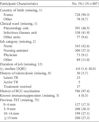
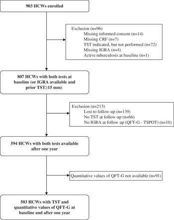
Overall, 903 HCWs agreed to participate and were enrolled in the study, with a median of 56 HCWs (range, 24–127) per participating hospital. A total of 21 HCWs were subsequently excluded because of missing data, and 75 HCWS were further excluded because of missing test at baseline (n=74) or active TB diagnosed at baseline (n=1). Characteristics of the 807 participants are described in Table 1. Among 734 HCWs with a previous known TST, 200 (27.2%) and 199 (27.1%) had an induration ≥15 mm and 10–14 mm, respectively. Nearly all HCWs (n=786, 97.4%) had been vaccinated with BCG.
Baseline Characteristics of HCWS Included in the Study

Note. IQR, interquartile range; BCG, bacille Calmette-Guerin; TST, TB skin testing; IGRA, interferon-γ release assay.
Baseline Results
QFT-G was positive in 113 of the 807 (14.0%) HCWs, negative in 687 (85.1%), and indeterminate in 7 (0.9%). Among the 7 HCWs with indeterminate QFT-G, a T-SPOT TB was negative in 4 HCWs and not performed in 3. Overall, the IGRA was positive in 113 (14.0%) HCWs and negative in 691 (85.6%) (Table 2).
Association between Quantitative TST and IGRA Results in 807 Participants at Baselinea

NOTE. TST, TB skin test; IGRA, interferon-γ release assay
a P<.0001, Cochran-Armitage test.
TST was performed in 607 of 807 HCWs who had no previous known TST induration ≥15 mm. Among them, an induration ≥15 mm and 10–14 mm was found in 82 (13.5%) and 164 (27.0%) HCWs, respectively (Table 2). Considering prior and current TST together, an induration ≥15 mm was present in 282 (34.9%) of the 807 HCWs. Using a cut-off of 10 mm to define TST positivity, TST was positive in 446 (55.3%) of the 807 HCWs.
Among the 807 HCWs with both QFT-G and TST available at baseline, there existed an association between a positive IGRA and induration of TST (Table 2). In the 216 HCWs with an induration <5 mm, the IGRA was positive in 6 (2.8%). In contrast, in the 282 HCWs with prior or baseline TST induration ≥15 mm, the IGRA was positive in 71 (25.2%).
A positive IGRA and TST were associated with age and duration of current function. Mean (SD) age and mean (SD) duration in the current job were higher in HCWs with positive IGRA, at age 42.9 years (SD, 9.8) and a duration of 9.1 years (SD, 8.9 years), than in HCWs with negative IGRAs, at age 38.1 years (SD, 9.8) and a duration of 6.2 years (SD, 6.8 years) (P<.0001 and P<.001). Mean (SD) age and mean (SD) duration of current function were higher in HCWs with prior or present TST ≥15 mm, at age 41.6 years (SD, 9.4 years) and a duration of 7.6 years (SD, 7.9 years), than in HCWs with present TST<15 mm, at age 37.2 years (SD, 9.9 years) and a duration of 6.1 years (SD, 6.7 years) (P<.0001 and P=.01).
Annual Incidence of LTBI
Among the 807 HCWs, 594 had both tests available at 1 year (Figure 1). Their median time (IQR) from baseline to follow-up was 13.3 months (range, 12.4–14.6 months). Analysis of the annual incidence of LTBI was thus restricted to this subgroup of subjects.
Flow chart. Footnote: the sum of specific reasons for exclusion may be higher than the total number of excluded HCWs due to multiple reasons for exclusion.

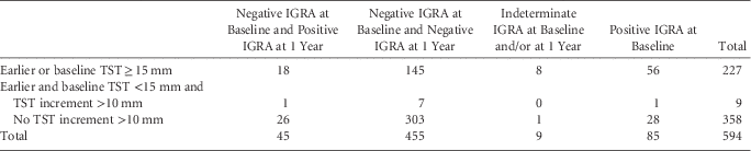
Among these 594 subjects, 85 had a positive IGRA at baseline, 500 had a negative one, and 9 had an indeterminate IGRA. Among the 500 subjects with a negative IGRA at baseline, 45 were positive at 1 year, for an IGRA-based annual conversion rate of 9.0% (Table 3). At baseline, 227 HCWs had prior or baseline TST≥15 mm and 367 TST<15 mm. Among these 367 HCWs, 9 showed conversion, for a TST-based annual conversion rate of 2.5% (Table 3). Only 1 HCW had conversion according to both the IGRA and TST (κ score, −0.002; P<.0001).
Conversion of TST and IGRA after 1 Year

Quantitative Values of QFT-G
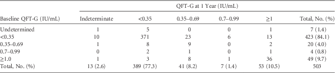
In 503 HCWs, quantitative values of QFT-G were recorded at baseline and at 1 year (Table 4). Among the 423 HCWs with a negative QTF-G at baseline, 371 (87.7%) still had a QFT-G negative at 1 year, 10 (2.4%) had an indeterminate test and 42 (9.9%) had a positive QFT-G at 1 year. The quantitative value of the QFT-G at 1 year in these 42 HCWs ranged between 0.35 and 0.69 UI/mL, 0.7–0.99 IU/mL and ≥1 IU/mL in 23, 6 and 13 HCWs, respectively.
Conversion and Reversion of QFT-G in 503 Participants at 1 Year

Among the 73 HCWs with a positive QFT-G at baseline, 58 still had a positive QFT at 1 year, 2 had an indeterminate result, and 13 (17.8%) a negative QFT-G. The quantitative value of these 13 positive QFT-G at baseline ranged mainly from 0.35 to 0.69 IU/mL (8 of 13). Thus, for HCWs with a baseline QTF between 0.35 and 0.69 IU/mL, the reversion rate was 40.0% (8 of 20). This rate was not significantly different from HCWs with a baseline QFT-G between 0.7 and 0.99 IU/mL at baseline (2 of 4, 50.0%, P=1.0) but was higher than in HCWs with a baseline QFT-G≥1 IU/mL (3 of 49, 6.1%, P=.001).
DISCUSSION
Screening of HCWs for TB is an important component of infection control in occupational health. Because of the limited sensitivity of TST and low specificity in BCG-vaccinated populations, the introduction of IGRAs was considered to represent major progress. However, several studies showed that conversion and reversion of IGRA were possible without evidence of LTBI.Reference Ringshausen, Schablon and Nienhaus 7 , Reference Zwerling, Hof, van den, Scholten, Cobelens, Menzies and Pai 8
Our results can be summarized as follows. In a population of BCG-vaccinated exposed HCWs, the prevalence of positive TST was higher than that of positive IGRA. The incidence of LTBI at 1 year was higher with the IGRA than with TST, with poor agreement between the 2 tests. Conversion and reversion of IGRA were frequent, mainly for values of IGRA close to the threshold of 0.35 IU/mL.
Cut-off defining a positive TST is usually set at 10 mm. However, in countries with a high rate of BCG vaccination, the cut-off of 15 mm may be used. In our study, the prevalence of TST positivity using a 15 mm cut-off at baseline or during a previous TST was 34.9%. Using a cut-off of 10 mm to define TST positivity, the prevalence was 55.3%. This high prevalence of positive TST has been previously noted in other countries with a low incidence of TB.Reference Zwerling, Hof, van den, Scholten, Cobelens, Menzies and Pai 8 , Reference Drobniewski, Balabanova, Zakamova, Nikolayevskyy and Fedorin 9 These findings may be ascribable to the low TST specificity in mandatory-vaccinated HCWs and to high exposure to TB in the study population, as shown by the longer job duration in high-risk units and, presumably, longer exposure to TB than HCWs without TST positivity.Reference Drobniewski, Balabanova, Zakamova, Nikolayevskyy and Fedorin 9 – Reference Lien, Hang and Kobayashi 12
To assess IGRA positivity, we used a combination of QFT-G and T-SPOT TB. QFT-G was used as the first test, and T SPOT TB was used if QFT-G was indeterminate. At study baseline, only 7 of the 807 QFT-G (0.9%) were indeterminate, 4 of whom had negative results with the T-SPOT TB, while T-SPOT TB was not performed for the last 3 HCWs. Combining QFT-G and T-SPOT TB, none of the 804 available IGRAs remained indeterminate. Combining the 2 tests can reduce the number of indeterminate results to a very low proportion in this population of healthy HCWs.
The prevalence of positive IGRA at study baseline was 14.0%, lower than the 34.9% positivity with TST. Our results are in line with a recent review comparing TST and QFT-G that showed that most studies reported a lower prevalence of positive IGRA than positive TST.Reference Zwerling, Hof, van den, Scholten, Cobelens, Menzies and Pai 8 Our results suggest that TST may not be an appropriate marker of LTBI, since only 25.2% of HCWs with a ≥15 mm positive TST had a positive IGRA. Other authors, however, have found a higher prevalence of positive QFT-G in HCWs with a TST≥15 mm.Reference Campo, del, Fouad, Solís-Bravo, Sánchez-Uriz, Mahíllo-Fernández and Esteban 13 – Reference Torres Costa, Sá and Cardoso 15 Three studies performed on a population with a high proportion of BCG vaccination showed a strong correlation between a positive QFT-G and a positive TST when the cut-off was set at 15 mm.Reference Hotta, Ogura and Nishii 16 – Reference Vinton, Mihrshahi, Johnson, Jenkin, Jolley and Biggs 18 As in other studies,Reference Demkow, Broniarek-Samson and Filewska 14 , Reference Torres Costa, Sá and Cardoso 15 , Reference Vinton, Mihrshahi, Johnson, Jenkin, Jolley and Biggs 18 the prevalence of IGRA at baseline increased with TST positivity, from 2.8% in HCWs with a negative TST to 25.2% in HCWs with a TST≥15 mm. Nonetheless, in the absence of a gold standard for LTBI, neither test is able to clearly show which HCW has LTBI.
After 1 year, the IGRA conversion rate (9.0%) was higher than the TST conversion rate (2.5%). Use of the IGRA conversion rate as the gold standard for diagnosing LTBI and initiating antibiotic treatment would lead to treatment of a large percentage of HCWs. In addition, there was a poor correlation between TST conversion and IGRA conversion. When using TST on non-vaccinated HCWs, a yearly 1% incidence rate is generally observed for HCWs in hospitals in countries that have high infection control standards.Reference Larsen, Biddle, Sotir, White, Parrott and Blumberg 19 Our 2.5% TST conversion rate in high-risk units was in line with these data, and appeared more reliable than the IGRA-defined conversion rate.
As in other studies,Reference Fong, Tomford and Teixeira 20 most conversions with IGRA in our study occurred with low positive values of QFT-G; 23 of 42 (55%) conversions occurred with QFT-G values between 0.35 and 0.69 IU/mL, and an additional 14% occurred with a QFT-G value between 0.7 and 0.99 IU/mL. To decide whether or not IGRA conversion represents true LTBI, prolonged surveillance using IGRA should be performed.
We observed a high reversion rate of 17.8% of QFT-G at 1 year. As in other studies, the reversion rate was higher for baseline values of QFT-G between 0.35 and 0.69 IU/mL (40.0%) and between 0.70 and 0.99 (50.0%) than for baseline QFT-G values≥1 IU/mL (6.1%).Reference Fong, Tomford and Teixeira 20 – Reference Ringshausen, Nienhaus, Schablon, Schlösser, Schultze-Werninghaus and Rohde 23 When considering both conversion and reversion rates, our results confirm the existence of an uncertainty zone between 0.35 and 1 IU/mL.
Our study had several limitations. Because of its insertion in a real-life situation without dedicated research personnel, a substantial proportion of HCWs were lost to follow-up and participants did not receive all tests planned. In addition, technical problems may have occurred due to different operators performing blood drawing and laboratory analysis, thus explaining some false-positive and false-negative IGRA tests. Secondly, as in all studies evaluating tests for LTBI, there is no gold standard for diagnosis. Our data demonstrate that both TST and IGRA tests have advantages and limitations.
The major strength of our study was the prospective multicenter design in a real-life setting study and the homogeneous population of HCWs with high occupational exposure to TB in a country with a low incidence of the disease. Furthermore, a combination of TST and IGRA in routine occupational health services without dedicated research personnel for performing tests enabled us to make conclusions than can be generalized.
In conclusion, our data confirm the high frequency of discordance between TST and IGRA. Since there is no gold standard for LTBI diagnosis, the value of QFT-G in addition to TST in exposed HCWs remains to be determined. Nevertheless, the IGRA has practical advantages. If used, the presence of an uncertainty zone between 0.35 and 1 IU/mL with QFT-G suggests that IGRA should be repeated in case of conversion, and relevant clinical information together with TST should be considered before offering chemotherapy. Additional research is needed to establish the biological basis of IGRA conversions and reversions to optimize test accuracy.
Acknowledgments
We thank both the personnel from the departments of occupational health of the 14 participating hospitals, who included and followed healthcare workers, and the biology laboratories that performed IGRA tests.
Financial support: The study was supported by research from the French Ministry of Health (STIC 2007, IC070411).
Potential conflicts of interest: None of the authors have any conflict of interest associated with the work presented in this manuscript.