I. INTRODUCTION
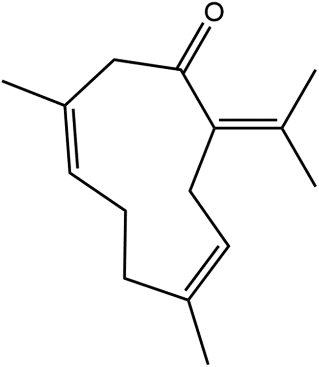
Germacrone was first isolated in 1927 from the essential oil of Geranium macrorrhizum (Rovesti, Reference Rovesti1927). Germacrone is a sesquiterpene, a terpene that consists of three isoprene units, and exhibits a range of pharmacological activities, including anti-inflammatory, anticancer, antiviral, anti-androgenic, antioxidant, antimicrobial, antifungal, neuroprotective, and also shows insecticidal activities (PubChem; Kim et al., Reference Kim, Chen, Cheng, Gindulyte, He, He, Li, Shoemaker, Thiessen, Yu, Zaslavsky, Zhang and Bolton2019). The systematic name (CAS Registry Number 6902-91-6) is (3E,7E)-3,7-dimethyl-10-propan-2-ylidenecyclodeca-3,7-dien-1-one. A two-dimensional molecular diagram is shown in Figure 1.
The 2D molecular structure of germacrone.

A crystal structure of germacrone has been reported, herein referred to as Type I (Clardy and Lobkovsky, Reference Clardy and Lobkovsky1998; CSD Refcode FIQLOG). The structure of a related molecule, isogermacrone, has also been reported (Jacobsson et al., Reference Jacobsson, Norin, Weber and Tsankova1985; CSD Refcode DICDOI). The FIQLOG structure is in space group Pc, and apparently is an example of a chiral structure formed by a cycloalkene molecule, which does not contain chiral centers (Barrero et al., Reference Barrero, Mar Herrador, Arteaga and Catalán2008).
This work was carried out as part of a project (Kaduk et al., Reference Kaduk, Crowder, Zhong, Fawcett and Suchomel2014) to determine the crystal structures of large-volume commercial pharmaceuticals, and include high-quality powder diffraction data for them in the Powder Diffraction File (Gates-Rector and Blanton, Reference Gates-Rector and Blanton2019).
II. EXPERIMENTAL
Germacrone was a commercial reagent, purchased from TargetMol (Lot #114635), and was used as-received. The white powder was packed into a 1.5 mm diameter Kapton capillary and rotated during the measurement at ~50 Hz. The powder diffraction pattern was measured at 295 K at beamline 11-BM (Antao et al., Reference Antao, Hassan, Wang, Lee and Toby2008; Lee et al., Reference Lee, Shu, Ramanathan, Preissner, Wang, Beno, Von Dreele, Ribaud, Kurtz, Antao, Jiao and Toby2008; Wang et al., Reference Wang, Toby, Lee, Ribaud, Antao, Kurtz, Ramanathan, Von Dreele and Beno2008) of the Advanced Photon Source at Argonne National Laboratory using a wavelength of 0.458133(2) Å from 0.5 to 50° 2θ with a step size of 0.001° and a counting time of 0.1 s per step. The high-resolution powder diffraction data were collected using 12 silicon crystal analyzers that allow for high angular resolution, high precision, and accurate peak positions. A silicon (NIST SRM 640c) and alumina (SRM 676a) standard (ratio Al2O3:Si = 2:1 by weight) was used to calibrate the instrument and refine the monochromatic wavelength used in the experiment.
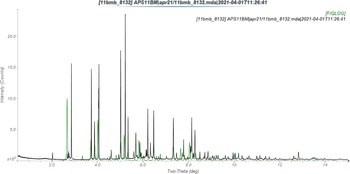
The observed powder pattern does not correspond to that calculated from the room-temperature FIQLOG crystal structure (Figure 2), but the sample does seem to contain the Type I phase as an impurity. The pattern collected in this study was difficult to index. The peaks having I rel > 4% could be indexed on a C-centered monoclinic unit cell with a = 26.01319, b = 9.84676, c = 10.53950 Å, β = 95.77°, V = 2685.98 Å3, and Z = 8 using JADE Pro (MDI, 2021). The suggested space group was C2/c, which was confirmed by successful solution and refinement of the structure. A reduced cell search in the Cambridge Structural Database (Groom et al., Reference Groom, Bruno, Lightfoot and Ward2016) yielded 2 hits, but no structures of germacrone derivatives. Additional unindexed non-FIQLOG peaks suggested the presence of at least one additional crystalline impurity.
Comparison of the observed synchrotron powder diffraction pattern (black) of germacrone to the pattern calculated from the Form I FIQLOG structure (green).

A germacrone molecule was extracted from the FIQLOG structure using Materials Studio (Dassault, 2021), and saved as a *.mol2 file. The new structure (Type II germacrone) was solved using Monte Carlo simulated annealing techniques as implemented in EXPO2014 (Altomare et al., Reference Altomare, Cuocci, Giacovazzo, Moliterni, Rizzi, Corriero and Falcicchio2013). Attempts to solve the structure in space group Cc using two independent molecules yielded significantly worse residuals.
Rietveld refinement was carried out using GSAS-II (Toby and Von Dreele, Reference Toby and Von Dreele2013). Only the 1.0–25.0° portion of the pattern was included in the refinement (d min = 1.058 Å). FIQLOG (Type I) was included as a second phase; its concentration refined to 1.3 wt.%. All non-H bond distances and angles were subjected to restraints, based on a Mercury/Mogul geometry check (Bruno et al., Reference Bruno, Cole, Kessler, Luo, Motherwell, Purkis, Smith, Taylor, Cooper, Harris and Orpen2004; Sykes et al., Reference Sykes, McCabe, Allen, Battle, Bruno and Wood2011). The Mogul average and standard deviation for each quantity were used as the restraint parameters. The restraints contributed 5.1% to the final χ 2. The hydrogen atoms were included in calculated positions, which were recalculated during the refinement using Materials Studio (Dassault, 2021). A single U iso was refined for the C and O atoms. The U iso for the H atoms were fixed at 1.3× the U iso of the heavy atoms to which they are attached. The peak profiles were described using an isotropic microstrain model. The background was modeled using a 3-term shifted Chebyshev polynomial.
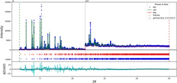
The final refinement of 59 variables using 23 237 observations and 37 restraints yielded the residuals R wp = 0.2051 and GOF = 4.02. The residuals are high because the peaks due to the Type I FIQLOG phase (and presumably also the other impurity peaks) exhibit significant overlap with those of the major phase. The largest peak (0.16 Å from C4) and hole (1.77 Å from O1) in the difference Fourier map were 0.60 and −0.59(15) eÅ−3, respectively. The largest errors in the difference plot (Figure 3) are in the shapes of some of the strong peaks and at the (ignored) impurity peaks.
The Rietveld plot for the refinement of germacrone. The blue crosses represent the observed data points, and the green line is the calculated pattern. The cyan curve is the normalized error plot. The vertical scale has been multiplied by a factor of 10× for 2θ > 12.0°. The row of blue tick marks indicates the calculated reflection positions for germacrone Form II, and the row of red tick marks indicates the form I FIQLOG peak positions.

The crystal structure was optimized using VASP (Kresse and Furthmüller, Reference Kresse and Furthmüller1996) (fixed experimental unit cell) through the MedeA graphical interface (Materials Design, 2016). The calculation was carried out on 16 2.4 GHz processors (each with 4 GB RAM) of a 64-processor HP Proliant DL580 Generation 7 Linux cluster at North Central College. The calculation used the GGA-PBE functional, a plane wave cutoff energy of 400.0 eV, and a k-point spacing of 0.5 Å−1 leading to a 1 × 2 × 2 mesh, and took ~5.8 h. A single-point density functional calculation (fixed experimental cell) and population analysis were carried out using CRYSTAL17 (Dovesi et al., Reference Dovesi, Erba, Orlando, Zicovich-Wilson, Civalleri, Maschio, Rerat, Casassa, Baima, Salustro and Kirtman2018). The basis sets for the H, C, and O atoms in the calculation were those of Gatti et al. (Reference Gatti, Saunders and Roetti1994). The calculations were run on a 3.5 GHz PC using 8 k-points and the B3LYP functional, and took ~2.1 h.
III. RESULTS AND DISCUSSION
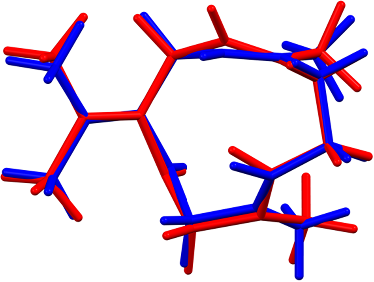
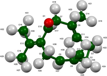
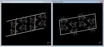
The root-mean-square (rms) Cartesian displacement between the Rietveld-refined and DFT-optimized structures is 0.184 Å (Figure 4), within the normal range for correct structures (van de Streek and Neumann, Reference van de Streek and Neumann2014). This discussion concentrates on the DFT-optimized structure. The asymmetric unit (with atom numbering) is illustrated in Figure 5, and the crystal structure is presented in Figure 6. The crystal structure (Figure 6) indicates discrete molecules, and no particularly strong intermolecular interactions are apparent.
Comparison of the Rietveld-refined (red) and VASP-optimized (blue) structures of the germacrone molecule in Form II. The rms Cartesian displacement is 0.184 Å.

The asymmetric unit of germacrone Form II, with the atom numbering. The atoms are represented by 50% probability spheroids.

The crystal structure of germacrone Form II, viewed down the b-axis.

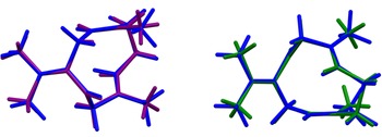
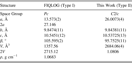
The rms Cartesian displacements between the germacrone molecule in this C2/c structure and the two independent molecules in the Form I FIQLOG Pc structure are 0.079 and 0.79 Å (Figure 7). The ADDSYM module of PLATON (Spek, Reference Spek2009) suggests that the symmetry of FIQLOG is P2/c, and checkCIF yields many alerts for this structure. The FIQLOG cell is about half the volume of the current one, but the angles differ (Table I). The structures exhibit many similarities (Figure 8), but the powder patterns (Figure 2) clearly show that they are different. VASP geometry optimizations for both this polymorph and FIQLOG indicate that this new polymorph is 0.55 kcal mol−1 cell−1 lower in energy, but this difference is within the expected error of such calculations. The two forms must be considered equivalent in energy.
Comparison of the germacrone molecule in Form II (blue) with molecule 1 in the FIQLOG structure (purple, left) and molecule 2 (green, right).

Comparison of the Form I FIQLOG and Form II crystal structures of germacrone.

Lattice parameters of germacrone polymorphs

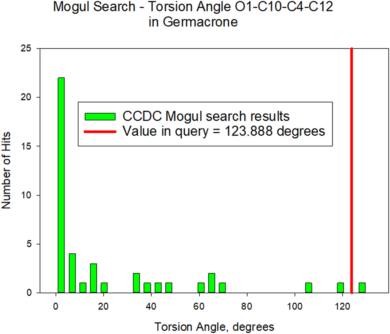
All of the bond distances and angles fall within the normal ranges indicated by a Mercury/Mogul geometry check (Macrae et al., Reference Macrae, Sovago, Cottrell, Galek, McCabe, Pidcock, Platings, Shields, Stevens, Towler and Wood2020). The O1–C10–C4–C5 and O1–C10–C4–C12 torsion angles (−56.2 and 123.9°, respectively) are flagged as unusual, and they are truly unusual. The unusual value is illustrated for O1–C10–C4–C12 (the two torsion angles are not independent) in Figure 9. These angles reflect the arrangement of the carbonyl and dimethylvinylidene groups, which are indeed an unusual pairing.
Comparison of the O1–C10–C4–C12 torsion angle in germacrone to the distribution of similar torsion angles indicated by a Mogul search.

Quantum chemical geometry optimization of the germacrone molecule (DFT/B3LYP/6-31G*/water) using Spartan ‘18 (Wavefunction, 2020) indicated that the observed conformation is only 0.9 kcal mol−1 higher in energy than the local minimum, and that the molecule is in the global minimum-energy conformation in the solid state.
Analysis of the contributions to the total crystal energy of the structure using the Forcite module of Materials Studio (Dassault, 2021) suggests that torsion distortion terms dominate the intramolecular deformation energy, though bond and angle distortion terms are also significant. The intermolecular energy is dominated by van der Waals attractions and electrostatic attractions, which in this force field analysis also include hydrogen bonds. The hydrogen bonds are better analyzed using the results of the DFT calculation.
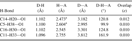
There are no traditional hydrogen bonds in the structure, but the oxygen atom O1 acts as an acceptor in four C–H⋯O hydrogen bonds (Table II). Two of these bonds are intramolecular. The Mulliken overlap populations indicate the presence of many, even weaker, intermolecular interactions.
Hydrogen bonds (CRYSTAL17) in germacrone

a Intamolecular.
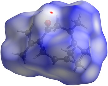
The volume enclosed by the Hirshfeld surface (Figure 10; Hirshfeld, Reference Hirshfeld1977; Turner et al., Reference Turner, McKinnon, Wolff, Grimwood, Spackman, Jayatilaka and Spackman2017) is 330.17 Å3, 98.41% of 1/8 the unit cell volume. The packing density is thus fairly typical. The only significant close contact (red in Figure 10) involves a hydrogen bond. The volume/non-hydrogen atom is larger than normal, at 20.9 Å3.
The Hirshfeld surface of germacrone Form II, calculated using CrystalExplorer17 (Turner et al., Reference Turner, McKinnon, Wolff, Grimwood, Spackman, Jayatilaka and Spackman2017). Intermolecular contacts longer than the sums of the van der Waals radii are colored blue, and contacts shorter than the sums of the radii are colored red. Contacts equal to the sums of radii are white.

The Bravais–Friedel–Donnay–Harker (Bravais, Reference Bravais1866; Friedel, Reference Friedel1907; Donnay and Harker, Reference Donnay and Harker1937) morphology using Mercury (Macrae et al., Reference Macrae, Sovago, Cottrell, Galek, McCabe, Pidcock, Platings, Shields, Stevens, Towler and Wood2020) suggests that we might expect fairly isotropic morphology for germacrone. No preferred orientation model was necessary for this rotated capillary specimen. The powder pattern of Type II germacrone from this synchrotron data set has been submitted to ICDD for inclusion in the Powder Diffraction File.
IV. DEPOSITED DATA
The supplementary material for this article, which includes the Crystallographic Information Framework (CIF) files containing the results of the Rietveld refinement (including the raw data) and the DFT geometry optimization was deposited with the ICDD. The data can be requested at info@icdd.com.
ACKNOWLEDGEMENTS
The use of the Advanced Photon Source at Argonne National Laboratory was supported by the U.S. Department of Energy, Office of Science, Office of Basic Energy Sciences, under Contract No. DE-AC02-06CH11357. This work was partially supported by the International Centre for Diffraction Data. We thank Lynn Ribaud and Saul Lapidus for their assistance in the data collection, and Andrey Rogachev for the use of computing resources at IIT.
CONFLICTS OF INTEREST
The authors have no conflicts of interest to declare.