INTRODUCTION
Plasmodium malariae infection comprises only a small portion of malaria global disease burden. However, this parasite is unique among the plasmodium species in that subclinical parasitaemia may persist for decades and illness may occur more than 40 years after the last possible exposure (Vinetz et al. Reference Vinetz, Li, McCutchan and Kaslow1998). Nephrotic syndrome in children had been linked in the 1960s to chronic infections with P. malariae Gilles and Hendrickse, Reference Gilles and Hendrickse1960 and a few cases have been recently associated with Plasmodium vivax (Barsoum, Reference Barsoum2000). The disease was subsequently attributed to immune complex basement membrane nephropathy (Gilles and Hendrickse, Reference Gilles and Hendrickse1960; Abdurrahman et al. Reference Abdurrahman, Greenwood, Narayana, Papaoye and Edington1981).
Studies of malaria parasite infections in experimental animal models have provided most of what is currently known about malaria. The infection of non-human primates with human malaria parasites offers interesting possibilities that could be compared with human infections. Although over 25 species of primate malaria parasites have been described, few have been extensively used as experimental models. Plasmodium inui is one of the seven malaria parasite species currently known to naturally infect Asian macaque monkeys that are capable of infecting humans (Coatney et al. Reference Coatney, Collins, Warren and Contacos1971), and has been experimentally transmitted to humans, and Aotus monkeys. Many different isolates of P. inui have been adapted to Macaca mulatta monkeys with over a dozen different strains available (Barnwell, 2007). It is quite possible that humans diagnosed as having P. malariae in Southeast Asia could be in fact naturally infected with P. inui because of their morphological similarity.
The immunological mechanisms contributing to the pathogenesis of renal involvement during malarial infections include acute transient immune-complex glomerulonephritis with reversible proteinuria that may lead to renal failure associated with falciparum malaria in Southeast Asia, India and sub-Saharan Africa (Barsoum, Reference Barsoum1998). The other process is a chronic and progressive immune complex glomerulonephritis with irreversible nephrotic syndrome specifically associated with chronic quartan malaria infections that affects mainly African children, usually 4–8 years old (Wing et al. Reference Wing, Hutt and Kibukamusoke1972; Barsoum and Sitprija, Reference Barsoum1996; Olowu et al. Reference Olowu, Adelusola, Adefehinti and Oyetunji2010).
Quartan malaria nephropathy (QMN) is a particular form of nephrotic syndrome, which is a persistent and progressive proteinuric kidney disorder due to P. malariae-induced glomerular damage (Hendrickse and Gilles, Reference Gilles and Hendrickse1963; Kibukamusoke et al. Reference Kibukamusoke, Hutt and Wilks1967; Adeniyi et al. Reference Adeniyi, Hendrickse and Houba1970; Hendrickse et al. Reference Hendrickse, Adeniyi, Edington, Glasgow, White and Houba1972). In one study, quartan malaria was the aetiological agent in 81·0% of all cases of childhood-onset nephrotic syndrome (Hendrickse et al. Reference Hendrickse, Adeniyi, Edington, Glasgow, White and Houba1972). Human QMN shows glomerular capillary wall thickening involving the subendothelial aspect of the basement membrane under the light microscope, and granular deposits of IgG, IgM and C3 complement along the glomerular basement membrane in immunofluorescence microscopy (Olowu et al. Reference Olowu, Adelusola, Adefehinti and Oyetunji2010). Renal immunopathology has been demonstrated in several malaria animal models as well as in acute falciparum malaria in man. Primate malaria species including the monkey quartan malaria parasites (P. inui and Plasmodium brasilianum) have strong similarities to human malaria parasites. The renal disease in rhesus monkey caused by chronic infections with P. inui is similar to known chronic nephrosis caused by P. malariae infection in human and non-human primates (Nimri and Lanners, Reference Nimri and Lanners1994).
Few animal models for human quartan malaria nephrotic syndrome have been proposed; none have shown pathological findings that are similar to those seen in humans. The current study examined the histopathology of the kidney of P. inui-infected rhesus monkeys by light and electron microscopy in order to develop a suitable animal model for human quartan malaria.
METHODS
All protocols were reviewed and approved by the Delta Regional Primate Research Center Animal Care and Use Committee in accordance with animal ethics guidelines described in the US Public Health Service Policy, 1986.
Experimental animals
Ten healthy adult rhesus monkeys (M. mulatta) weighing 5·2–10·4 kg were used in this study and were housed at the Delta Regional Primate Research Center (Covington, LA). They had no previous Plasmodium infection or any immune complex disease. They had been infected with P. inui for periods of 29–63 weeks. The infection was monitored by frequent examination and observing the parasite in Giemsa-stained blood films. The housing conditions and course of infection were as previously described (Nimri and Lanners, Reference Nimri and Lanners1994).
Monkey B147, a splenectomized monkey, was inoculated intravenously with cryopreserved blood cells infected with P. inui, in 1 mL of phosphate-buffered saline (pH 7·2) to establish the infection. When infection was patent (>1% parasitaemia), this animal became a source of parasites for the infection of the other monkeys.
Clinical chemistry and haematology
Several haematologic parameters including red blood cell counts, haemoglobin and haematocrit were tested for all monkeys before and after infection. The average range of normal baseline values for these monkeys were recorded and compared with values recorded after infection for the same tests.
Blood chemistry tests were: total proteins, albumin, globulin, blood urea nitrogen (BUN), creatinine, serum glutamic-oxaloacetic transaminase (SGOT) and serum glutamic-phosphate transaminase (SGPT).
Routine urine analysis was done several times for all monkeys before and during the course of infection.
Anti-streptolysin O titres (ASO)
Serum samples were tested for this factor using a commercial ASO latex-agglutination kit (Baxter Co., USA) according to the manufacturer's instructions to exclude the streptolysin O antigen as a cause of any renal change that might be found after infection.
Renal biopsies
Open renal biopsies were performed twice on all monkeys. The first series of renal biopsies were performed 2 weeks before infection as a baseline control. The second series of renal biopsies were performed 3 months after the first infection was patent.
Microscopy
Light microscopy
Kidney tissue biopsies were fixed in 10% buffered formaldehyde and embedded in paraffin. Thin tissue sections, 5 μm, were cut, sectioned and stained with hematoxylin-eosin, periodic acid Schiff and Jone's silver methenamine stain.
Transmission electron microscopy (TEM)
One-millimetre cubes of kidney tissue were fixed in 2% glutaraldehyde buffered with 0·1 m cacodylate-HCL buffer (pH 7·3), were post fixed for 75 min in 1·5% buffered osmium tetroxide (OsO4) at 4 °C, and embedded in Spurr's medium as described previously (Voller et al. Reference Voller, Davies and Hutt1973). Semi-thin sections were stained with 1% toluidine blue and examined by light microscope to identify areas of interest. Ultra-thin sections (70–80 nm) were cut from blocks and were picked up on copper grids, and stained with uranyl acetate and lead citrate. Multiple grid squares were examined for each monkey and photographed under a JEOL-JEM 1200 EX 11 transmission electron microscope.
Pathological features in the kidney tissues of infected monkeys were examined, with special reference to the glomerular capillaries and basement membranes, peritubular capillaries and blood vessels. Glomerular capillaries were observed for thickening and the presence of any electron-dense deposits in the glomerular basement membrane either in the mesangium or peripheral capillary loops that correspond to immune complex deposition. The morphology of glomerular epithelial cell foot processes was assessed to observe if foot process fusion (effacement) occurred, which may indicate proteinuria.
RESULTS
Facial oedema (swelling of the face) that has been documented as the first sign of nephrotic syndrome in children could not be detected in any monkey, either because it was not pronounced or because of the difficulty of diagnosis in monkeys.
Clinical chemistry
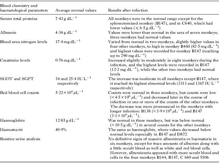
Blood chemistry and haematological values were compared with the baseline values recorded in the normal rhesus monkeys before infection (Table 1).
Different clinical parameters of all monkeys with average normal values before infection and the changes after infection
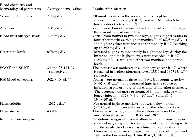
Serum total proteins were all in the normal range (average normal value 7·43 g dL−1) except for the splenectomized monkey (B 147), and in C640, both had lower values (⩽6·5 g dL−1).
Albumin was lower than normal (average normal value 4·34 g dL−1) in the sera of seven monkeys and normal in three monkeys.
BUN levels varied from normal (17·4 mg dL−1) in two monkeys, slightly higher values in four other monkeys, to high in monkey B460 (82·5 mg dL−1) and highest values were recorded for monkey B147 (reaching up to 290 mg dL−1).
Creatinine levels (normal value: 0·76 mg dL−1) also increased slightly to moderately in eight monkeys during the infection, and the highest levels were recorded in B147 (12·2 mg dL−1), while the other two monkeys had normal levels.
The increase in SGOT and SGPT (normal values: 34 and 25·4 IU L−1 respectively) were moderate in all monkeys except B147, where it reached its highest abnormal levels (3311 and 1367 IU L−1 respectively).
Red blood cell counts were normal (5·22×106/μL) in three monkeys, but counts were low (<4·5×106 μL−1) and decreased later in the course of infection in one or more of the counts of the other monkeys. The decrease was more pronounced in the monkeys with longer infection as B138 (<3·9×106 μL−1), and B147 (3·1×106 μL−1).
Haemoglobin was normal (average normal value 12·85 g dL−1) in three monkeys, but was below normal (<10·5 g dL−1) in several counts for the other monkeys. The same was true for haematocrit (normal value: 40·9%), but values decreased below normal levels especially in B147 and D872.
Routine urine analysis did not show definitive signs of massive albuminuria or haematuria in six monkeys, except for trace amounts of albumin along with a little occult blood as well as white and red blood. However, albuminuria appeared with more occult blood and cells in the other four monkeys; B144, B147, C 640 and 9306.
Anti-streptolysin O titres
All monkeys were negative except for B460, which had one positive result, but later became negative. There was no history of any recent streptococcal infection in this monkey.
Histopathological analysis
The infection in the intact (non-splenectomized) monkeys was mild, characterized by low parasitaemia (<2%).
Light microscopy
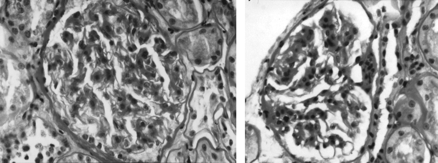
The stained histological slides of the renal biopsies taken after infection revealed morphological changes consistent with malaria-induced pathologies. Nuclei in mesangial regions were counted to assess any glomerular changes. The presence of more than three nuclei/region was considered an increase in mesangial cells. Although proliferation was not a marked feature, a mild focal segmental increase in mesangial cells and matrix was observed in all monkeys with variable degrees. Pronounced hypercellularity and membranoproliferative pattern with diffuse mesangial proliferation was observed more in infected monkeys A074, D893 (Fig. 1) and 9306 (Fig. 2).
Section of the kidney of P. inui-infected monkey D893 showing membranoproliferative pattern with diffuse mesangial proliferation (H & E, 600×).

Section of the kidney of P. inui-infected monkey D893 (left) and 9306 (right) showing mesangial hypercellularity (H & Eosin, 600×).
The changes varied from one glomerulus to another and normal areas could be seen within the same glomerulus. An image of normal glomerular membrane before infection is shown (Fig. 3)
Normal glomerular membrane in uninfected monkey.
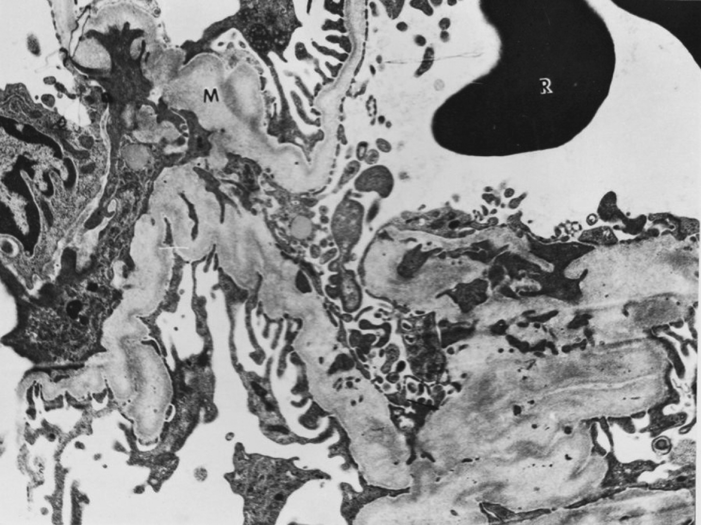
Silver methenamine-stained sections did not show thickening of the glomerular basement membrane or of the peripheral capillary loops. However, a collapse in the capillary loops in infected monkey 9306 was observed indicating segmental sclerosis that was noticed also in the electron microscope (Fig. 4).
Transmission electron micrograph of the glomerulus of monkey 9306 showing a collapse of the capillary loop associate with segmental sclerosis. M: mesangium, R: red blood cell (12 500×).
None of the monkeys had ‘splitting’ or ‘spike’ formation, crescent formation or hyalinosis, and capillary loops were thin and normal. Tubular changes were absent or slight as in the case of monkey B876.
Transmission electron microscopy (TEM)
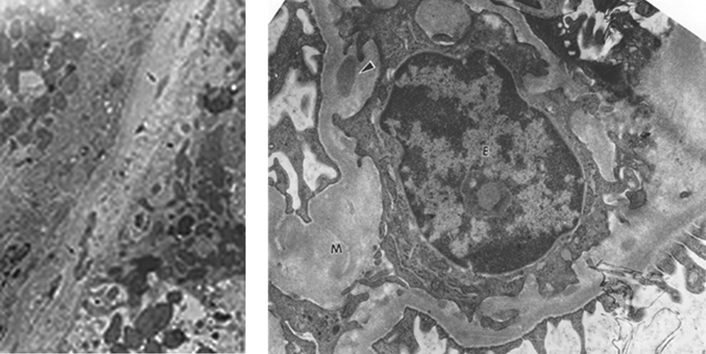
Abnormalities could be seen in all monkeys after infection with an increase in mesangeal cell cytoplasm as the principal change. These changes coincided with parasitaemia levels (<2%), peaks of the complement levels and an increase in the level of circulating immune complexes above normal values in infected monkeys (except B144) (Nimri and Lanners, Reference Nimri and Lanners1994). The matrix showed areas of altered electron density and discrete deposits of small or medium size, which were irregular, uniformly distributed, or both as in D872, but were not uniformly distributed in the mesangeal area. Discrete medium-size deposits were also observed in the tubular basement membrane of infected monkey C640 (Fig. 5), but were more diffused than appeared in the mesangium. More diffused deposits were also observed in the tubular basement membrane of other infected monkeys i.e. B876 (Fig. 5). The epithelial cells appeared normal, however, in monkey C640 they contained proteinaceous bodies and cytoplasmic fragments that might be related to the parasites. No thickening or discrete deposits were observed in the glomerular basement membrane, but there were altered electron dense areas alongside the membrane, especially on the sub-epithelial side. There was also a segmental fusion of the foot processes in some monkeys as in B147 (Fig. 6) and B876. Other non-specific abnormalities such as the presence of tubule-reticular inclusions were also observed in the tubular basement membrane of four monkeys; B147, C640, B876 and D893.
Transmission electron micrograph of the glomerulus of P. inui-infected monkeys, Right, B876 showing diffused immune complex deposits (arrows) in the tubular basement membrane (TB) (7500×). Left, monkey C640 showing medium-size discrete immune complex deposits in the mesangium (arrowhead), E: epithelial cell, M: mesangium (20 000×).
Transmission electron micrograph of the glomerulus of the splenectomized monkey B147 showing fusion of the foot processes (arrows) (7500×).
DISCUSSION
Plasmodium inui infections similar to P. malariae infections in humans are characterized by being chronic, often lasting for the lifetime of the host, despite lacking a relapse mechanism (The Plasmodium Writing Group, 2007). However, in both species infection, recrudescent parasitaemia does occur. Chronic infections of P. inui cause renal disease in rhesus monkeys, same as P. malariae is known to cause chronic nephropathies in humans and non-human primates (Nimri and Lanners, Reference Nimri and Lanners1994).
There have been rare cases of natural infections or accidental laboratory infections by several Plasmodium species (Herwaldt, Reference Herwaldt2001) including P. inui and P. brasilianum (Collins and Barnwell, Reference Collins and Barnwell2009). Severe systemic symptoms and fever have been described, but no severe complications or deaths have been reported.
The current study investigated the renal pathological changes in 10 P. inui- infected M. mulatta monkeys by light and electron microscopy.
Blood chemistry results in the infected monkeys showed normal serum total proteins except in two monkeys that had lower values. Albumin values were lower than normal in seven monkeys and normal in three monkeys. There were moderate increases in BUN, creatinine and serum transaminases (SGOT and SGPT) in most monkeys. Haematological results showed decreases in the red blood cell counts, haemoglobin and haematocrit in most monkeys. The correlation between these results and the observed histopathology is not definitive, since the timing of the blood samples tested could not be monitored in relation to the onset of glomerular disease because of the limitations of the number of renal biopsy samples that could be taken.
The hypoalbuminaemia and the albuminuria observed in monkeys B144, B147, C640 and 9306, are suggestive of the development of nephrotic syndrome in these monkeys.
The morphological changes in renal biopsies taken after infection observed by light microscopy revealed a mild focal segmental increase in mesangial cells and matrix in all monkeys with variable degrees. Hypercellularity was more pronounced in monkeys A074, D893 and 9306, with mild kidney lesions. The mild kidney lesions might not indicate a proliferative membranous glomerulonephritis as reported in some human cases (Gilles and Hendrickse, Reference Gilles and Hendrickse1963; Kibukamusoke et al. Reference Kibukamusoke, Hutt and Wilks1967; Adedoyin et al. Reference Adedoyin, Gbelee and Adeniyi2001), and in Aotus monkeys infected with P. malariae (Voller et al. Reference Voller, Draper, Shwe and Hutt1971). However, the glomerular pathology seen in monkeys A074, D893 and 9306 was of the membranoproliferative type and showed close similarities to reported changes in human nephrotic syndrome cases associated with chronic cases of P. malariae infections (Kibukamusoke et al. Reference Kibukamusoke, Hutt and Wilks1967; Ward and Kibukamusoke, Reference Ward and Kibukamusoke1969; Houba et al. Reference Houba, Allison, Adeniyi and Houba1971; Houba, Reference Houba1975). A proliferative lesion with prominent mesangial involvement seems to parallel the changes reported in human adult cases (Barsoum, Reference Barsoum1998) and in Aotus monkeys infected with P. malariae (Voller et al. Reference Voller, Draper, Shwe and Hutt1971, Reference Voller, Davies and Hutt1973). In addition, changes observed in the kidneys of P. malariae-infected Aotus monkeys showed that the nephrotic syndrome developed in these monkeys was consistent with what is reported in humans (Aikawa et al. Reference Aikawa, Broderson, Igarashi, Jacobs, Pappaioanou, Collins and Campbell1988).
The segmental sclerosis observed in monkey 9306 has also been reported in humans (Kibukamusoke, Reference Kibukamusoke1968; Sitprija and Boonpucknavig, Reference Sitprija, Boonpucknavig, Tisher and Brenner1994; Hedelius et al. Reference Hedelius, Fletcher, Glass, Susanti and Maguire2011), in Nigerian children who had quartan malarial infection (Hendrickse et al. Reference Hendrickse, Adeniyi, Edington, Glasgow, White and Houba1972; Hendrickse and Adeniyi, Reference Hendrickse and Adeniyi1979; Van Marck et al. Reference van Marck, Abdurrahman, Edington and Aliyu1983) and in Senegal (Morel-Maroger et al. Reference Morel-Maroger, Saimot, Sloper, Woodrow, Adam, Niang and Payet1975). In early human cases mesangial sclerosis affects only few glomeruli in a segmental manner, which becomes diffused and leads to progressive glomerular damage and secondary tubular atrophy (Hendrickse et al. Reference Hendrickse, Adeniyi, Edington, Glasgow, White and Houba1972).
Abnormalities in the kidney tissues of all the infected monkeys were observed by electron microscopy. The finding of several electron-dense discrete mesangial deposits of various sizes, as well as an increase in mesangial cells and matrix, were also associated with the morphological changes of the mesangium observed by light microscope. These changes coincided with parasitaemia levels (<2%), peaks of the complement levels and an increase in the level of circulating immune complexes above normal values in infected monkeys (except for B144) (Nimri and Lanners, Reference Nimri and Lanners1994). The evidence of immune complex deposition and endothelial cell damage was reported previously supported by the unique and consistent immunofluorescence patterns of the IgG, 1gM, C3, C4, fibrinogen and albumin. This histological pattern is consistent with a membranoproliferative glomerulonephritis type similar to that reported in humans infected with P. malariae (Sitprija and Boonpucknavig, Reference Sitprija, Boonpucknavig, Tisher and Brenner1994; van Velthuysen and Florquin, Reference van Velthuysen and Florquin2000). In addition, similar changes were reported in Aotus monkeys infected with P. malariae or P. brasilianum (Houba et al. Reference Houba, Lambert, Voller and Soyanwo1976), but were most marked in the animal with chronic infection, which developed the nephrotic syndrome (Voller et al. Reference Voller, Davies and Hutt1973) and in Aotus monkeys infected with P. brasilianum and Plasmodium falciparum (Aikawa et al. Reference Aikawa, Broderson, Igarashi, Jacobs, Pappaioanou, Collins and Campbell1988). The localization of some of these deposits in the mesangium only in some monkeys might be due to the size of the immune complexes, which could not penetrate the glomerular basement membrane and were retained in the mesangium.
There were discrete deposits in monkey C640 and diffused deposits in the tubular basement membrane in B876. These results are consistent with earlier studies of renal biopsies from patients with P. malariae-associated nephropathies showing electron-dense deposits localized within the glomerular basement membrane, usually on the epithelial side whereas masses of dense material distributed throughout the basement membrane were observed in progressive cases (Houba, Reference Houba1975; Hedelius et al. Reference Hedelius, Fletcher, Glass, Susanti and Maguire2011).
The epithelial cells appeared normal, but some contained proteinaceous bodies and cytoplasmic fragments as in C640 that might be related to the presence of the parasite. Abnormal appearance in the endothelial cells was observed in monkey B147.
Electron microscopy showed fusion of the foot-process of the epithelial cells in several monkeys that might indicate proteinuria, and was more pronounced in monkeys B147 and B876. The same results were reported in Nigerian children (van Marck et al. Reference van Marck, Abdurrahman, Edington and Aliyu1983), and in Aotus monkeys infected with P. malariae, which developed nephrotic syndrome (Voller et al. Reference Voller, Davies and Hutt1973).
Increased secretion of urine albumin, also called albuminuria, was observed in B147, B876, B144, C640 and 9306, which may indicate damage to the kidneys. The appearance of immune complex deposits in the immuno-fluorescence test (Nimri and Lanners, Reference Nimri and Lanners1994) and electron microscopy observed in monkeys 9306, B147, B144, C640 and B876, listed according to severity, could be the early stage of disease that might progress later to nephrotic syndrome (Houba et al. Reference Houba, Allison, Hendrickse, de Petris, Edington, Adeniyi, Bonomo and Turk1970). Although the clinical symptoms presented by the blood chemistry tests were severe in the splenectomized monkey B147, the histological changes in the light and electron microscopy were more pronounced in 9306.
The histology results of the monkeys B138 and B460 that had relatively longer duration of infection did not differ from the other monkeys. On the contrary, these monkeys had relatively smaller and fewer deposits than the monkeys that were infected later. The removal of immune deposits from kidney tissue was reported in human cases, whereas the majorities had only a transient spontaneously resolving nephritis (Voller, Reference Voller1974). It occurs either spontaneously by phagocytic activity of the mesangial cells or after treatment (WHO, 1972).
Although it is unlikely that any single model will reproduce the complexity and spectrum of disease and immunity observed in human malaria infections, there are parallels between certain human cases and animal presentations of malaria infections (Craig et al. 2012).
In conclusion, our results strongly support that the P. inui-infected rhesus monkey develops an immune-complex-mediated glomerulonephritis in the course of the infection. Therefore, the P. inui/rhesus experimental model represents a useful tool to better understand the different parameters and the consequences of quartan malaria-associated chronic renal failure in children and adults. In addition, it might be a suitable model to investigate possible inflammatory processes in chronic disease and treatment and of vaccine efficacy studies.
ACKNOWLEDGEMENTS
We are grateful to Dr Patrick Walker for his assistance with pathology analysis of the kidney tissue samples. Special thanks to Maryjane Dodd for technical assistance in the parasite counts and collection of serum specimens. Special thanks to Mr Norwood Meiners for his assistance in electron microscopy and photography.