INTRODUCTION
It is estimated that 350 million people in 88 countries are at risk of leishmaniasis, with an annual incidence of 500 000 for the lethal visceral form of the disease and 1·5 million for cutaneous forms (WHO, 2004). The interaction between Leishmania and macrophages plays a central role in the pathogenesis of the infection. Initiation of infection is related to the ability of the parasite to bind and enter the host cell and the mechanisms involved in the inhibition of macrophage activity are largely known (de Almeida et al. Reference de Almeida, Vilhena, Barral and Barral-Netto2003; Gregory and Olivier, Reference Gregory and Olivier2005). Once an amastigote has established itself and replicated inside the host cell, the next step is for it and its descendants to spread to other macrophages. Given the modest number of parasites inoculated during natural transmission, on average 1000 (Rogers et al. Reference Rogers, Ilg, Nikolaev, Ferguson and Bates2004), the spread of amastigotes to uninfected macrophages is crucial to the development of the disease. This ‘silent stage’ following infection (Belkaid et al. Reference Belkaid, Mendez, Lira, Kadambi, Milon and Sacks2000) is associated with the interaction of a distinct group of parasite molecules consisting of invasive determinants which establish the infection but do not activate immune effector mechanisms (Chang and McGwire, Reference Chang and McGwire2002; Chang et al. Reference Chang, Reed, McGwire and Soong2003). Parasites therefore must spread to uninfected cells without causing an immune response.
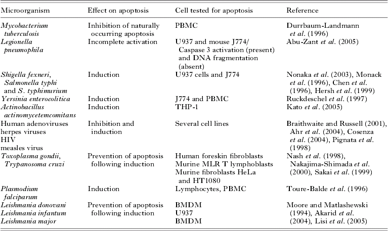
Since apoptosis is a mechanism through which cells are phagocytosed by macrophages without eliciting an inflammatory response (Savill et al. Reference Savill, Gregory and Haslett2003), we suggest that this could be the mechanism used by Leishmania parasites to spread ‘silently’. Previous studies on the effects of intracellular microorganisms have shown both negative and positive modulation of host cell apoptosis (Table 1). Studies on L. donovani, L. infantum and L. major suggested that Leishmania parasites are able to rescue host cells from undergoing apoptosis and that this might play a role in both host invasion and in the persistence of parasites inside host cells. In each case parasites were able to rescue murine-macrophages after apoptosis was induced by either deprival of (M)-CSF or treatment with an apoptosis inducer. Although intracellular parasites might find it beneficial to increase the life span of the host cells they might find it just as beneficial to induce apoptosis in order to spread into another macrophage without induction of inflammation (Savill, Reference Savill1997; Savill et al. Reference Savill, Fadok, Henson and Haslett1993).
The effect of various infectious organisms on their host apoptosis

The aim of this work was to determine whether 3 Old World species causing cutaneous leishmaniasis were able to interfere with apoptosis in healthy host macrophages and to establish if apoptosis induction could affect parasite spread.
MATERIALS AND METHODS
Leishmania
Promastigotes of L. aethiopica (MHOM/ET/72/L100), L. major (MHOM/SU/73/5ASKH) and L. tropica (MHOM/SU/58/OD) were grown in DMEM/F12 (Sigma, UK) supplemented with 10% heat-inactivated foetal calf serum and 2 mm Glutamine (Sigma, UK) at neutral pH. Promastigotes were inoculated at a concentration of 5×10⁵ cells/ml into 25 ml tubes (Sterilin, UK) and placed in a cooling incubator at 23°C. The promastigotes were passaged once a week. In control experiments promastigotes were killed with 2% PBS-formalin followed by thorough washing and used to infect U937 cells.
Cell lines of human monocytes
Two human cell lines and peripheral blood-derived human macrophages were chosen to study the interaction between Leishmania parasites and human cells. The cell lines were chosen because they have been reported to support Leishmania infection and multiplication (Ogunkolade et al. Reference Ogunkolade, Colomb-Valet, Monjour, Rhodes-Feuillette, Abita and Frommel1990) and have been repeatedly used as models for infection and for screening anti-leishmanial drugs (Gebre-Hiwot et al. Reference Gebre-Hiwot, Tadesse, Croft and Frommel1992). The cell lines, THP-1 (8808120) and U937 (85011440), were obtained from the European Collection of Cell Cultures (ECACC) and were cultured in DMEM/F12 supplemented with 10% foetal calf serum and 2 mm glutamine in a humidified 5% CO2 atmosphere at 37°C. Cells were passaged twice a week at a density of 3×10⁵ cells/ml.
Macrophage purification from peripheral blood
Blood-derived human macrophages were purified from the same subject for all experiments. The blood sample was immediately diluted in medium containing heparin and centrifuged on a density gradient (Lymphoprep®, Sigma-Aldrich, UK) and the isolation carried out as previously described (Davis, Reference Davis2002). Briefly, the mononuclear cells that lie over the lymphoprep were gently removed with a sterile Pasteur pipette and transferred to a clean tube while erythrocytes and granulocytes were aggregated by polysucrose and rapidly sedimented.
The cells were washed of residues of lymphoprep through sequential centrifugations in PBS (5 min at 800 g) and diluted in fresh medium to a concentration of 106 cells/ml. Cells were transferred into 24-well plates (NUNC, Denmark) and incubated overnight in a 5% CO2 atmosphere at 37°C. Non-adherent cells were washed out and fresh medium added to adherent macrophages. The number of adherent cells was counted using a 40× objective on an inverted microscope. A minimum of 50 fields of view from each of the 3 wells were counted and the average taken to calculate the total number.
The purified adherent macrophages were infected after 24 h and tested 24 h after infection.
Infection and apoptosis
THP-1 cells were transformed into non-adherent macrophages by incubation for 72 h with 1 μm retinoic acid (RA, Sigma-Aldrich, UK) and U937 cells by incubation with phorbol 12-myristate 13-acetate (PMA, Sigma, UK, 10 μg/ml) for 24 h. The transformed THP-1 and U937 cells were washed ×3 with PBS to eliminate residual RA and PMA and infected with live stationary-phase promastigotes of Leishmania at a ratio of 10:1 and maintained for 48 h at 37°C and 5% CO2 in a humidified incubator. A negative control for each species was left untreated and a positive control was treated with 3 μm of a known apoptosis inducer, camptothecin B (Sigma-Aldrich, UK). A further control was prepared by adding zymosan (Sigma-Aldrich, UK) to the cells at a ratio of 10:1. Following incubation the percentage of apoptotic cells was determined. The same ratio of promastigotes was added to blood-derived human macrophages.
The early stages of apoptosis were detected by microscopical analysis using an Annexin-V-Cy3 apoptosis kit containing 6-carboxyfluorescein diacetate as vital stain (Sigma, UK), BD Apoalert™ Mitochondrial Membrane Sensor Kit (BD Biosciences, UK), MitoGLO™ and Caspase FLICA assay (Imgenex, US; distributed by Cambridge BioScience, UK). They were used according to the manufacturers' instructions for cells detected under fluorescence microscopy. In both cases a minimum of 100 cells per sample were counted under the microscope and the percentage of apoptotic cells recorded. The fluorescence signals produced by the various kits are easily distinguished by fluorescence microscopy and flow cytometry. Microscopic analysis was chosen to avoid a source of errors due to the possible presence of non-internalized apoptotic parasites in the samples. Samples were analysed 48 h after infection and caspase activity was further checked 72 h after infection.
A later stage of apoptosis was checked through flow cytometry detection of DNA fragmentation. The cellular DNA content was measured following cell permeabilization with detergents and fixation with alcohol (Darzynkiewicz et al. Reference Darzynkiewicz, Bruno, Del Bino, Gorczyca, Hotz, Lassota and Traganos1992). The cells were stained with propidium iodide (PI, Sigma-Aldrich). PI staining solution plus RNaseA was added as described by Darzynkiewicz et al. (Reference Darzynkiewicz, Bruno, Del Bino, Gorczyca, Hotz, Lassota and Traganos1992). Following incubation for 30 min in the dark at room temperature, the samples were analysed by flow cytometry (Partec CA-III flow cytometer Partec GmbH, Munster, Germany) with a 100 W high-pressure mercury lamp, KG1, BG38, UG1 filters, TK420 dichroic mirror and a GG435 barrier filter. Just before analysis, clumped cells were carefully separated by passing them through a 25 g hypodermic needle.
Effect of apoptosis on infection
Terminally differentiated U937 cells were infected with live promastigotes as described above. Following 24 h incubation for each sample infected with a different species, a negative control was left untreated and one was treated with an apoptotic inducer (camptothecin). After a further 24 h the cells were stained with Giemsa stain (BDH Laboratory Supplies, UK) in water buffered to pH 6·8 as previously described (Lillie, Reference Lillie1977). The slides were observed under a light microscope and the number of infected macrophages counted.
Statistical analysis
Data represent the mean of at least 3 experiments carried out in duplicate and are reported ±s.e. The distribution of each set of data was confirmed to follow a normal distribution by Kolmogorov-Smirnov and Shapiro-Wilk tests. The increase in apoptotic cells in the presence of each Leishmania species' infection was compared with that in the absence of infection using a one-way ANOVA. When homogeneity of variances could be assumed (Levene's test) a post-hoc Dunnett's modification (>Control) was used. When population variances were different, Games-Howell procedure was used. The effect of apoptosis on infection and differences between apoptotic cells at 48 and 72 h from infection were analysed with Student's t-test. Values of P<0·05 were considered to be statistically significant.
RESULTS
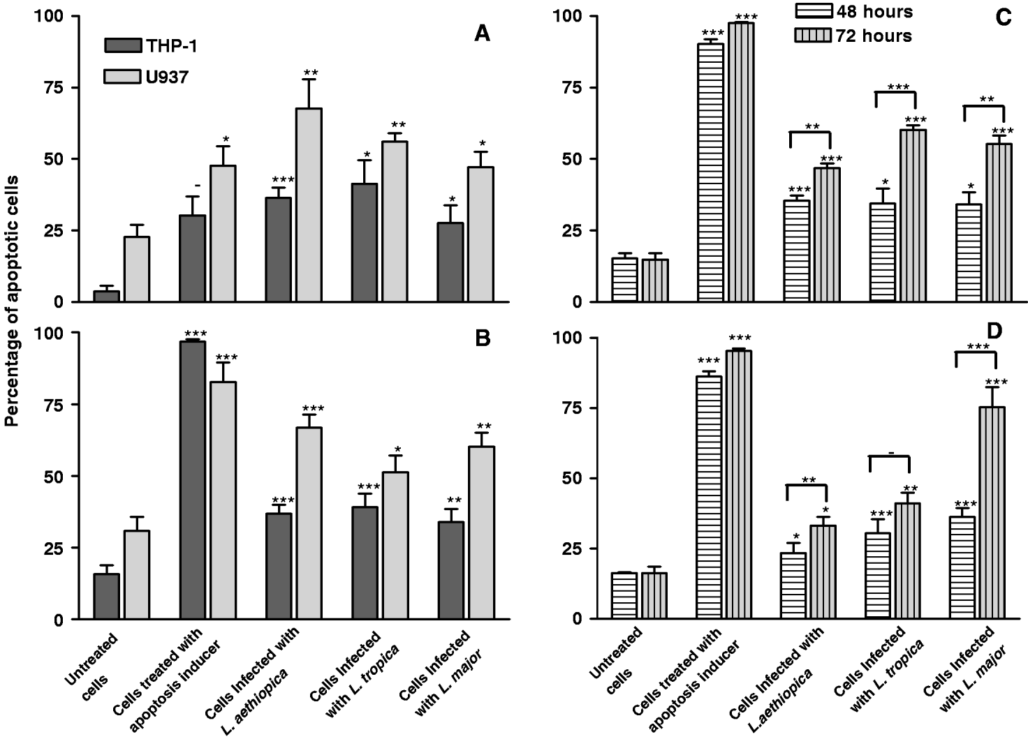
In preliminary experiments apoptosis was checked 24 h after infection (Annexin V assay in THP-1 cells) and a small increase in apoptosis was detected, with less than 10% of the cells showing apoptotic features following infection with any of the 3 species tested (0·5% were apoptotic in the untreated control). A higher percentage of apoptotic cells was detected with early apoptosis tests (Annexin V and/or Mitosensor kit) in both cell lines and blood-derived macrophages 48 h after infection. Differences between untreated control and cells infected with 2 of the 3 Leishmania species were significant when checked with the Mitosensor kit (P<0·04; Fig. 1A) and with all 3 species when checked with Annexin V (P<0·04, Fig. 1B). In both cases infected U937 cells showed a higher sensitivity to apoptosis induction than THP-1 cells.
Effect of infection on THP-1 and U937 host cell apoptosis. One sample was left uninfected and untreated as negative control, while one sample was treated with an apoptosis inducer, camptothecin B, and used as positive control. (A) Mitosensor Kit in two terminally differentiated cells lines (THP-1 and U937). (B) Annexin V assay in two terminally differentiated cells lines (THP-1 and U937). (C) MitoGlo assay in THP-1 cells 48 and 72 h following infection. (D) FLICA assay in THP-1 cells 48 and 72 h following infection. The P values were calculated using a one-way ANOVA test with post-hoc Dunnett's modification (>Control) or Games-Howell. Uninfected cells were used as control. Differences between apoptotic cells at 48 and 72 h from infection were analysed with Student's t-test (* P<0·05; ** P<0·01; *** P<0·001). Each experiment was conducted in duplicate and repeated at least 3 times. Bars represent standard errors.

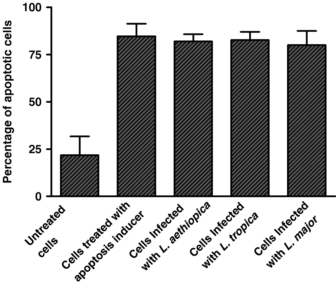
In order to confirm that apoptosis induction is not a consequence of the phagocytic process and that it is dependent on the action of Leishmania parasites, possibly following differentiation in amastigotes, control experiments with zymosan and dead promastigotes were carried out. Phagocytosis of control zymosan did not cause a significant increase in the percentage of apoptotic cells compared with the untreated control. Moreover, infection with dead parasites showed that a significantly smaller amount of apoptosis was induced (P<0·0001) when compared with cells infected with live parasites (results not shown). The results obtained were confirmed in peripheral blood-derived macrophages (PBDM) as these are the closest in vitro representation of the in vivo infection. The level of apoptosis induced by infection in blood-derived macrophages was much higher than that found in the cell lines with an almost 200% increase of apoptotic cells between infected and uninfected macrophages (P<0·01, Fig. 2). There was no significant difference in the degree of apoptosis induced by the 3 species tested when early stages of apoptosis were checked.
Effect of infection on peripheral blood-derived human macrophages. Annexin V assay in blood-derived human macrophages (BDHM). The P values were calculated using a one-way ANOVA test with post-hoc Dunnett's modification (>Control). Uninfected cells were used as control (** P<0·01). Each experiment was conducted in duplicate and repeated at least 3 times. Bars represent standard errors.

The data obtained only give us indications on early stages of apoptosis so the presence of later stages was also investigated. Late apoptotic cells are characterized by DNA fragmentation and loss and can be identified on the basis of their DNA content via flow cytometry analysis of PI-stained cells (Fig. 3). The numbers of events (cells counted) was reported for each channel (quantity of DNA) in a histogram that identifies in each peak the amount of cells characterized by a certain amount of DNA. In the control (Fig. 3A) two peaks were present, the first one representing the expected diploid population and a second one due to the presence of cells that stick together and therefore show a double DNA content. When apoptosis was induced (Fig. 3B) both peaks broadened due to the presence of cells that had partially lost their DNA and were therefore read at lower channels values. Following infection with any of the 3 species, peak broadening was not detected (Fig. 3C and D). Therefore we can conclude that DNA fragmentation was not detected 48 h from infection.
Flow cytometry analysis of fragmented DNA. Histogram with 1000 channels, diploid peak at 125, low level set at 40. A minimum of 36 000 cells were analysed in each sample. DNA-related fluorescence is reported on the X-axes and number of cells on the Y-axes. (A) Negative control, uninfected and untreated THP-1 cells. (B) Positive control, camptothecin-treated THP-1 cells. (C) THP-1 cells infected with Leishmania aethiopica. (D) THP-1 cells infected with L. major.

Early signs of apoptosis were maintained by the host cells at 72 h following infection (Fig. 1C) and coincided with caspase activation (Fig. 1D). The number of cells presenting signs of mitochondrial permeabilizaton and caspase activation showed that the highest percentage of apoptotic cells was detected 72 h after infection according to both FLICA and MitoGlo assays. Differences between untreated control and cells infected with each of the 3 Leishmania species were significant (Fig. 1C and D) both at 48 h and 72 h after infection (P<0·05), confirming that late stages of infection produce an increase in the number of host cells showing apoptotic features.
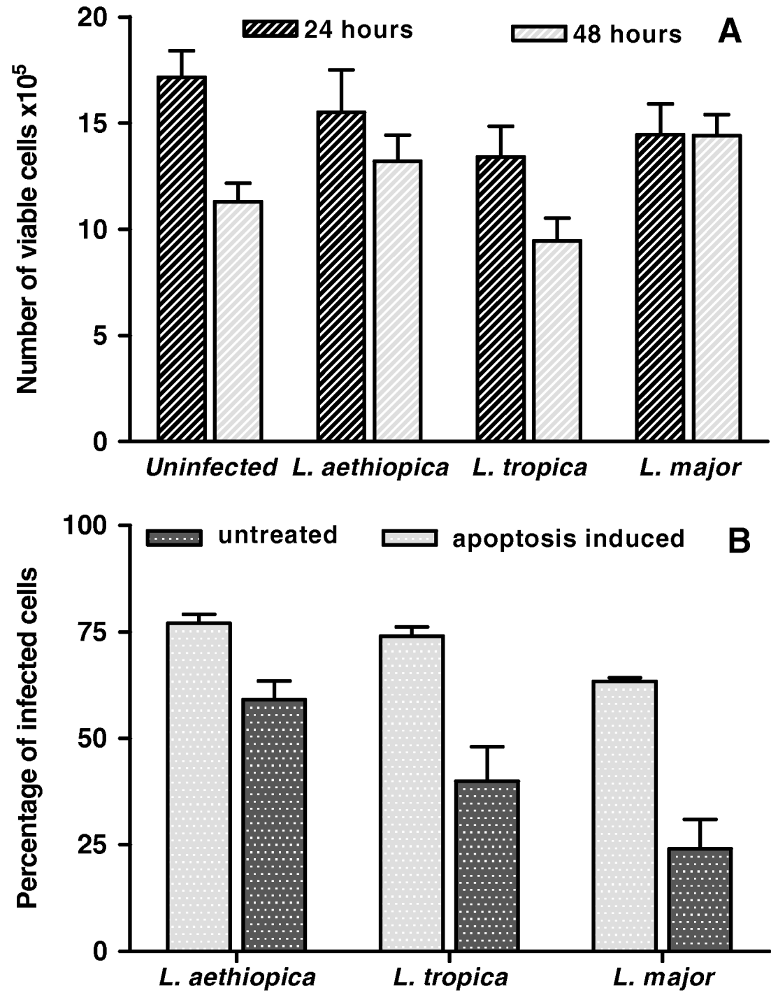
Evidence of the involvement of apoptosis in the infection process was obtained by the increases in infected cells following apoptosis induction. Specifically, U937 macrophages were infected with each of the 3 species studied and the percentage of infection was compared between untreated cells and the same cells following induction of apoptosis (Fig. 4B). In each case, induction of apoptosis significantly increased the percentage of infection when compared with untreated infected macrophages (P<0·05). During the course of the experiment induction of apoptosis had no effect on cell density (Fig. 4A), suggesting that the difference in the number of infected cells was related to apoptosis induction and not to loss of viable adherent cells.
Infection in relation to host apoptosis and viability. (A) Number of viable cells at 24 and 48 h after infection. One-way ANOVA analysis with post-hoc Dunnett's modification (>Control) showed no significant difference (P>0·05) between the number of viable cells in any of the infected populations when compared with the uninfected control. (B) Percentages of infected cells were counted 24 h following infection plus 24 h with and without treatment with the apoptosis inducer camptothecin B in terminally differentiated U937 cells. Differences between treated and untreated cells were analysed with Student's t-test. Each experiment was conducted in duplicate and repeated at least 3 times. Bars represent standard errors.

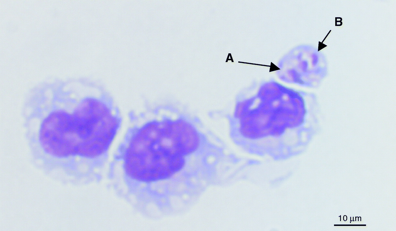
The infection was monitored during each experiment via microscopical observation of Giemsa stained cells. In Fig. 5 an interesting view of 2 amastigotes enclosed within an apoptotic body-like cellular membrane outside the host cell is shown. This view is very rare and its rarity can be explained by the fact the phagocytic cells such as terminally differentiated THP-1 cells are very fast in phagocytosing apoptotic bodies.
Leishmania aethiopica-infected THP-1 cells. Following 48 h of infection with L. aethiopica, THP-1 cells were Giemsa stained and examined under a light microscope at 40× magnification. In this image 2 amastigotes enclosed within a cellular membrane but outside the host cell are shown.

DISCUSSION
The results clearly indicate that host cell apoptosis significantly increases following 48 and 72 h of infection with all 3 species of Leishmania. This effect was dependent on the presence of viable promastigotes and was significantly reduced in experiments with dead parasites and absent with the control zymosan. The data obtained with the control confirmed that the process of phagocytosis itself is not the cause of apoptosis induction. Parasites need to be alive to comprehensively interfere with the apoptotic pathways of the host cells. Little increase of apoptosis was detected 24 h after infection compared with 48 h after infection; at the later stage a higher number of Leishmania parasites will have differentiated into amastigote forms and started to multiply inside the parasitophorous vacuole of the host macrophages. It is therefore likely that molecular determinants specific to the promastigote stage are not sufficient to cause apoptosis.
The results showed that 3 main features of apoptosis are induced by the parasites; mitochondria permeabilization, phosphatidyl serine (PS) exhibition and caspase activation. PS is normally present on the macrophage membranes but is asymmetrically distributed on the cytosolic side by the action of specific enzymes (Daleke, Reference Daleke2003). Since externalization of PS is a well-known induction signal for phagocytosis (Grimsley and Ravichandran, Reference Grimsley and Ravichandran2003) it could also represent a signal for uninfected cells to phagocytose infected ones and therefore parasites. A second advantage of this is that phagocytosis of apoptotic cells does not result in macrophage activation (Cocco and Ucker, Reference Cocco and Ucker2001).
Macrophage recruitment to the site of infection is an important feature of disease progression. Since very few macrophages are initially present it is important to recruit more to the site as well as facilitate amastigote phagocytosis. Apoptotic cells have been reported to recruit macrophages (Lauber et al. Reference Lauber, Bohn, Krober, Xiao, Blumenthal, Lindemann, Marini, Wiedig, Zobywalski, Baksh, Xu, Autenrieth, Schulze-Osthoff, Belka, Stuhler and Wesselborg2003) and induction of host cell apoptosis could therefore represent a mechanism by which Leishmania recruits uninfected macrophages to the infection site and uses them to spread.
The intracellular amastigote, possibly after multiplication, could induce the mitochondrial apoptotic pathway. Consequently the apoptotic cascade would start from mitochondrial permeabilization of the host cells, as confirmed by the results presented here. Interestingly, late apoptosis (DNA fragmentation) was not found during infection in any of the species studied. According to Nagata (Reference Nagata2000) apoptosis can occur independently of DNA fragmentation and apoptosis-related features can be present in the absence of nuclear degeneration. Moreover, the production of apoptotic bodies, which would play a central role in our model, is not always related to DNA fragmentation (Zhang et al. Reference Zhang, Zhang, Wu, Tashiro, Onodera and Ikejima2004). Incomplete activation of macrophage apoptosis has been recently described during intracellular replication of other pathogens such as Legionella pneumophila (Abu-Zant et al. Reference Abu-Zant, Santic, Molmeret, Jones, Helbig and Abu Kwaik2005). Our study shows that Leishmania induces incomplete apoptosis in the infected host cell, during which mitochondrial permeabilization, caspase activation and PS expression are not followed by DNA fragmentation.
The link between apoptosis and spreading of infection was confirmed by the finding that inducing apoptosis after infection increased the number of infected macrophages. As shown in Fig. 4, following camptothecin B treatment the percentage of infected cells in the tested population significantly increased compared with the number of infected cells present in the same infected but untreated population.
Previous reports have shown that both visceralizing and cutaneous species are able to interfere with host cell apoptosis. Specifically, Leishmania is able to rescue bone marrow-derived macrophages deprived of macrophage-colony stimulating factor, M-CSF (Akarid et al. Reference Akarid, Arnoult, Micic-Polianski, Sif, Estaquier and Ameisen2004; Moore and Matlashewski, Reference Moore and Matlashewski1994) or treated with M-CSF and the chemical apoptosis inducer, staurosporin (Akarid et al. Reference Akarid, Arnoult, Micic-Polianski, Sif, Estaquier and Ameisen2004; Moore and Matlashewski, Reference Moore and Matlashewski1994). Moreover, L. infantum was reported to prevent induction of apoptosis in U937 cell lines following treatment with actinomycin D (Lisi et al. Reference Lisi, Sisto, Acquafredda, Spinelli, Schiavone, Mitolo, Brandonisio and Panaro2005). These results are only apparently contradictory with the ones reported in this paper when the differences in the experimental conditions are taken into consideration. In previous studies, parasitic infection was limited to actively apoptotic cells. Under these circumstances Leishmania inhibited the already initiated apoptotic process. Moreover previous studies analysed the beginning of the infection process (within 24 h after infection), while our work described later stages of infection (48–72 h). Taken together the data suggest that Leishmania infection affects the host cells' apoptosis differently at different stages of infection. Promastigotes and amastigotes at early stages of infection (within 24 h) showed an inhibitory effect on induced apoptosis while intracellular amastigotes showed partial induction of apoptosis at late stages of infection (48–72 h). It is therefore possible that Leishmania exercise a tight control on this vital function of the host cell. Parasites can both prevent apoptosis in a population under stress and partially induce it when the host cells are in a healthy state.
The parasite would benefit by prolonging the life span of its host during the first stages of infection as a healthy host is the ideal environment for the parasite to undergo transformation into amastigotes and multiply. At later stages of infection, on the other hand, amastigotes need to leave the infected macrophages to spread to uninfected hosts. Induction of host cell apoptosis could play an active part at this stage. Amastigotes could leave the apoptotic host cells encapsulated in apoptotic bodies and therefore maintain the host protection. Apoptotic bodies are quickly phagocytosed by neighbouring cells which recognize PS on the membrane surface without inducing inflammation.
In summary, the present study suggests that host cell apoptosis is an important part of the survival of Leishmania parasites inside the human host and is therefore controlled by the parasite itself. Moreover this could be the mechanism through which cutaneous leishmaniasis-inducing species spread from macrophage to macrophage during the first silent phase of the infection.
The authors wish to thank Kevin Clough and Duncan Kennedy for technical support, Mody Mullassery for his contribution to the experiment with live and dead parasites and Oluwatoyin Omowumi Oyalana for her contribution with the FLICA and mitoGLO kit experiments and Dr Jatinder Ahluwalia for critical reading of the manuscript. This work was supported by a University of Greenwich HEFCE bursary to G.T.M.G. and a Nuffield summer bursary to O.O.O.