




No CrossRef data available.
Article contents
Solitary trunk from the right ventricle with a cleft mitral valve simulating the trifoliate left valve of an atrioventricular septal defect with common atrioventricular junction
Published online by Cambridge University Press: 21 January 2005
Abstract
We report a case of a true cleft in the anterior leaflet of the mitral valve. The cleft, however, is directed toward the ventricular septum, and the left ventricular papillary muscles have the same arrangement as seen in the setting of a common atrioventricular orifice. The atrioventricular septum is intact. This appearance reflects the presence, in this patient, of right ventricular origin of a solitary arterial trunk, so that there was no outflow tract within the left ventricle to interpose between the mitral valve and the septum.
Keywords
Information
- Type
- Brief Report
- Information
- Copyright
- © 2004 Cambridge University Press
A characteristic of hearts with an atrioventricular septal defect in the setting of a common atrioventricular junction is that the left side of the effectively common atrioventricular valve is a trifoliate structure,1, 2 with the left ventricular papillary muscles abnormally located in superior and inferior positions along the posterior ventricular wall.3 In this setting, the zone of apposition between the superior and inferior bridging leaflets is sometimes described, erroneously, as a cleft. The true cleft of the mitral valve is found in its aortic leaflet, and is directed toward the subaortic outlet.4 With this arrangement, the left ventricular papillary muscles are normally orientated, being positioned infero-anteriorly and supero-posteriorly. We describe here a patient with a true cleft in the mitral valve, but with the cleft directed toward the septum, with the left ventricular papillary muscles having the same arrangement as anticipated for a common atrioventricular junction. In this patient, however, the atrioventricular septum was intact. The arrangement reflects the fact that there was also a solitary arterial trunk, which took its origin exclusively from the right ventricle.
Case report
The patient was a female infant, who was born in 1994 at 40 weeks gestation. She was first seen at the Isaac Walton Killam Health Centre at the age of 5 months, when the diagnosis was made of Fallot's tetralogy with pulmonary atresia, with additional absence of the intrapericardial pulmonary arteries. Pulmonary arterial supply was via three aortopulmonary collateral arteries, two running to the right lung, and one to the left lung. Severe subglottic stenosis was discovered when tracheal intubation was performed for an investigative cardiac catheterisation. A tracheostomy was performed. Surgery was delayed until the age of 7 months because of recurrent respiratory infections. At operation, via a right thoracotomy, the right pulmonary arteries were unifocalised using pericardium and Gore-Tex, and a modified right Blalock–Taussig shunt of 4 mm diameter was inserted. The right-sided collateral arteries were then ligated. At the end of the procedure, the patient suffered a cardiac arrest, from which she could not be resuscitated.
Autopsy findings
There were no significant congenital anomalies other than the cardiac anomaly. There was the usual arrangement of the thoraco-abdominal organs, and the spleen was single and left-sided. The heart was located in the left chest, with its apex pointing to the left. There was usual atrial arrangement, with concordant atrioventricular connections, and right hand ventricular topology. The ventriculo-arterial connection was single outlet from the morphologically right ventricle via a solitary arterial trunk. The systemic and pulmonary venous connections were normal, as was the coronary sinus. The flap valve of the oval fossa remained probe patent at its superior margin. The atrioventricular septal sandwich was intact. The tricuspid valve was morphologically normal. The right ventricle was hypertrophied, and there was a large subarterial ventricular septal defect. A solitary arterial trunk arose from the right ventricle, and there was discontinuity between the leaflets of the solitary arterial valve and the mitral valve. The valve guarding the orifice of the solitary trunk was trifoliate and tricommissural, with the right coronary artery arising from the left-sided anterior sinus, and the left coronary artery arising from a diminutive posterior sinus. The left coronary artery was of normal size, but its origin appeared to be compromised by the diminutive valvar leaflet. A probe of 1 mm diameter could be passed retrogradely, with difficulty, from the left coronary artery into the arterial trunk. The aortic arch was left-sided, and branched in normal fashion. There were no pulmonary arteries within the pericardial cavity. The left lung was supplied by one large aortopulmonary collateral artery, while the right lung was supplied by two collateral arteries, one of which had been ligated. All the collateral arteries arose from the proximal descending thoracic aorta.
The pulmonary venous return was normal. The mitral valve was supported by paired papillary muscles positioned superiorly and inferiorly within the right ventricle, and there was a cleft in its anterior leaflet, which pointed toward the ventricular septum, the edges of the cleft being attached to the septum (Fig. 1a). The cleft could be visualised from the left atrium (Fig. 1b).

Figure 1. Opening the left ventricle (a) reveals two papillary muscles positioned superiorly (SPM) and inferiorly (IPM). There is a cleft (arrow) in the anterior leaflet (AL) of the mitral valve, with attachment of the leaflet to the ventricular septum. This, with the location of the mural leaflet (ML), gives the valve a trifoliate appearance. The interventricular communication (VSD) leads to the subarterial part of the right ventricle. The left atrial view of the mitral valve (b) shows the cleft in the AL.
The right-sided Blalock–Taussig shunt was patent and had been connected to the unifocalised right pulmonary artery. The echocardiogram was reviewed, and with the aid of hindsight, it proved possible to recognise the cleft in the anterior leaflet of the mitral valve (Fig. 2).

Figure 2. A series of echocardiograms taken in parasternal short axis view showing the arrangement of the so-called “cleft” as seen in an atrioventricular septal defect (a), a true cleft of the aortic leaflet of the mitral valve (b), and the cleft as found in our patient (c). The dotted lines in (b) represent the subaortic outflow tract.
Discussion
A characteristic of hearts with an atrioventricular septal defect in the setting of a common atrioventricular junction, irrespective of whether the junction is guarded by common valve or separate right and left atrioventricular valves, is that the left-sided component of the effectively common valve is a trifoliate structure.1, 2 It is made up of the left ventricular components of the superior and inferior bridging leaflets, along with the mural leaflet. The left ventricular papillary muscles are abnormally positioned, being positioned superiorly and inferiorly along the posterior wall of the left ventricle.3 The mural leaflet is thus parallel to the inferior part of the ventricular septum, while the superior and inferior bridging leaflets are perpendicular to the septum. In consequence, the zone of apposition between these two bridging leaflets, which is sometimes erroneously referred to as a cleft, points toward the inlet portion of the ventricular septum.5 This “cleft”, in reality the zone of apposition between the bridging leaflets, is readily seen in the parasternal short axis echocardiogram (Fig. 2a).
The true cleft of the mitral valve is found in its aortic leaflet, and is directed toward the subaortic outlet.4 In this condition, the left ventricular papillary muscles are normally orientated, being positioned supero-posteriorly and infero-anteriorly within the left ventricle, although they are usually described, incorrectly, as being antero-medial and postero-lateral. This reflects the fact that, in clinical practice, it is still usual to describe the heart as seen in the autopsy room rather than as it lies within the body. Irrespective of the terminology, the echocardiogram in this setting will show a true cleft of the mitral valve directed toward the outlet portion of the septum (Fig. 2b).
In our patient, there was a true cleft in the mitral valve. Surprisingly, it was directed toward the inlet part of the muscular septum, and the left ventricular papillary muscles were positioned superiorly and inferiorly within the left ventricle. In other words, the arrangement was anticipated for an atrioventricular septal defect with a common atrioventricular junction. In our patient, however, the atrioventricular septum was unequivocally intact, and the left atrioventricular junction was a discrete and separate structure. The reason for this finding was the associated presence of a solitary arterial trunk arising exclusively from the morphologically right ventricle. Because of this ventriculo-arterial connection, there was no outflow tract within the left ventricle to lift the mitral valvar orifice away from the septum. Hence, the anterior leaflet of the valve was related to the inlet part of the muscular ventricular septum, with the mural leaflet guarding the posterior part of the left atrioventricular junction.
References
Carpentier A. Surgical anatomy and management of the mitral component of the atrioventricular canal defects. In: Anderson RH, Shinebourne EA (eds). Pediatric Cardiology. Churchill Livingstone, Edinburgh, 1978, pp 477–490.
Anderson RH, Zuberbuhler JR, Penkoske PA, Neches WH. Of clefts, commissures and things. J Thorac Cardiovasc Surg 1985; 90: 605–610.Google Scholar
Chin AJ, Bierman FZ, Sanders SP, Williams RG, Norwood WI, Castaneda AR. Subxyphoid 2-dimensional echocardiographic identification of left ventricular papillary muscle anomalies in complete common atrioventricular canal. Am J Cardiol 1983; 51: 1695–1700.Google Scholar
Sigfuson G, Ettedgui JA, Silverman NH, Anderson RH. Is the cleft in the anterior leaflet of an otherwise normal mitral valve an atrioventricular canal malformation? J Am Coll Cardiol 1995; 26: 508–515.Google Scholar
Smallhorn JF, Tommasini G, Anderson RH, Macartney FJ. Assessment of atrioventricular septal defects by two-dimensional echocardiography. Br Heart J 1982; 47: 109–121.Google Scholar

Opening the left ventricle (a) reveals two papillary muscles positioned superiorly (SPM) and inferiorly (IPM). There is a cleft (arrow) in the anterior leaflet (AL) of the mitral valve, with attachment of the leaflet to the ventricular septum. This, with the location of the mural leaflet (ML), gives the valve a trifoliate appearance. The interventricular communication (VSD) leads to the subarterial part of the right ventricle. The left atrial view of the mitral valve (b) shows the cleft in the AL.
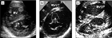
A series of echocardiograms taken in parasternal short axis view showing the arrangement of the so-called “cleft” as seen in an atrioventricular septal defect (a), a true cleft of the aortic leaflet of the mitral valve (b), and the cleft as found in our patient (c). The dotted lines in (b) represent the subaortic outflow tract.
 You have
Access