INTRODUCTION
Saponins – biologically active compounds found in some plant species, which can be present in animal food – are known to affect several processes in animals (Francis et al. Reference Francis, Kerem, Makkar and Becker2002; Wina et al. Reference Wina, Muetzel and Becker2005). In ruminants, saponins can influence rumen microorganisms and modify their activity (Patra & Saxena Reference Patra and Saxena2009; Cieslak et al. Reference Cieslak, Szumacher-Strabel, Stochmal and Oleszek2013). Indirectly, they contribute to increased feed efficiency (Mader & Brumm Reference Mader and Brumm1987; Wang et al. Reference Wang, Ye and Liu2012), improve animal growth (Mader & Brumm Reference Mader and Brumm1987; Hu et al. Reference Hu, Liu, Wu, Guo and Ye2006), milk (Anantasook et al. Reference Anantasook, Wanapat, Cherdthong and Gunun2015) and wool production (Navas-Camacho et al. Reference Navas-Camacho, Laredo, Cuesta, Anzola and Leon1993), and may also affect the quality of milk components (Francis et al. Reference Francis, Kerem, Makkar and Becker2002). Saponins from Quillaja saponaria have been shown to increase the efficiency of protein synthesis by rumen microorganisms in vitro and reduce digestibility of proteins (Makkar & Becker Reference Makkar, Becker, Waller and Yamasaki1996; Hoffmann et al. Reference Hoffmann, Selje-Assmann and Becker2008). Moreover, saponins from Yucca schidigera, Q. saponaria and Salvia officinalis were reported to decrease methane production in vitro (Holtshausen et al. Reference Holtshausen, Chaves, Beauchemin, McGinn, McAllister, Odongo, Cheeke and Benchaar2009; Szczechowiak et al. Reference Szczechowiak, Szumacher-Strabel, Stochmal, Nadolna, Pers-Kamczyc, Nowak, Kowalczyk and Cieslak2013). It was also reported that saponin-induced modifications in the rumen may also influence metabolic parameters and metabolism regulating hormones, food intake, reproduction and lactation (Francis et al. Reference Francis, Kerem, Makkar and Becker2002).
Although many studies concerning the effects of saponins (supplied with diet or via rumen fistula) have been performed, data regarding some aspects of their activity in ruminants are rather scarce. Little is known about saponin influence on processes not related directly to changes in the rumen. It was decided to investigate the influence of S. officinalis root powder, containing triterpenoid saponins, on some metabolic parameters and fatty acid composition in blood and concentrations of hormones regulating metabolic processes in dairy cows.
MATERIALS AND METHODS
Animal care
The experiment was carried out according to The Care and Use of Agricultural Animals in Research and Teaching (FASS 2010) guidelines. All procedures involving live animals were approved by the Institutional Animal Care and Use Committee at Poznan University of Life Sciences (no. 9/2010).
Saponaria officinalis root powder
Dry powder of S. officinalis root was obtained from a commercial source (Herbapol Cracov, Poland). The chemical composition of S. officinalis root powder (SO) was as follows (in g/kg dry matter, DM): organic matter (917 ± 3·3), crude ash (83 ± 3·3), crude protein (CP) (104 ± 1·0), crude fat (5 ± 1·9) and neutral detergent fibre (NDF) (206 ± 14). Concentration of bioactive components in S. officinalis root powder was analysed according to the protocol described by Cieslak et al. (Reference Cieslak, Zmora, Stochmal, Pecio, Oleszek, Pers-Kamczyc, Szczechowiak, Nowak and Szumacher-Strabel2014). The following compound fractions were identified: polysaccharides (310 g/kg DM), saponins (91 g/kg DM) and polyphenols (17 g/kg DM).
Experimental design, cows and treatments
The current experiment was designed as a balanced 3 × 3 Latin square for carryover effects, using three dairy Polish Holstein-Friesian cows. The cows, of mean body weight of 603 ± 5 kg, were enrolled in the experiment at the 4th month of lactation. Each of the three experimental cycles lasted for 26 days, including a 23-day adaptation period (adapting the animals and their rumen microflora to the experimental diets) and a 3-day sample collection period. The three rumen-fistulated cows were part of a larger experiment to investigate the effect of S. officinalis root powder on basic rumen parameters, milk production and CH4 production. During the first 8 days of each adaptation period, the cows were fed ad libitum and S. officinalis root powder was introduced gradually to avoid any negative impact on DM intake. From day 9 onwards, the cows were fed with 95% of the average daily voluntary DM intake they received from days 3 to 8, which consequently allowed for restricting feed consumption by particular cows to avoid the confounding effects of DM intake on milk production. The Latin square design was applied due to a high amount of additive used, and hence the possibility of a negative impact on animal welfare. Control diets were balanced and formulated according to The Institut National de la Recherche Agronomique (INRA) standards to contain 1595 units of truly digestible protein in the small intestine and 16·0 units for milk production (INRA 1993). For calculations, INWAR software version 1.0 and INRAtion program version 4.01 (1998) were used. Diets were prepared daily as follows: the control diet consisted of a total mixed ration (TMR) based on maize silage (7·4 kg DM), ensiled brewer's grains (1·2 kg DM), ensiled beet pulp (0·4 kg DM), meadow hay (1·3 kg DM), rapeseed meal (1·6 kg DM) and a commercial concentrate (6·1 kg DM; 19 g of crude protein/100 g concentrate). The experimental diets were as follows: SO1 diet, which was the control diet supplemented with 440 g/day of S. officinalis root powder, and SO2 diet, which was the control diet supplemented with 660 g/day of S. officinalis root powder. Animals were fed twice a day (05.00 and 17.00 h) in individual box feeders and had free access to water and a mineral saltlick.
Sampling and chemical analysis of feed
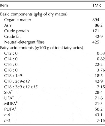
Representative samples of the TMR and S. officinalis root powder were collected three times during each sample collection period and stored at −20 °C until analysis. Samples of the TMR and S. officinalis root powder were analysed according to the AOAC (2007) for DM (method No. 934·01) and ash (method No. 942·05). Crude protein was determined using a Kjel-Foss Automatic 16 210 analyser (method No. 976·05). Crude fat was determined using the Soxtec System HT analyser (method No. 973·18). Neutral detergent fibre and acid detergent fibre were determined using the method of Van Soest et al. (Reference Van Soest, Robertson and Lewis1991). Organic matter was calculated as the difference between DM and ash contents. Fatty acid composition in the TMR was analysed according to the protocol described by Cieslak et al. (Reference Cieslak, Machmüller, Szumacher-Strabel and Scheeder2009). The chemical composition and fatty acid contents of TMR are presented in Table 1.
Chemical composition and fatty acid contents of the total mixed ration (TMR)

* SFA, saturated fatty acids (C12 : 0; C14 : 0; C15 : 0; C16 : 0; C18 : 0; C21 : 0; C22 : 0; C23 : 0).
† UFA, unsaturated fatty acids (cis-9 C16 : 1; cis-9 C18 : 1; cis-11 C18 : 1; cis-9, cis-12 C18 : 2; cis-9, cis-12, cis-15 C18 : 3; C22 : 2).
‡ MUFA, monounsaturated fatty acids (cis-9 C16 : 1; cis-9 C18 : 1; cis-11 C18 : 1).
§ PUFA, polyunsaturated fatty acids (cis-9, cis-12 C18 : 2; cis-9, cis-12, cis-15 C18 : 3; C22 : 2).
Blood sampling and analyses
Blood samples were collected from the jugular vein into tubes containing clot accelerator and granule serum separator (serum separation, Deltalab, Barcelona, Spain) from 24th to 26th day of the experiment, 3 h after morning feeding. Blood was centrifuged at 4000 rpm for 15 min at 10 °C to obtain serum, which was stored at −20 °C until analysed for metabolites and fatty acids.
Concentration of glucose was analysed photocolorimetrically by means of an enzymatic method with peroxide-phenol-aminophenazone (Trinder Reference Trinder1969). Free fatty acid concentrations in serum was determined according to Duncombe (Reference Duncombe1964) and triglyceride concentration by a colorimetric method by Megraw et al. (Reference Megraw, Dunn and Biggs1979). Total cholesterol was assayed by an enzymatic method developed by Trinder (Reference Trinder1969) with cholesterol esterase and oxidase. High-density lipoprotein cholesterol (HDL) and low-density lipoprotein cholesterol (LDL) were determined using commercially available Liquid autoHDL and autoLDL cholesterol reagent sets obtained from Pointe Scientific Inc. (Warsaw, Poland). Fatty acid composition in blood samples was analysed based on Cieslak et al. (Reference Cieslak, Machmüller, Szumacher-Strabel and Scheeder2009). A gas chromatograph (Varian Star CP 3800, Walnut Creek, CA, USA) fitted with a flame ionization detector and a 100-m fused silica capillary column (internal diameter 0·25 mm) coated with 0·2 µm of CP-Sil 88 (CHROMPACK, Varian, Rheinfelden, Germany) was used. Observed peaks were identified by comparison of retention times with appropriate fatty acid methyl ester standards (37 FAME Mix and conjugated LA, Supelco, Poole, UK). Moreover, the C18 : 12c9-t11 peaks were identified by comparison with retention times of the reference standard (conjugated LA methyl esters, a mixture of cis- and trans- 9,11- and -10,12-octadecadienoic acid methyl esters; Sigma-Aldrich, St. Louis, MO, USA). Results are expressed as g/100 g of total fatty acids. The atherogenicity index was calculated according to Chilliard et al. (Reference Chilliard, Ferlay, Rouel and Lamberet2003), whereas the thrombogenic index was calculated as described in Ulbricht & Southgate (Reference Ulbricht and Southgate1991). Erythrocytes were counted under a light microscope (100×) in a Bürker chamber (0·1 mm depth, Blau Brand, Wertheim, Germany).
Concentrations of insulin, glucagon and leptin were determined radioimmunologically using radioimmunoassay (RIA) kits from Millipore (St. Charles, MO, USA). Triiodothyronine (T3) and thyroxine (T4) were assayed by RIA kits from Cisbio Bioassays (Codolet, France), and the ratio of T3 to T4 calculated.
Statistical analysis
Data were analysed with polynomial contrasts to describe linear (L) and quadratic (Q) responses to the level of S. officinalis root powder supplementation. In all analyses, significant effects were declared at P ⩽ 0·05. All analyses were performed by SAS software version 9.3 (SAS Institute Inc., Cary, NC, USA).
RESULTS
Metabolic parameters and hormone concentration in blood
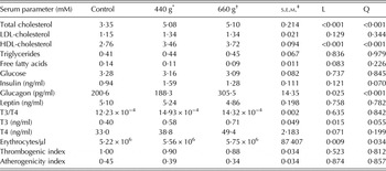
Results presented in Table 2 indicated that total cholesterol was significantly elevated when the diet was supplemented with increasing doses of S. officinalis root powder (L < 0·001, Q < 0·001). Similarly, HDL-cholesterol was augmented (L < 0·001, Q < 0·001), whereas LDL did not change when animals were fed with SO (Table 2). The experimental diets did not affect other metabolic parameters in blood serum, i.e., triglycerides, free fatty acids (FFA) and glucose.
Effect of Saponaria officinalis root administered with diet on metabolic parameter concentrations in blood serum and number of erythrocytes and blood thrombogenic and atherogenicity index (n = 3)

* SO1, 440 g of Saponaria officinalis root supplementation.
† SO2, 660 g of Saponaria officinalis root supplementation.
‡ s.e.m., standard error of the mean.
Increasing the level of SO in cows’ diets resulted in a pronounced rise of T3 (Table 2; L = 0·015). The T3/T4 ratio remained unchanged, which reflected a lack of change in extrathyroidal deiodination of T4. Glucagon concentration was substantially increased due to SO administration (L = 0·025, Q < 0·001) and insulin concentrations also increased after the consumption of experimental diets (Q = 0·07). Leptin concentration remained unaltered.
Erythrocyte number, thrombogenic and atherogenicity indexes
The SO increased erythrocyte numbers in animals (Table 2, L = 0·009, Q = 0·034); however, thrombogenic and atherogenicity indexes were unchanged after saponin ingestion (Table 2, L = 0·523, Q = 0·812 and L = 0·874, Q = 0·857, respectively).
Fatty acid composition in blood
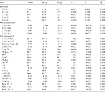
Concentration of C20 : 4n-6 increased linearly after SO supplementation (L = 0·017, Table 3), but the level of analysed monounsaturated fatty acids decreased (C16 : 1c9, C18 : 1t10, C18 : 1t11, C18 : 1c9) as well as saturated fatty acids such as C16 : 0 (L = 0·014).
Effect of Saponaria officinalis root administered with diet on the blood serum fatty acid composition, g/100 g of fatty acids (n = 3)

* SO1, 440 g of Saponaria officinalis root supplementation.
† SO2, 660 g of Saponaria officinalis root supplementation.
‡ s.e.m., standard error of the mean.
§ SFA, saturated fatty acids (C6 : 0; C8 : 0; C10 : 0; C12 : 0; C14 : 0; C15 : 0; C16 : 0; C17 : 0; C18 : 0; C20 : 0; C21 : 0; C22 : 0; C23 : 0; C24 : 0).
# UFA, unsaturated fatty acids (cis-9 C14 : 1; cis-10 C15 : 1; cis-9 C16 : 1; cis-10 C17 : 1; trans-5 C18 : 1; trans-6-8 C18 : 1; trans-9 C18 : 1; trans-10 C18 : 1; trans-11 C18 : 1; trans-12 C18 : 1; trans-15 C18 : 1; cis-9 C18 : 1; cis-11 C18 : 1; cis-12 C18 : 1; cis-13 C18 : 1; cis-14 C18 : 1; cis-15 C18 : 1; cis-9.cis-12 C18 : 2; cis-9.cis-15 C18 : 2; cis-9.trans-11 C18 : 2; trans-10.cis-12 C18 : 2; cis-9.cis-12.cis-15 C18 : 3; cis-6.cis-9.cis-12 C18 : 3; C20 : 1 n-9; C20 : 3 n-3; C20 : 4 n-6; C24 : 1; C22 : 1 n-9; C22 : 2; C20 : 5 n-3; C22 : 6 n-3).
|| MUFA, monounsaturated fatty acids (cis-9 C14 : 1; cis-10 C15 : 1; cis-9 C16 : 1; cis-10 C17 : 1; trans-5 C18 : 1; trans-6-8 C18 : 1; trans-9 C18 : 1; trans-10 C18 : 1; trans-11 C18 : 1; trans-12 C18 : 1; trans-15 C18 : 1; cis-9 C18 : 1; cis-11 C18 : 1; cis-12 C18 : 1; cis-13 C18 : 1; cis-14 C18 : 1; cis-15 C18 : 1; C20 : 1 n-9; C24 : 1; C22 : 1 n-9).
¶ PUFA, polyunsaturated fatty acids (cis-9.trans-11 C18 : 2; trans-10.cis-12 C18 : 2; cis-9.cis-12 C18 : 2; cis-9.cis-15 C18 : 2; cis-9.cis-12.cis-15 C18 : 3; cis-6.cis-9.cis-12 C18 : 3; C20 : 3 n-3; C20 : 4 n-6; C22 : 2; C20 : 5 n-3; C22 : 6 n-3).
DISCUSSION
In the current experiment, a significant increase of total cholesterol in blood serum was observed in both experimental groups of animals, which were administered S.officinalis root powder in their diet, when compared with the control group. The current results contradicted some results obtained in non-ruminants (Shim Reference Shim2010; Wu et al. Reference Wu, Leung, Kwan, Sham, Tang, Wang, Wan, Lee and Chan2013; Weng et al. Reference Weng, Yu, Cui, Zhu, Guo, Wei, Duan, Yin, Guan, Wang, Yang, Xi and Wen2014); however others confirm a lack of saponin effects on cholesterol concentration (Calvert et al. Reference Calvert, Blight, Illman, Topping and Potter1981) or even increased cholesterol synthesis after dietary saponin administration (Jenkins & Atwal Reference Jenkins and Atwal1994). Many studies on monogastric animals have revealed that saponins decreased total blood cholesterol and lowered LDL-cholesterol (Shim Reference Shim2010; Wu et al. Reference Wu, Leung, Kwan, Sham, Tang, Wang, Wan, Lee and Chan2013; Weng et al. Reference Weng, Yu, Cui, Zhu, Guo, Wei, Duan, Yin, Guan, Wang, Yang, Xi and Wen2014). In those studies, the metabolism of cholesterol after saponin treatment was augmented. One of the reasons for this effect was the formation of large mixed micelles by saponins with bile acids (Oakenfull & Sidhu Reference Oakenfull and Sidhu1990), which were excreted, so serum cholesterol concentration was decreased (Oakenfull & Sidhu Reference Oakenfull and Sidhu1990). Moreover, in the intestine, saponins could interact with cholesterol and decrease intestinal cholesterol absorption with no change in bile absorption (Harwood et al. Reference Harwood, Chandler, Pellarin, Bangerter, Wilkins, Long, Cosgrove, Malinow, Marzetta, Pettini, Savoy and Mayne1993). However, these kinds of interactions of saponins are rather implausible in ruminants, because of rumen metabolism. Several studies have demonstrated that saponins are transformed by microbes in the rumen (Gutierrez & Davis Reference Gutierrez and Davis1962; Meagher et al. Reference Meagher, Smith and Wilkins2001; Flåøyen et al. Reference Flåøyen, Wilkins and Sandvik2002) and undergo hydrolysis, epimerization and hydrogenation (Gutierrez & Davis Reference Gutierrez and Davis1962; Meagher et al. Reference Meagher, Smith and Wilkins2001). The derivatives of saponin degradation in the rumen, sapogenins, can manifest other properties concerning interaction with cholesterol. Lorent et al. (Reference Lorent, Le Duff, Quetin-Leclercq and Mingeot-Leclercq2013) studied the interactions of triterpenoid monodesmosidic saponins (α-hederin, a derivative that lacks one sugar, δ-hederin and another derivative, an aglycone form, i.e. hederagenin with cholesterol) and their effect on membrane permeabilization. They found that these interactions are highly dependent on sugars linked to the saponin aglycone form (Lorent et al. Reference Lorent, Le Duff, Quetin-Leclercq and Mingeot-Leclercq2013) and α-hederin was the most effective, whereas hederagenin manifested the smallest activity (Lorent et al. Reference Lorent, Le Duff, Quetin-Leclercq and Mingeot-Leclercq2013). Thus, in the current experiment, the effect of saponins on serum cholesterol concentration, different to that observed in non-ruminants, could be partly the result of their rumen modification and increased sapogenin production. After post-ruminal absorption, the interaction of sapogenins with cholesterol is less active and therefore their influence on blood concentration of LDL-cholesterol could be weaker. The concentration of LDL-cholesterol concentration was observed to be unchanged after saponin administration. However, total cholesterol was increased, probably as a result of augmented HDL-cholesterol, since LDL-cholesterol remained unchanged. It has been reported that elevated blood cholesterol in cattle is related to increased concentration of HDL-cholesterol (Grummer & Carroll Reference Grummer and Carroll1988; Hocquette & Bauchart Reference Hocquette and Bauchart1999). A probable reason for elevated HDL-cholesterol after saponin treatment in the current experiment will be discussed below.
There are many other factors modifying blood cholesterol concentration. Thyroid hormones are responsible for cholesterol metabolism and limit its concentration in blood (Ness Reference Ness1991). Insufficient level of thyroid hormones in blood leads to hypercholesterolaemia (Roberts & Ladenson Reference Roberts and Ladenson2004; Duntas & Wartofsky Reference Duntas and Wartofsky2007). In the current experiment, the increased blood cholesterol was independent of thyroid hormones, because their concentrations after saponin treatment were increased.
As for the other active compounds, fatty acids, depending on the number of saturated bonds and isomeric form, can improve or impair cholesterol metabolism due to its concentration in blood (Fernandez & West Reference Fernandez and West2005; Ascherio Reference Ascherio2006; Aronis et al. Reference Aronis, Khan and Mantzoros2012). Unsaturated fatty acids improve both cholesterol metabolism and its elimination from blood (Fernandez & West Reference Fernandez and West2005). Although the concentrations of FFA were unaltered in the current experiment, their profile – proportions and composition – varied significantly, suggesting an important influence of saponins on fatty acid synthesis/distribution in the organism. However, other than volatile fatty acid (VFA) modification in the rumen (Wina et al. Reference Wina, Muetzel and Becker2005), this effect has not so far been studied in animals. The only report concerns the effect of Q. saponaria L. saponin on meat fatty acid composition in lambs – an increase in C20 : 4n-6 amount was observed, as well as reduced ratio between α-linolenic and linoleic acids (Brogna et al. Reference Brogna, Nasri, Salem, Mele, Serra, Bella, Priolo, Makkar and Vasta2011). It was established in in vivo (Wallace et al. Reference Wallace, Arthaud and Newbold1994) and in vitro (Makkar et al. Reference Makkar, Sen, Blümmel and Becker1998) experiments that saponins can modify the balance of rumen microorganisms through their antiprotozoal activity and due to this fact an increase of rumen bacteria was observed. These microbes participate in dietary fatty acid metabolism. Considering the above data, it can be assumed that altered concentrations of fatty acids in the current experiment could be the result of changes in rumen microflora after saponin action.
Unsaturated fatty acids improved both cholesterol metabolism and its elimination from the blood (Fernandez & West Reference Fernandez and West2005). Dietary saturated fatty acids (especially C16 : 0), as well as trans isomers of unsaturated fatty acids, evoked deleterious effects on cholesterol metabolism (Fernandez & West Reference Fernandez and West2005; Brouwer et al. Reference Brouwer, Wanders and Katan2010; Ooi et al. Reference Ooi, Ng, Watts and Barrett2013), increasing concentrations of LDL-cholesterol and lowering HDL-cholesterol. Moreover, there is no doubt that in monogastric animals and humans, dietary polyunsaturated fatty acids (PUFA), especially n-3 but also n-6, favour the metabolism of cholesterol augmenting HDL-cholesterol and reducing LDL-cholesterol blood concentrations (Fernandez & West Reference Fernandez and West2005). In cattle, dietary PUFA undergo biohydrogenation in the rumen (Demeyer & Doreau Reference Demeyer and Doreau1999), however, this process can be limited by various factors (Petersen & Jensen Reference Petersen and Jensen2014). Therefore, these fatty acids can be absorbed from the rumen into blood and then be present in milk. Taking into account the data above, the elevated blood concentration of n-6 - C20 : 4n-6 observed in the current experiment after saponin administration at the higher dose could be one of the reasons for elevated HDL-cholesterol concentration in blood. Hocquette & Bauchart (Reference Hocquette and Bauchart1999) found that diets enriched in PUFA can stimulate synthesis of HDL and may contribute to higher blood cholesterol concentration. This effect may be considered as protective due to prevention of atherogenesis. Other parameters of significant health importance evaluated in the current experiment – atherogenic or thrombogenic indices – were unchanged, suggesting rather harmless saponin action.
The substantial elevation of serum T3 and numerically increased T4 were observed after saponin administration. So far, there is no evidence confirming any possible effect of saponins on the thyroid gland and thyroid hormone secretion in dairy cows. However, as was mentioned above, numerous reports from in vivo and in vitro studies have indicated that saponins, through inactivation of protozoa in rumen, contribute to increases in the bacterial population (Makkar et al. Reference Makkar, Sen, Blümmel and Becker1998). Therefore, an increase of microbial protein synthesis is observed (Makkar & Becker Reference Makkar, Becker, Waller and Yamasaki1996; Wang et al. Reference Wang, McAllister, Newbold, Cheeke and Cheng1997; Patra & Yu Reference Patra and Yu2014), as well as bacterial nitrogen flow to the duodenum. Thus, the elevation of serum thyroid hormone concentration observed in the current experiment may result from improved protein turnover after saponins’ action. It was reported by Hammond et al. (Reference Hammond, Huntington and Bitman1984) that an increase in T4 concentration in growing steers was related to an increased protein intake. Moreover, Gerrits et al. (Reference Gerrits, Decuypere, Verstegen and Karabinas1998) found elevated T3 and T4 in blood after increased protein intake in calves. According to Gerrits et al. (Reference Gerrits, Decuypere, Verstegen and Karabinas1998) the augmented T3 concentration resulted from the increased T4 concentration (Gerrits et al. Reference Gerrits, Decuypere, Verstegen and Karabinas1998). There is also a report about a stimulatory effect of soyasaponins on thyroid hormone concentration in mice (Ishii & Tanizawa Reference Ishii and Tanizawa2006).
Besides thyroid hormones, glucagon plays a pivotal role in lactating ruminants (Danfaer et al. Reference Danfaer, Tetens and Agergaard1995). Glucagon concentration in portal blood can be increased as a consequence of elevated amino acids or propionate, and their availability seems to be an important factor limiting the process (Danfaer et al. Reference Danfaer, Tetens and Agergaard1995). In the present experiment, saponins administered at a higher dose evoked a substantial rise in glucagon concentration. The glucagon increase after saponin treatment could be related to their action in the rumen. It is commonly known that the major effect of saponins on rumen fermentation is an increased production of propionate and decreased acetate to propionate ratio (Wina et al. Reference Wina, Muetzel and Becker2005). Moreover, Budan et al. (Reference Budan, Bellenot, Freuze, Gillmann, Chicoteau, Richomme and Guilet2014) recently observed in an in vitro experiment that S. officinalis root extract increased total VFA concentration in the rumen and markedly decreased acetate to propionate ratio. In an earlier experiment, Cieslak et al. (Reference Cieslak, Zmora, Stochmal, Pecio, Oleszek, Pers-Kamczyc, Szczechowiak, Nowak and Szumacher-Strabel2014) obtained similar results using S. officinalis root powder in in vitro ruminal fermentation. Although in the present experiment the exact content of VFA in serum was not analysed, it can be supposed that elevated propionate production in rumen could be one possibility for the increased glucagon concentration.
Unlike glucagon, blood insulin concentration was only slightly increased after feeding with S. officinalis root powder. There are no other data concerning the effect of saponins on insulin concentration in ruminants; however, rodent studies revealed that saponins augmented insulin secretion (Lee et al. Reference Lee, Kao, Liu and Cheng2006; Park et al. Reference Park, Ha and Chung2008) and showed antidiabetic properties, decreasing disaccharidase activities in the small intestine (Lee et al. Reference Lee, Choi, Seo, Lee, Lee, Lee, Kim and Lee2014), stimulating insulin signalling (Kwon et al. Reference Kwon, Kim, Hong and Park2009) and lowering glucose concentration (Singh et al. Reference Singh, Farswan, Ali, Afzal, Al-Abbasi, Kazmi and Anwar2014; Weng et al. Reference Weng, Yu, Cui, Zhu, Guo, Wei, Duan, Yin, Guan, Wang, Yang, Xi and Wen2014).
Saponins are known to evoke some deleterious effects on blood cells. In many studies performed on rodents and humans, it was established that saponins exhibited a lytic action on erythrocyte membranes, probably connected to their affinity for membrane sterols (Bomford Reference Bomford1980). For human erythrocytes, triterpenoid saponins appeared to be highly haemolytic (Sindambiwe et al. Reference Sindambiwe, Calomme, Geerts, Pieters, Vlietinck and Vanden Berghe1998). In vitro ruminant studies also proved their haemolytic properties (Voutquenne et al. Reference Voutquenne, Guinot, Froissard, Thoison, Litaudon and Lavaud2005; Budan et al. Reference Budan, Tessier, Saunier, Gillmann, Hamelin, Chicoteau, Richomme and Guilet2013); however, in vivo ruminant studies did not confirm the detrimental effects of saponins on blood cell parameters (Hess et al. Reference Hess, Beuret, Lötscher, Hindrichsen, Machmüller, Carulla, Lascano and Kreuzer2004; Sliwinski et al. Reference Sliwinski, Kreuzer, Sutter, Machmuller and Wettstein2004). In dairy cows, no effects of Yucca saponin were observed on haematocrit readings or on haemoglobin concentration (Sliwinski et al. Reference Sliwinski, Kreuzer, Sutter, Machmuller and Wettstein2004). The lack of haemolytic effect was also noticed in sheep fed with S. saponaria (Hess et al. Reference Hess, Beuret, Lötscher, Hindrichsen, Machmüller, Carulla, Lascano and Kreuzer2004). In the current experiment, saponins exerted a different effect; the red blood cell number in dairy cows was significantly elevated. It is difficult to explain this result, particularly since literature data concerning this problem in ruminants are very scarce. Further experiments are needed to explain this phenomenon.
CONCLUSIONS
To summarize, saponins from S. officinalis root powder administered in the diet of lactating dairy cows evoked significant changes of parameters of much importance for metabolism. The current data allow the fact to be established for the first time that saponins from S. officinalis can alter blood parameters in ruminants. These effects seem to result from saponin-induced changes in the rumen. Hitherto, saponins were known as important factors favourably modifying rumen fermentation and methanogenesis, improving ruminant growth and production. The results of the current experiments prompt one to suppose, rather, that they do not exert any detrimental effects on animal welfare. However, the dose and the type of saponins used in ruminant nutrition, as well as their plant source, must be considered due to the possibility of some health disturbances. Further studies demonstrating the effects of saponins on reproductive processes are also needed to ensure that they are safe for dairy cows, because no data concerning this issue were published.
This study was supported by Ministry of Science and Higher Education No. NN311476339 to Adam Cieslak.