Two-dimensional versus three-dimensional cell culture for tissue engineering
The direct integration of fully functional, natural cell-based materials as a therapeutic for damaged endogenous tissue dates back to 2500 bc when Susruta excised skin from one person and sewed it onto another. Reference Chick1 Tissue engineering aims to develop functional tissue constructs from cultured cells that can be directly implanted into damaged tissue, either for local repair or for full replacement. Modern regeneration techniques lie between the two extremes as a means of integration into living organisms—“top-down” assembly of individual cells and “bottom-up” assembly of pre-assembled cells. The top-down approach involves seeding cells into three-dimensional (3D) scaffolds that guide attachment and proliferation. The bottom-up approach involves aggregating cells from simple two-dimensional (2D) culture to achieve functional tissue without the need for an intermediate scaffold.
These extremes exist because functional tissue cannot be regenerated simply by mixing the requisite cells and allowing them to assemble into structures that possess the higher-order organization of native tissue with respect to both the spatial distribution of the cells and the extracellular matrix (ECM). The ECM is a collection of noncellular components that is formed during tissue development, providing both biochemical and structural support for the cells. Consequently, some sort of templating must exist to at least drive initial organization. The simplest approach to organizing cells is 2D cell culture. This was first achieved in 1910, when a fragment of tadpole nerve cord was placed into a droplet of frog lymph and new nerve fibers were observed. Reference Harrison2 However, 2D culture does not provide the necessary guidance to engender a specific tissue architecture with the level of complexity that replicates developing embryos, wound healing, or the regeneration activities of living animals. Reference Pampaloni, Reynaud and Stelzer3 This is not entirely surprising, as 2D environments (at least those imposed by the ubiquitous petri or agar dishes) are not representative of the cellular environment found in physiological tissues.
As a result, there has been significant research into 3D cell culture. The first known example was demonstrated in 1972, when fetal lung fibroblasts were cultured on hydrated collagen lattices. Reference Elsdale and Bard4 Novel cellular activity was immediately observed. Cells attached and extended within the lattices unlike their behavior on soft agar. It is now commonly accepted that 3D cell culture has several advantages over 2D culture with respect to cell proliferation, orientation, cooperation, and gene expression. For example, 3D cultured cells can up- and down-regulate genes at levels similar to those found in living organisms. A good example is the 2D and 3D culture of mammary epithelial cells; 2D culture leads to higher than normal expression of cell adhesion proteins, whereas 3D culture regulates expression levels more closely associated with those found in native tissues. Reference Delcommenne and Streuli5
Despite the plethora of research into 3D cell culture, there remains a lack of standards or universally accepted approaches for achieving functional tissue. The general concept behind most strategies includes seeding individual cells into degradable scaffolds. Reference Knowlton, Cho, Li, Khademhosseini and Tasoglu6 The cells are expected to attach and proliferate while simultaneously secreting ECM as the scaffold degrades. Reference Heydrick, Roberts, Kim, Emani and Wong7 Scaffolds, in general, have a wide range of designs from the simple to the complex, where morphology, porosity, interconnectivity, and stiffness are explicitly controlled. Scaffolds can also have well-controlled variations in properties (including morphological, chemical, and mechanical) for the engineering of anisotropic tissues.
Even with this diversity in scaffold designs, clinical successes of top-down engineering are largely limited to thin tissues such as skin or avascular tissues such as cartilage—for thick tissues, the major obstacle is mass transport via vascularization. Reference Heydrick, Roberts, Kim, Emani and Wong7 The reason for this is that while cells populate the scaffold architecture, they do so without forming a vascular network. Thus, all nutrients and waste must be transported to and from the cells by diffusion. Reference Wei, Dai and Yin8 Diffusion only provides adequate metabolite exchange for constructs less than approximately 200-µm thick. Reference Carmeliet and Jain9 Just as importantly, the scaffold itself competes for space and limits the growth of cell-dense tissues.
It could be argued that vascularization and cell density are matters related to properly designing the scaffold. However, it could also be argued that scaffolding strategies are too wide ranging, and in some cases, too complicated, to be universally adopted. To address this complexity, cell sheet engineering may offer several advantages over 3D cell culture.
Cell sheet engineering promises simplicity of 2D cell culture with benefits of 3D cell culture
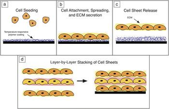
Cell sheet engineering is a type of scaffoldless tissue engineering that involves building functional tissue from cell-dense monolayers with well-formed cell–cell junctions and the ECM. Cells are initially cultured in two dimensions and then combined layer by layer to construct 3D tissue (as depicted in Figure 1 ). Reference Okano, Yamada, Sakai and Sakurai10 Since each layer is cultured as a 2D sheet, vascularization, at least initially, is unnecessary. Artificial scaffolds are not used in this approach, rendering both their complex manufacturing procedures and associated complications regarding cell growth moot. While the limitations of 2D cell culture have previously been stated, the stacking of the cell sheets creates a 3D environment with preformed ECM that potentially reduces the gap between engineered and native tissue.
Cell sheet engineering. Cells are initially seeded on a (a) “releasable” cell culture dish with a temperature-responsive polymer coating that guides (b) attachment, spreading, and extracellular matrix (ECM) secretion. (c) By changing temperature, the intact cell monolayer is released, which can be subsequently stacked with (d) similar or dissimilar cell sheets to form a 3D cellular construct.

In order for any type of layer-by-layer stacking process to be successfully implanted, the cells must be removed from the cell culture substrate as an intact sheet. In traditional cell culture, separating cells from substrates requires digestive enzymes such as trypsin, which cleave surface proteins and disrupt proliferation or differentiation of cells. Reference Huang, Hsing, Lai, Chen, Lee, Chan, Lyu, Wu, Lu, Lin, Lin, Lai, Chang, Chou and Chan11 Further, the scavenging of divalent cations with agents such as ethylenediaminetetraacetic acid (EDTA) breaks cell junctions, separating constructs into individual cells. Hence, traditional methods cannot be used to harvest intact cell sheets. Reference Sumide, Nishida, Yamato, Ide, Hayashida, Watanabe, Yang, Kohno, Kikuchi, Maeda, Watanabe, Okano and Tano12
Okano et. al. first showed that culturing cells on a thin responsive polymer coating that switches between a hydrophobic and a hydrophilic state could provide a mechanism for releasing sheets of cells with both ECM and cell–cell junctions intact to overcome such difficulties. Reference Okano, Yamada, Sakai and Sakurai10 The most widely used material for this purpose is a thermally responsive polymer known as poly(N-isopropylacrylamide) (pNIPAAm), which switches from the hydrophobic to the hydrophilic state at approximately 32°C.
At cell culture temperatures (37°C), pNIPAAm takes on a hydrophobic state to which suspended cells will attach and grow to confluency, the point at which cells cover the entire culture surface completely. A variety of cell types can be patterned onto a single cell sheet with a high degree of control over spatial orientation. After cells have reached confluency, the temperature is lowered below 32°C, which increases the water content of the pNIPAAm coating. In this process, the cultured cells start to lose their flattened appearance and become rounded, which indicates an alteration of the cytoskeletal stress. Reference Yamato, Okuhara, Karikusa, Kikuchi, Sakurai and Okano13,Reference Ingber, Dike, Hansen, Karp, Liley, Maniotis, McNamee, Mooney, Plopper, Sims and Wang14 This occurs slowly, suggesting an adenosine triphosphate (ATP)-dependent process like cytoskeletal rearrangement. After a period of time, the contiguous sheet of cells and matrix is released under gentle agitation. Staining indicates that cell–matrix junctions are maintained when the cells are released from the polymer; but, the mechanism of cell sheet separation form the pNIPAAm surface is not well understood. Further, the role of metabolic processes is not clear. Reference Hirose, Kwon, Yamato, Kikuchi and Okano15 However, the fact that a gentle agitation dislodges the sheet suggests that the interface between the sheet and the polymer is weakened by the hydration of the grafted polymer layer. The released cell sheet with its cells and intact matrix allows direct adhesion to other cell sheets in vitro or to tissues in vivo without the use of sutures. Moreover, cellular activity of these cell sheets is reported to be similar to that of native tissues. Reference Hirose, Kwon, Yamato, Kikuchi and Okano15
Other methods have also been demonstrated for the release of intact cell sheets, though they are far less studied and have not been developed sufficiently for implantation purposes. For instance, magnetic fields can be used to control attachment and release of magnetically labeled cells. Reference Ito, Hayashida, Honda, Hata, Kagami, Ueda and Kobayashi16 Electric fields have also been shown to release cells from surfaces with electroactive ligands. Reference Yeo and Mrksich17 Sacrificial substrates such as tyramine-conjugated polymers of carboxymethyl cellulose and alginate can be degraded by enzymes that catalyze hydrolysis for detachment. Reference Heydrick, Roberts, Kim, Emani and Wong7 The hydrophobicity of titanium and zinc oxide can be controlled by UV light to trigger cell release. Reference Hong, Yu, Weng, Cheng, Wang and Lin18 Spiropyran, a photochromatic molecule, also changes hydrophobicity when exposed to cell-safe 365-nm UV light. It can be grafted to both poly(methyl methacrylate) and pNIPAAm to develop photonically triggered responsive polymers for cell sheet detachment. Research into the integrity of cell junctions and ECM through these approaches have yet to be undertaken.
A strain-based method has also been reported to release cells. Akintewe et al. exploited the swelling response of surface arrays of high-aspect-ratio pNIPAAm microbeams. Reference Akintewe, DuPont, Elineni, Cross, Toomey and Gallant19 Similar to Okano’s method, cells attach and grow to confluency at cell culture temperatures. As temperature is reduced below 32°C, the microbeams undergo significant lateral swelling, leading to tension on the cell sheet. If the strain associated with the swelling exceeds a certain threshold, intact cell sheets can be released on the order of 1 s without the need for agitation. The speed of this action suggests that it is not an ATP-dependent process. In fact, it was shown that even cell sheets that had their metabolism blocked could still be released, in contrast to the standard cell sheet release method of Okano. Reference Yamato, Konno, Kushida, Hirose, Utsumi, Kikuchi and Okano20
There are potential advantages to the high-aspect-ratio microbeam array approach over other thin, low-aspect-ratio coatings. First, secondary chemicals are not needed for patterning as this is built into the microbeam culture surface. Second, cell sheet shapes are determined by contact guidance of the microbeams edges, which, if the microbeams are on the order of cellular dimensions, can be grown with a high degree of spatial organization. Reference Akintewe, DuPont, Elineni, Cross, Toomey and Gallant19 Last, the rapid detachment minimizes alignment loss and morphological changes due to normal metabolic activity of the cells.
Clinical successes of cell sheet engineering
The goal of any one of these release methods is the ability to fabricate scaffold-free, functional cell-dense tissue for direct implantation. Cell sheet technology has been successfully tested in numerous therapeutic applications using the following general procedure (as denoted in Figure 2 ). Essentially, if an organ is damaged, autologous cells of that organ can be seeded onto specially designed cell-release culture dishes. The cells are cultured to confluency, at which time, release is triggered. This sheet can then be transferred or stacked on another creating a 3D cellular construct. Cell sheets quickly adhere to each other without extra additives. Reference Hirose, Kwon, Yamato, Kikuchi and Okano15 Currently, most constructs are three to five layers thick due to the vascularization limitations previously discussed. Several clinical successes of cell sheet technology are highlighted next.
Example of cell sheet engineering for corneal reconstruction. First, isolated epithelial cells from a patient are seeded onto a poly(N-isopropylacrylamide coated cell culture dish and incubated for two weeks at 37°C. After the two-week period, the temperature is reduced to 20°C for 30 min, which triggers release of the cell sheet from the temperature-responsive cell culture with intact cell–cell junctions and extracellular matrix. The cell sheet can then be transplanted directly onto the diseased eye without the need for sutures. Note: The cornea is approximately 11.5 mm in humans. Reprinted with permission from Reference Reference Nishida, Yamato, Hayashida, Watanabe, Yamamoto, Adachi, Nagai, Kikuchi, Maeda, Watanabe, Okano and Tano22. © 2004 Massachusetts Medical Society.

Stem cell deficiencies in the corneal limbus can lead to vision loss. Reference Iwata, Washio, Yoshida, Ishikawa, Ando, Yamato and Okano21 Without these stem cells, regeneration of the corneal epithelial lining (outermost layer of cells covering the front of the cornea) is impaired. However, cell sheet technology has proven successful in repairing this type of damage. Autologous oral mucosal cells, which are extremely easy to harvest and quite similar to native ocular tissue, were seeded onto thin-film pNIPAAm modified dishes and cultured for 14 days. A cell sheet construct was then grown and transplanted directly into the eye without sutures. Reference Nishida, Yamato, Hayashida, Watanabe, Yamamoto, Adachi, Nagai, Kikuchi, Maeda, Watanabe, Okano and Tano22 It was suggested that the construct was able to stabilize in the eye without sutures due to the presence of ECM secreted in 2D culture. This procedure was performed on four patients; all showed complete re-epithelization of corneal surfaces and had their vision restored. A similar successful study was conducted with 25 patients and 360-day follow-ups in France. No complications ensued. Reference Burillon, Huot, Justin, Nataf, Chapuis, Decullier and Damour23
In another example, a patient who was in need of a left ventricular assist system in the heart to pump blood was successfully treated with cell sheet technology. This time, autologous cardiac muscle cells from the patient’s thigh were seeded onto thin-film pNIPAAm modified culture dishes. Several tissue constructs were fabricated from stacking four cell sheets and implanted onto the diseased part of the patient’s heart. Reference Sawa, Miyagawa, Sakaguchi, Fujita, Matsuyama, Saito, Shimizu and Okano24 After eight weeks, the patient’s left ventricle ejection fraction, or the volume of blood ejected from the ventricle during systole (contraction) divided by the volume of blood in the ventricle at the end of diastole (relaxation), increased from 26% to 46% (normal ranges are between 50% and 75%), and the overall function of the heart improved. The patient was discharged and no longer needed the assist device.
Animal studies have also shown high levels of success with cell sheet implants. Autologous oral mucosal epithelial cell sheets have proven to be effective treatments to alleviate the postoperative effects of cancerous esophageal biopsies in dogs. Reference Ohki, Yamato, Murakami, Takagi, Yang, Namiki, Okano and Takasaki25 Two cell sheets were individually implanted one at a time at the ulceration site. No sutures were used. The cell sheets were first attached to a polyvinylidene difluoride support membrane when harvested from the cell-release culture dish. The membrane and the cell sheet were then maneuvered into position using an endoscope. Pressure was applied and after 10 min, the membrane was removed and the cell sheet remained in place. This was repeated again with the second sheet. Canines receiving the cell sheets recovered completely in four weeks compared to control animals that had only healed partially. Human clinical trials for this procedure have begun. 26
Similarly, chondrocyte (cartilage) cell sheets have proven effective in repairing the partial thickness defects associated with osteoarthritis, chronic degeneration of the joints, in rabbits. Although drilling techniques are currently used to induce differentiation of marrow cells into chondrocytes, cartilage produced from this approach is fibrous, which is both visually and structurally different from the hyaline cartilage generally found in joints. Reference Wakitani, Imoto, Yamamoto, Saito, Murata and Yoneda27 Further, while top-down tissue engineering of bone-marrow-derived mesenchymal stem cells (stem cells that turn into different cells) can aid in the regeneration of full thickness defects, they fail to repair partial defects. Reference Kaneshiro, Sato, Ishihara, Mitani, Sakai and Mochida28 Cell sheet engineering has demonstrated the potential to repair these defects. Three-layer sheet constructs composed of human and rabbit chondrocytes were attached to defective areas with nylon sutures. Both human and rabbit constructs retained their cartilaginous phenotype after implantation and acted as a barrier to preventing further protein degradation.
Finally, cell sheet constructs can also demonstrate rather sophisticated levels of functionality similar to native tissues, though these functionalities have yet to be applied to clinical issues. For example, cell sheet constructs composed of neonatal rat cardiomyocytes have been cultured and demonstrated to spontaneously beat upon the stacking of four layers. Reference Shimizu, Sekine, Isoi, Yamato, Kikuchi and Okano29 Three-layer sheets have been transplanted subcutaneously in rats and they maintained a pulse for over one year. Moreover, multilayer hepatocyte (liver cell) sheets were subcutaneously implanted in mice where they formed a large 3D liver-like mass with native tissue morphology. Reference Ohashi, Yokoyama, Yamato, Kuge, Kanehiro, Tsutsumi, Amanuma, Iwata, Yang, Okano and Nakajima30 The mass was stable for 140 days.
Vascularization remains a significant challenge in cell sheet engineering
While there has been success in the repair of damaged tissue with cell sheet constructs comprising five or less layers, the creation of thicker constructs with preformed vasculature is still unresolved. However, as previously stated, thin constructs can survive on the metabolite exchange supplied by diffusion. Thus, significant work has been conducted in attempts to vascularize thinner constructs that can survive on diffusive transport before they are “stacked” together into a larger one.
For example, human umbilical vein endothelial cells (HUVECs) are known to promote vascularization. Endothelial cells line the interior of blood vessels. Sasagawa et al. cultured dense myoblast cell sheets and sparse HUVEC cell sheets. Reference Sasagawa, Shimizu, Sekiya, Haraguchi, Yamato, Sawa and Okano31 The cell sheets were alternately layered (up to five myoblast cell sheets); preliminary capillary networks were observed to form after four days of in vitro culture. Subsequent implantation of this construct into a rat showed that the capillary networks merged with the rat’s circulatory system.
Another vascularization technique is polysurgery, which refers to the technique of implanting thin constructs into an animal, waiting for vascularization to occur, and then implanting another one. Ten separate three-layer myoblast cell constructs were successfully implanted and vascularized in this manner. The surgeries were performed on a single rat, waiting one to two days between each surgery. A vascularized construct 1-mm thick was produced. Reference Shimizu, Sekine, Yang, Isoi, Yamato, Kikuchi, Kobayashi and Okano32 Times between surgeries cannot be extended, however, as too many connective tissue forms and the layers will beat asynchronously.
Finally, cell sheets cultured on a perfusion bioreactor (which provides an actively flowing nutrient feed) produced cell sheets 110-μm thick. Reference Sakaguchi, Shimizu, Horaguchi, Sekine, Yamato, Umezu and Okano33 Here, three-layer cardiomyocyte cell sheets interspersed with endothelial cells were placed on the bioreactor. Nutrients were transported through the collagen channels underneath. Endothelial cells from within the construct migrated through the collagen forming a vascular network to the nutrients. Additional three-layer constructs were stacked on top at five-day intervals, eventually producing a viable 12-layer cell sheet with a vascular network.
Conclusion and outlook
Cell sheet engineering provides a simple way to fabricate functional cell-based materials from 2D cultured cells that (1) behave similarly enough to native tissue to allow direct implantation in certain cases and (2) do not require vascularization to survive. While 2D cultured cells lack the cellular environment found in living organisms, the ability to harvest the cultured cells as sheets with intact ECM to fabricate 3D constructs in a layer-by-layer fashion may ultimately render native-like tissues that can be directly integrated into living systems. Thin constructs have already proven successful in several surgical applications, and vascularization of thick tissue constructs can be undertaken at the building-block level as opposed to the entire construct. This has been demonstrated by the techniques of polysurgery and perfusion induced vasculature formation where smaller constructs can survive indefinitely until vascularization occurs. Reference Shimizu, Sekine, Yang, Isoi, Yamato, Kikuchi, Kobayashi and Okano32,Reference Sakaguchi, Shimizu, Horaguchi, Sekine, Yamato, Umezu and Okano33
In the larger context, however, cell sheet engineering is not as well studied as scaffold-based, top-down engineering approaches. There are three unresolved questions: (1) What is the extent to which stacked 2D cultured cells mimic physiologically relevant mechanical and biochemical cues or induce proper cell–cell signaling between sheets? (2) What is the effect of cell sheet release, if any, on the behavior and properties of the cultured cells and surrounding ECM? (3) How can vascularization be established that connects to native tissue upon implantation? It is envisioned that the further development of cell-release culture platforms and stacking schemes (and perhaps in combination with scaffolding technology) will greatly expand the repertoire of fabrication strategies for fully functional tissue constructs.
Acknowledgment
N.G. and R.T. gratefully acknowledge partial support from NSF-CMMI 1538727.

Nicholas Baksh is a doctoral candidate in mechanical engineering at the University of South Florida. He received his bachelor’s degrees in mechanical engineering and management from the Massachusetts Institute of Technology. He was a partner at Oxy-Care Inc. for 10 years. His research interests include degradable medical implants for bone regeneration, cytoplasmic streaming, and biomimetic engineering designs. Baksh can be reached by phone at 813-685-5693 or by email at nbaksh@mail.usf.edu.

Nathan D. Gallant has been an associate professor in the Department of Mechanical Engineering at the University of South Florida since 2008. He received his BS degree in 1999, MS degree in 2004, and PhD degree in 2004, all in mechanical engineering, from the Georgia Institute of Technology. He was a National Research Council postdoctoral fellow at the National Institute of Standards and Technology in the Polymers Division. His Cellular Mechanotransduction and Biomaterial Laboratory was established to analyze the mechanical and biochemical interactions between cells and their microenvironment in order to control cell behavior. Gallant can be reached by phone at 813-974-5840 or by email at ngallant@usf.edu.

Ryan G. Toomey has been an associate professor in the Department of Chemical and Biomedical Engineering at the University of South Florida since 2005. He received his bachelor’s degree in chemical engineering from the University of California, Berkeley, in 1995, and his PhD degree in chemical engineering at the University of Minnesota in 2002. His research focuses on polymers and gels, particularly the physics of polymers that deform under external stimuli, including electric, magnetic, and thermal cues. He is the recipient of a 2007 NSF CAREER Award. Toomey can be reached by phone at 813-974-9164 or by email at toomey@usf.edu.