Introduction
During fertilization, sperm entry triggers an increase in egg cytosolic Ca2+, which in mammals is followed by sustained oscillations in intracellular Ca2+ concentration ([Ca2+]i) (Whitaker & Swann, Reference Whitaker and Swann1993; Swann & Ozil, Reference Swann and Ozil1994). In lower species, such as echinoderms and frogs, a single transient occurs that traverses the egg in a wave-like fashion (Nuccitelli, Reference Nuccitelli1991). These transient increases in [Ca2+]i are essential for cortical granule release, meiosis resumption and pronuclear formation (Schultz & Kopf, Reference Schultz and Kopft1995). A similar effect is brought about by treatment with several agents, which elevate intracellular calcium levels. Mimicking the fertilization calcium transients and oscillations have been widely used to achieve the artificial activation of oocytes in nuclear transplantation (Collas et al., Reference Collas, Fissore, Robl, Sullivan and Barnes1993; Rickords & White, Reference Rickords and White1993).
The source of Ca2+ for the initial transient originates mainly from intracellular stores located in specialized regions of the endoplasmic reticulum (Miyazaki et al., Reference Miyazaki, Shirakawa, Nakada and Honda1993; Deguchi et al., Reference Deguchi, Osanai and Morisawa1996).
Despite the importance of Ca2+ in oocyte activation, little is known about the mechanisms by which sperm causes Ca2+ release from intracellular stores. The most accepted theories involve two families of intracellular calcium channels: the inositol trisphosphate receptor (IP3R), which is universal and mediates IP3-induced Ca2+ release (IICR) and the species-specific ryanodine receptor (RyR), which takes part in Ca2+-induced Ca2+ release (CICR). The way in which these two pathways interact in eggs expressing both receptors is unsolved. Functional IP3Rs are present in the eggs of all studied species (Clapper & Lee, Reference Clapper and Lee1985; Carroll & Swann, Reference Carroll and Swann1992; Fissore et al., Reference Fissore, Dobrinsky, Balise, Duby and Robl1992; Swann, Reference Swann1992; Fissore & Robl, Reference Fissore and Robl1993; Galione et al., Reference Galione, McDougall, Busa, Willmott, Gillot and Whitaker1993a; Rickords & White, Reference Rickords and White1993; White et al., Reference White, Bunch, Reed, Wang and Yue1993) and appear to play a crucial role in egg activation. These receptors have been characterized in both sea urchin (Lee, Reference Lee, Aarhus and Walseth1993) and Xenopus eggs (Parys et al., Reference Parys, Sernett, DeLisle, Sayder, Welsh and Campbell1992, Reference Parys, McPherson, Matthews, Campbell and Longo1994). Functional RyR/channel complexes appear to be present in the eggs of some species such as sea urchins, mice, bovines and humans (Swann, Reference Swann1992; Galione et al., Reference Galione, McDougall, Busa, Willmott, Gillot and Whitaker1993a; Ayabe et al., Reference Ayabe, Kopf and Schultz1995; Yue et al., Reference Yue, White, Reed and Bunch1995; Herbert et al., Reference Herbert, Gillespie and Murdoch1997), even if their role in Ca2+ signalling at fertilization is unclear (Galione et al., Reference Galione, McDougall, Busa, Willmott, Gillot and Whitaker1993a; Ayabe et al., Reference Ayabe, Kopf and Schultz1995; Berridge, Reference Berridge1996). Surprisingly, RyRs have not been previously detected in Xenopus laevis oocytes (Erickson et al., Reference Erickson, Mooren, Moore-Nichols and Dunn2004), although their presence in oocytes from both lower and higher order species has been reported.
The effect of thimerosal on IP3R-mediated Ca2+ release is well documented. Studies on several cell types demonstrate that thimerosal sensitizes IP3Rs by increasing affinity for IP3 binding (Bootman et al., Reference Bootman, Taylor and Berridge1992; Bird et al., Reference Bird, Burgess and Putney1993; Hilly et al., Reference Hilly, Pietri-Rouxel, Coquil and Mauger1993). However, this action is specific only when low doses of thimerosal are used. At higher doses and during prolonged exposure, it inhibits Ca2+-ATPase pumps. This action causes a passive movement of free Ca2+ into the cytoplasm that can, in turn, trigger oscillations through a CICR mechanism (Herbert et al., Reference Herbert, Gillespie and Murdoch1997). Heparin is a well known IP3R antagonist, widely used in search of IP3Rs in many species.
RyR agonists include exogenous ryanodine and caffeine, and endogenous cyclic ADP-ribose (c-ADP), a CICR modulator that has been proposed as second messenger (Berridge, Reference Berridge1993; Galione et al., Reference Galione, White, Willmott, Turner, Potter and Watson1993b). RyR antagonists include ruthenium red and procaine.
The aim of this work was to investigate the existence of these receptors in in vitro matured Bufo arenarum oocytes and their probable role in triggering egg activation events.
Materials and methods
Animals
Adult specimens of Bufo arenarum were collected in the northwestern area of Argentina and kept at 15 °C until use, which generally took place 7 days after collection. Experimental manipulation and oocyte culture were conducted at room temperature (22–25 °C) using amphibian Ringer solution (AR): 6.6 g NaCl/l, 0.15 g KCl/l, 0.15 g CaCl2/l, containing penicillin G-sodium (30 mg/l), streptomycin sulphate (50 mg/l) and 0.005 M Tris–HCl buffer (pH 7.4).
Hormones and reagents
Progesterone (Sigma) was dissolved in ethanol and added directly to the culture medium to give a final concentration of 1 μg/ml. Caffeine solution 25% was from Fada and heparin sodium salt was from Rivero. Thimerosal (Lilly) was added directly to the culture medium. Ruthenium red (Ted Pella) was dissolved in ddH2O in order to obtain a suspension; then it was heated at 60 °C for 5 min while shaking and centrifuged at 1600 r.p.m. for 10 min. The supernatant resulted in a stock solution <1%.
All the compounds injected were diluted into calcium-free Tris-buffered saline solution containing 7.59 g NaCl/l, 2.40 g/l Tris–HCl pH 7.4.
Trypsin (Sigma) was dissolved in AR to give a final concentration of 0.001 g %.
Gametes collection
Fully grown ovarian oocytes (1.7–1.8 mm in diameter) were obtained from adult female specimens of Bufo arenarum. Oocytes were denuded by manually pulling off the follicle epithelium and theca layer using fine forceps under a stereoscopic microscope. Follicle cells were removed by shaking in AR for 5 min with gentle shaking (100 oscillations/min). Oocytes remained with only the vitelline envelope.
Sperm suspensions were obtained by gently disrupting the testes of adult male specimens in 4 ml AR and centrifuging at 200 r.p.m. for 30 to 60 s; then the supernatant was centrifuged at 1500 r.p.m. for 3 min. The pellet was resuspended in 2 ml AR.
In vitro maturation of oocytes
Hormonal maturation was induced by treatment of denuded oocytes with progesterone. Oocyte maturation was assessed 18 h after removing follicle cells or after hormone addition. Meiosis reinitiation was scored both by the presence of a transient white spot at the animal pole and by cytological examination. In our working conditions, oocytes reached metaphase II at 16 h after defolliculating or progesterone treatment.
Insemination of ovarian oocytes/in vitro fertilization
Trypsin-treated denuded oocytes matured in vitro with progesterone were inseminated in 10% AR containing diffusible factor (1:1) obtained according to Barbieri & Oterino (1972). Sperm suspension was added (final concentration 2 × 10⁶) and, after 10 min, oocytes were placed in 10% AR. The following were considered as parameters of activation: flattening of the animal pole, disappearance of the white spot, exocytosis of the cortical granules and formation of the fertilization envelope.
Microinjection procedures
Oocytes were microinjected using a Leitz Wetzlar Micromanipulator. Glass micropipettes (Humagen™ Fertility Diagnostics) were filled by suction of a microdrop (30 nl) containing either 0.5–10 μg/ml heparin, 1.25–10 mM caffeine, or 20–200 μM ruthenium red. All injections were carried out at room temperature in calcium-free Tris-buffered saline solution. Microinjections were performed at approximately 18 and 20 h post hormonal stimulation of oocytes.
Cytological preparations
Oocytes were fixed in Ancel and Vintemberger's solution (10% formaldehyde, 0.5% acetic acid and 0.5% NaCl), embedded in paraffin and sliced into 7 μm thick sections. Then they were stained with Alcian Blue at pH 2.5 and counterstained with eosin. This method allowed us to observe cortical granules.
Statistical analysis
Results are expressed as means + SEM. Comparisons among different treatments were carried out using Student's t-test at 5% level of significance.
Results
In order to determine the participation of both receptors in Bufo arenarum oocyte activation, we evaluated the ability of agonists to induce egg activation and the inhibitory effect of antagonists on sperm-induced oocyte activation.
Effect of thimerosal on the activation of in vitro matured oocytes
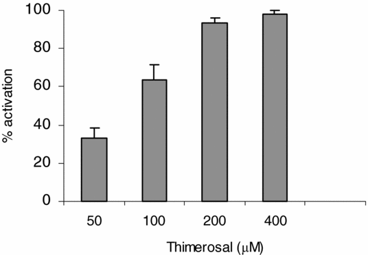
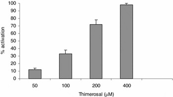
In order to determine the participation of IP3Rs in the activation process we evaluated the effect of low doses of thimerosal on egg activation. Denuded ovarian oocytes matured in vitro with progesterone (2.5 μM) were exposed to different doses of thimerosal (50–400 μM). Egg activation was scored after 20 min of culture.
Results indicate (Fig. 1) that thimerosal induced egg activation in a dose-dependent manner.
Dose–response curve for thimerosal-induced egg activation. Groups of 20 denuded fully grown ovarian oocytes matured in vitro with progesterone were incubated in AR with different doses of thimerosal (50–400 μM) and after 20 min activation parameters were evaluated. Values are the mean ± SEM of six experiments. Each experiment was performed on a different animal.
Effect of heparin on sperm-induced activation of in vitro matured oocytes
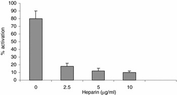
The role of IP3Rs in egg activation was also assayed using heparin, an IP3 receptor antagonist. Denuded fully grown ovarian oocytes matured in vitro with progesterone 2.5 μM were exposed to different heparin doses (2.5–10 μg/ml) for 30 min before insemination with an homologous sperm suspension (2 × 10⁶ sperm/ml).
Results show (Fig. 2) that heparin strongly inhibited activation in a dose-dependent manner.
Effect of heparin on sperm-induced egg activation. Groups of 20 denuded fully grown ovarian oocytes matured in vitro with progesterone 2.5 μM were preincubated for 30 min in AR (control) and AR with different doses of heparin (2.5–10 μg/ml) before insemination with a homologous sperm suspension (2 × 10⁶ sperm/ml). Activation was scored after 20 min of insemination. Values are the mean ± SEM of three experiments. Each experiment was performed on a different animal.

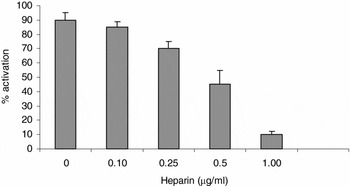
We also tested the effect of microinjection of low heparin doses on the sperm-induced activation of in vitro matured oocytes (Fig. 3).
Effect of heparin on sperm-induced egg activation. Groups of 20 denuded fully grown ovarian oocytes matured in vitro with progesterone 2.5 μM were microinjected with different heparin doses (0.10–1.00 μg/ml) before insemination with an homologous sperm suspension (2 × 10⁶ sperm/ml). Activation was scored after 20 min of insemination. Values are the mean ± SEM of three experiments. Each experiment was performed on a different animal.

We were able to verify the inhibitory effect of heparin on sperm-induced activation when low doses are microinjected into mature oocytes.
Effect of caffeine on the activation of in vitro matured oocytes
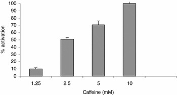
In order to determine the existence of RyRs, we evaluated the ability of caffeine to induce egg activation. Denuded ovarian oocytes matured in vitro with progesterone 2.5 μM were microinjected with different doses of caffeine (1.25–10 mM).
Data obtained (Fig. 4) show that caffeine induced egg activation in a dose-dependent manner.
Dose–response curve for caffeine-induced egg activation. Groups of 20 denuded fully grown ovarian oocytes matured in vitro with progesterone 2.5 μM were microinjected with different doses of caffeine (1.25–10 mM) and after 20 min activation parameters were evaluated. Values are the mean ± SEM of three experiments. Each experiment was performed on a different animal.

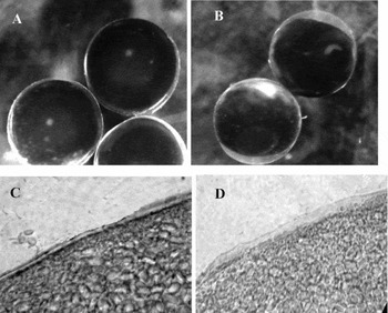
Results presented in Figure 5 show that caffeine injection was able to trigger activation events in in vitro matured oocytes with normal morphological features. Under a stereoscopic microscope, caffeine-treated oocytes exhibited flattening of the animal pole, disappearance of the white spot and modification of the vitelline envelope (Fig. 5B). In cytological preparations, caffeine-activated oocytes showed complete extrusion of the cortical granules (Fig. 5D).
(A) Oocytes matured in vitro with progesterone exhibit a white spot on the animal pole. Note the vitelline envelope, close to the plasma membrane. (B) Oocyte matured in vitro 15 min after caffeine injection. Note the disappearance of the white spot and the elevation of the vitelline envelope. (C) Cortex of matured oocytes: cortical granules are located in close proximity to the plasma membrane. (D) Cortex of in vitro matured oocytes after caffeine treatment: the cortical granules have disappeared.

The above data show that 100% oocyte activation was reached after RyRs stimulation with caffeine. The activation parameters tested are extremely similar to those observed in sperm-induced oocyte activation.
Effect of ruthenium red on sperm-induced activation of in vitro matured oocytes
The participation of RyRs in sperm-induced oocyte activation has been inferred from the effect of its specific antagonist, ruthenium red.
Denuded fully grown ovarian oocytes matured in vitro with progesterone 2.5 μM were preincubated for 30 min in AR (control) and AR with different doses of ruthenium red (20–100 μM) before insemination with an homologous sperm suspension (2 × 10⁶ sperm/ml)
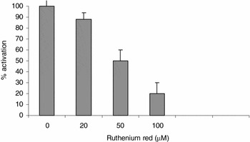
Ruthenium red, at concentrations of 20–100 μM, blocked sperm-induced oocyte activation in a dose-dependent manner (Fig. 6).
Effect of ruthenium red on sperm-induced egg activation. Groups of 20 denuded fully grown ovarian oocytes matured in vitro with progesterone were preincubated for 30 min in AR (control) and AR with different doses of ruthenium red (20–100 μM) before insemination with a homologous sperm suspension (2 × 10⁶ sperm/ml). Activation was scored after 20 min of insemination. Values are the mean ± SEM of three experiments. Each experiment was performed on a different animal.

In order to determine whether these different types of agonists used specific mechanisms to induce the apparition of characteristic signs of egg activation, we further evaluated the effect of specific antagonists on those agonists.
Effect of heparin on thimerosal-induced activation of in vitro matured oocytes
In order to confirm whether thimerosal-induced egg activation is mediated specifically by IP3Rs, we analyzed the effect of heparin (an IP3 receptor antagonist) injection before exposure to thimerosal.
Denuded fully grown ovarian oocytes matured in vitro with progesterone 2.5 μM were microinjected with heparin 1 μg/ml before treatment with thimerosal 200 μM. As control, oocytes were injected with Tris-buffered saline solution.
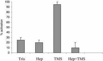
We found that 1 μg/ml heparin injection strongly inhibited the effect of 200 μM thimerosal (Fig. 7).
Effect of heparin on thimerosal-induced egg activation. Groups of 20 denuded fully grown ovarian oocytes matured in vitro with progesterone were microinjected with heparin 1 μg/ml (Hep) before treatment with thimerosal 200 μM (TMS) and after 20 min activation parameters were evaluated. Values are the mean ± SEM of three experiments. Each experiment was performed on a different animal.

Effect of ruthenium red on caffeine-induced activation of in vitro matured oocytes
To ascertain that caffeine induced egg activation by exerting its effect on RyR, we evaluated the effect of ruthenium red injection before exposure to caffeine.
Denuded fully grown ovarian oocytes matured in vitro with progesterone 2.5 μM were microinjected with 100 μM ruthenium red before treatment with caffeine 25 mM.
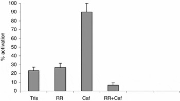
We found that previous microinjection of ruthenium red 100 μM completely inhibited 25 mM caffeine-induced activation (Fig. 8). These results show that caffeine-induced activation is mediated specifically by RyRs.
Effect of ruthenium red on caffeine-induced egg activation. Groups of 20 denuded fully grown ovarian oocytes matured in vitro with progesterone were microinjected with ruthenium red 100 μM (RR) before treatment with caffeine 25 mM (Caf). Values are the mean ± SEM of three experiments. Each experiment was performed on a different animal.

Interdependence between IP3Rs and RyRs
Interdependence between both classes of receptors was investigated through the ability of agonists of one type of receptor to revert the inhibitory effect of the antagonists of the other class.
Effect of heparin on caffeine-induced activation and effect of ruthenium red on thimerosal-induced activation
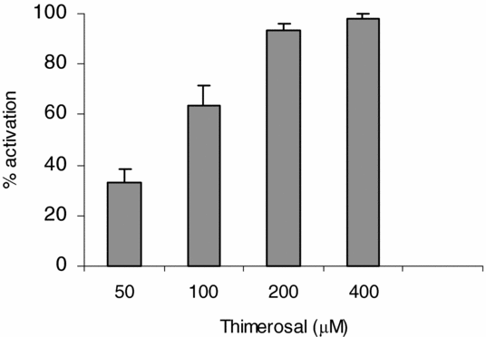
In order to establish whether the used agonists exerted their effect specifically on one type of receptor, in vitro matured oocytes were injected with 100 μM ruthenium red and then thimerosal (50–400 μM) was added to the culture medium.
Thimerosal overcame the inhibitory effect of ruthenium red (Fig. 9).
Effect of ruthenium red on thimerosal-induced egg activation. Groups of 20 denuded fully grown ovarian oocytes matured in vitro with progesterone were microinjected with ruthenium red 100 μM before treatment with different doses of thimerosal (50–400 μM). Values are the mean ± SEM of three experiments. Each experiment was performed on a different animal.

Ruthenium red, a known RyR antagonist, did not have any significant effect on IP3R as indicated by its inability to affect thimerosal-induced activation (Fig. 9).
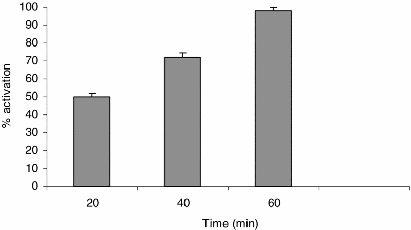
On the other hand, in vitro matured oocytes were microinjected with heparin 1 μg/ml before the addition of caffeine 25 mM to the culture medium (Fig. 10). The blockage of IP3Rs by heparin partially affected caffeine-induced activation. We found that prior injection of heparin 1 μg/ml delayed the response to 25 mM caffeine as compared with that for caffeine alone (20 ± 2 min for caffeine 25 mM versus 60 ± 2 min for heparin 1 μg/ml + caffeine 25 mM).
Effect of heparin on caffeine-induced egg activation. Groups of 20 denuded fully grown ovarian oocytes matured in vitro with progesterone were microinjected with heparin 1 μg/ml before treatment with caffeine 25 mM activation parameters were evaluated at different times. Values are the mean ± SEM of three experiments. Each experiment was performed on a different animal.
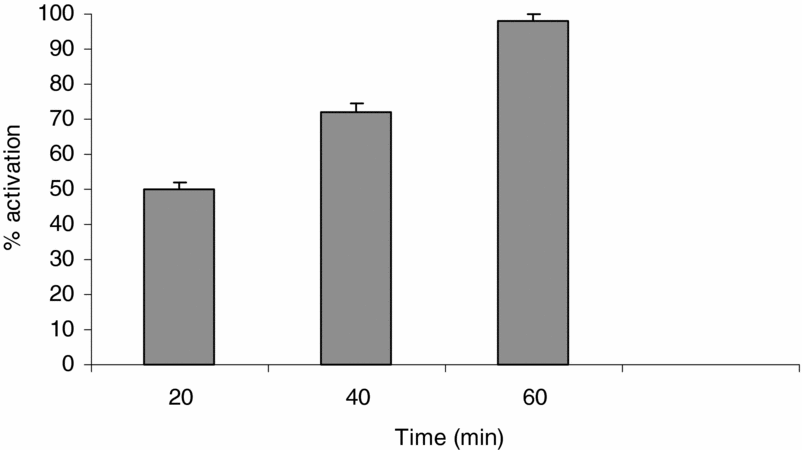
We previously tested the effect of the addition of different doses of caffeine to the culture medium. Exposure to caffeine 25 mM was enough to induce significant activation levels (86.5 + 2.5%) in 20 min (data not shown).
Discussion
Participation of IP3Rs and RyRs in oocyte activation
Calcium is considered the most important second messenger at fertilization. Transient release from intracellular stores is modulated through both agonist-gated channels: IP3Rs and RyRs, which can be found individually or together depending on the oocyte species.
The coexistence of these two families of intracellular calcium-release channels has been well documented in many cell types, including the oocytes of species such as sea urchins, mice, bovines and humans (Swann, Reference Swann1992; Galione et al., Reference Galione, McDougall, Busa, Willmott, Gillot and Whitaker1993a; Ayabe et al., Reference Ayabe, Kopf and Schultz1995; Yue et al., Reference Yue, White, Reed and Bunch1995; Herbert et al., Reference Herbert, Gillespie and Murdoch1997). The concept that both receptors are responsible for calcium changes associated with fertilization in sea urchin is firmly established (Galione, Reference Galione, McDougall, Busa, Willmott, Gillot and Whitaker1993a; Lee et al., Reference Lee, Aarhus and Walseth1993). In contrast, no RyRs have been detected using either type I or II RyR-specific antibodies in Xenopus and hamster oocytes. Additionally, RyR agonists have failed to induce changes in intracellular calcium in Xenopus and hamster oocytes. The relative contribution of these two receptors to intracellular calcium increase during fertilization still remains unresolved.
Both receptors have been generally detected by functional criteria. IP3 induces repetitive or single calcium release in oocytes of all species studied. RyR agonists include exogenous ryanodine and caffeine, and cyclic ADP-ribose, which is the only known endogenous CICR modulator and has been proposed as a new second messenger. RyR antagonists include ruthenium red and procaine. Previous studies in frog eggs did not reveal the existence of RyRs. Only functional IP3Rs were found in hamster eggs (Miyazaki et al., Reference Miyazaki, Yuzaki, Nakada, Shirakawa, Nakanishi, Nakade and Mikoshiba1992) and conflicting results have been obtained in mouse oocytes and eggs (Swan, Reference Swann1992; Kline & Kline, Reference Kline and Kline1994). Ryanodine injection into rabbit oocytes did not cause calcium release (Fissore & Robl, Reference Fissore and Robl1993). Independent IP3Rs and RyRs coexist in mature bovine oocytes (Yue, Reference Yue, White, Reed and Bunch1995).
In the present work, we evaluated the activator and blocker effects of these receptors on the activation of in vitro matured Bufo arenarum oocytes.
As with oocytes from other species, independent IP3Rs appear to be functional in Bufo arenarum oocytes. Exposure to thimerosal-induced oocyte activation in a dose-dependent manner and previous heparin injection significantly blocked that effect, clearly indicating that activation could be attributed to the effect of IP3R stimulation.
We found that caffeine, a well known specific RyR agonist, was able to trigger oocyte activation in a dose-dependent manner. Microinjection of 10 mM of caffeine showed 100% of oocytes exhibiting characteristic morphological criteria of egg activation. Ruthenium red, the specific RyR blocker, was able to inhibit oocyte activation induced either by sperm or caffeine. These findings allow us to infer the existence of RyRs in Bufo arenarum oocytes, but further studies are needed to verify this statement.
To a limited extent (about 20%), the activation can be due to mechanical stimulation caused by microinjection procedures.
Interdependence of IP3Rs and RyRs
Although intracellular calcium release has an essential role in fertilization, the physiological significance of each calcium-release mechanism in calcium intracellular increase during fertilization has not been fully elucidated.
Cyclic adenosine diphosphate-ribose (cADPR), an endogenous RyRs activator, is as potent as IP3 in mobilizing Ca2+ in sea urchin eggs. Experiments with antagonists of the RyRs and IP3Rs revealed that both Ca2+-mobilizing systems were activated during fertilization. Blockage of either of the systems alone was not sufficient to prevent the sperm-induced Ca2+ transient.
These dual receptors work in a backup manner during the fertilization of sea urchin eggs. The inhibition of either receptor does not abolish normal fertilization calcium transients. Therefore, IP3Rs may not play a major role in sea urchin fertilization (Galione et al., Reference Galione, McDougall, Busa, Willmott, Gillot and Whitaker1993a; Lee, Reference Lee, Aarhus and Walseth1993; Whitaker & Swan, Reference Whitaker and Swann1993).
It has been established that independent IP3Rs and RyRs coexist in mature bovine oocytes (Yue et al., Reference Yue, White, Reed and Bunch1995). The ryanodine receptor was observed uniformly localized in the periphery of mature oocytes, while a weak and discontinuous signal was observed in the germinal vesicle intact stage of bovine oocytes (Yue et al., Reference Yue, White, Reed and King1998) Although these two receptors may have different functional roles in the regulation of calcium release during meiotic maturation and fertilization, it appears that both IP3Rs and RyRs contribute to the significant increase in intracellular calcium during fertilization and activation in the bovine oocyte (Wang et al., Reference Wang, White, Reed and Campbell2005).
We further explored the relationship between IP3Rs and RyRs in Bufo arenarum oocytes by exposing them to the agonists of one class after injecting a blocker of the other class of receptor.
We found that thimerosal overcame the inhibitory effect of ruthenium red on oocytes activation, indicating that IP3Rs function as independent receptors. In contrast, previous injection of heparin delayed caffeine-induced calcium release, revealing a relative dependence of RyRs on functional IP3Rs. These results could be explained through a calcium-induced calcium-release mechanism (CICR), because of the structural relationship between both receptors. CICR provides a means for the amplification of microscopic initiation events into propagating calcium signals. Without calcium contribution originated in IP3Rs, caffeine-induced activation relies only on calcium released from RyRs and becomes delayed.
In conclusion, using the four commonly used compounds: thimerosal, caffeine, heparin and ruthenium red, we investigated the existence and interdependence of both IP3Rs and RyRs in mature Bufo arenarum oocytes. We found evidence of these two families of receptors. Our present findings provide the first reported evidence of the existence of RyR in frogs. Either microinjection of 10 mM caffeine or addition to the culture medium of 40 mM caffeine resulted in 100% oocyte activation with normal morphological features. We observed that IP3Rs were able to trigger oocyte activation by exerting their effect in an independent manner; in contrast, RyR-induced oocyte activation decreased when blocking IP3Rs with heparin, showing a relationship between both pathways, probably through a CICR mechanism. Both receptors play a role in Ca2+-release mechanisms although their relative contribution to the activation process is unclear. Knowledge of the existence and regulation of RyRs and IP3Rs is crucial for further understanding the calcium releasing mechanism in oocytes, not only in frogs but also in other species The effect of these dual receptors in the events associated with fertilization is currently under investigation.
Acknowledgements
This work was supported by a grant from the Science Council of the National University of Tucumán (CIUNT) and from the National Agency for Promotion of Science and Technology (ANPCYT).