The biological processes and molecular mechanisms underlying age-related physiological and behavioral changes are of special interest to a number of researchers in different areas of gerontology (Stuart-Hamilton, Reference Stuart-Hamilton2006). This might be at least partially related to the trajectory of demographic change occurring in most developed countries, which is due to the aging of their population. During the last century in the United States, there has been a substantial increase in the proportion of the population 65 years of age and older, from 4.1% of the population in 1900 and to 12.8% in 1995 (Malmgren, Reference Malmgren, Coffey and Cummings2000) to 19% in 2009, according to the US Census (http://factfinder.census.gov). This pattern in the aging of the population might be related to the rise of age-related disorders and to cognitive and behavioral changes.
Most of the behavioral changes associated with aging are related to noticeable declines in different domains of cognitive functioning (Kluger et al., Reference Kluger, Gianutsos, Golomb, Ferris, George and Franssen1997). A number of studies have shown a significantly decreased ability in specific functions such as attention, conceptual organization, creativity, general intelligence, and particularly fluid intelligence (Stuart-Hamilton, Reference Stuart-Hamilton2006). Age-related language decline involving difficulties with lexical understanding and speech production also have been reported (Barresi, Nicholas, Tabor Connor, Obler, & Albert, Reference Barresi, Nicholas, Tabor Connor, Obler and Albert2000; Barresi, Obler, An, & Albert, Reference Barresi, Obler, Au, Albert and Hamilton1999; Nicholas, Connor, Obler, & Albert, Reference Nicholas, Connor, Obler, Albert and Sarno1998; Nicholas, Obler, Albert, & Goodglass, Reference Nicholas, Obler, Albert and Goodglass1985). In addition, researchers have established an association between cognitive decline and age-related morphological changes in the central nervous system (CNS), such as structural changes in the frontal lobes of the brain (Band, Ridderinkhof, & Segalowitz, Reference Band, Ridderinkhof and Segalowitz2002; Rabbitt, Reference Rabbit1997) and axonal demyelination (Deary, Leaper, Murray, Staff, & Whalley, Reference Deary, Leaper, Murray, Staff and Whalley2003).
A number of studies of the morphology and biochemistry of developing and aging brains have provided evidence that age-related changes in the brain unfold in a systematic way, so that both the structure and function of the brain are modified. At birth most of the brain's cells are already formed; during early stages of development (a few years after birth) the brain is expanded intensively and extensively through the division and multiplication of glia (Dekaban & Sadowsky, Reference Dekaban and Sadowsky1978) and the establishment of many new connections among cells. By the age of 20–25 years, the brain has fully matured. Then, after middle adulthood, multiple aging-related changes occur in the brain; these changes are clearly detectable by the age of 50–60 years. Specifically, the weight and the volume of the brain decrease (Dekaban & Sadowsky, Reference Dekaban and Sadowsky1978; Skullerud, Reference Skullerud1985); the number of neurons diminishes, and their morphology changes (Kabaso, Coskren, Henry, Hof, & Wearn, Reference Kabaso, Coskren, Henry, Hof and Wearn2009; Terry, DeTeresa, & Hansen, Reference Terry, DeTeresa and Hansen1987); the number of synapses decreases (Masliah, Mallory, Hansen, DeTeresa, & Terry, Reference Masliah, Mallory, Hansen, DeTeresa and Terry1993; Peters, Sethares, & Luebke, Reference Peters, Sethares and Luebke2008); and the synthesis of neurotransmitters and the density of their receptors lessen (Amenta, Zaccheo, & Collier, Reference Amenta, Zaccheo and Collier1991; Kaiser, Schuff, Cashdollar, & Weiner, Reference Kaiser, Schuff, Cashdollar and Weiner2005; Ota et al., Reference Ota, Yasuno, Ito, Seki, Kozaki and Asada2006; Wong et al., Reference Wong, Wagner, Dannals, Links, Frost and Ravert1984). Functional outcomes of such age-related changes manifest in lower levels of neural conductivity (Miller & O'Callaghan, Reference Miller and O'Callaghan2003) and a loss of brain plasticity (Burke & Barnes, Reference Burke and Barnes2006; Kolb & Whishaw, Reference Kolb and Whishaw1988), which are clearly associated with a decline in intellectual functioning and memory (Albert & Stafford, Reference Albert, Stafford, Albert and Moss1988; Bakker et al., Reference Bakker, de Leeuw, de Groot, Hofman, Koudstaal and Breteler1999; Woodruff-Pak, Reference Woodruff-Pak1997). From the current point of view, most of the age-related changes in the brain listed above are induced by changes in the cell's structure, cellular homeostasis, and intercellular interactions, rather than by a diminution of the number of cells in the brain.
One of the main molecular mechanisms that control life support, development, metabolism, and the specific functioning of cells and tissues is gene expression. A complex orchestra of genes in a cell produces a set of RNA molecules that is called the transcriptome; these molecules are organized into an expression (also referred to as a coexpression) network. The transcriptome reflects the genome activity in a particular cell (or tissue) at a particular moment; the content and structure of the transcriptome are determined by the cell and tissue types under examination, as well as the stages of the cell cycle and organism development.
A number of studies have demonstrated that brain development and function depend on the precise regulation of gene expression, and that the changes in gene expression in the brain throughout the life-span correspond closely to the major stages of prenatal and neonatal development, and the maturing and aging of the brain (Colantuoni et al., Reference Colantuoni, Lipska, Ye, Hyde, Tao and Leek2011; Diaz, Reference Diaz2009; Johnson et al., Reference Johnson, Kawasawa, Mason, Krsnik, Coppola and Bogdanovic2009; Kang et al., Reference Kang, Kawasawa, Cheng, Zhu, Xu and Li2011; Lambert et al., Reference Lambert, Lambot, Bilheu, Albert, Englert and Libert2011; Somel et al., Reference Somel, Franz, Yan, Lorenc, Guo and Giger2009, Reference Somel, Guo, Fu, Yan, Hu and Xu2010). Thus, the rapid change of the transcriptome in the brain during the prenatal and neonatal stages of development is associated with major neurodevelopmental trajectories: genes related to neuronal differentiation, cell proliferation, and migration show the highest expression levels in the early fetal cortex, whereas genes associated with dendrite and synapse development dramatically increase their expression between the late midfetal period and late infancy (Colantuoni et al., Reference Colantuoni, Lipska, Ye, Hyde, Tao and Leek2011; Kang et al., Reference Kang, Kawasawa, Cheng, Zhu, Xu and Li2011). Dynamic changes in gene expression are registered during brain maturation before 20–25 years; the rate of change then slows, reaching a plateau by the age of 30. After middle age (~40–45 years) multiple changes in the transcriptome occur, which are clearly detectable by the age of 60–65 years (Lu et al., Reference Lu, Pan, Kao, Li, Kohane and Chan2004; Somel et al., Reference Somel, Franz, Yan, Lorenc, Guo and Giger2009). The main trajectory of these age-related changes is seen as a global decrease in levels of gene expression, with an increase in heterogeneity of expression that is widespread throughout the transcriptome. This effect was explained as an outcome of accumulating stochastic changes in the somatic cells, such as mutations, and cellular and DNA damage (Lu et al., Reference Lu, Pan, Kao, Li, Kohane and Chan2004; Somel, Khaitovich, Bahn, Paabo, & Lachmann, Reference Somel, Khaitovich, Bahn, Paabo and Lachmann2006; Somel et al., Reference Somel, Guo, Fu, Yan, Hu and Xu2010). Recent studies of transcriptome structure and its temporal changes in the CNS have formed a global view of gene-expression dynamics as the program running normal human brain development throughout life (Colantuoni et al., Reference Colantuoni, Lipska, Ye, Hyde, Tao and Leek2011; Kang et al., Reference Kang, Kawasawa, Cheng, Zhu, Xu and Li2011).
Despite insufficient knowledge about the relationship between gene expression in the brain and the realization of higher cognitive functions, a number of studies have reported that the transcriptome structure is associated with the anatomy/function structure of the CNS (Colantuoni, Purcell, Bouton, & Pevsner, Reference Colantuoni, Purcell, Bouton and Pevsner2000; Lockhart & Barlow, Reference Lockhart, Barlow, Chin and Moldin2001; Molyneaux, Arlotta, Menezes, & Macklis, Reference Molyneaux, Arlotta, Menezes and Macklis2007; Roth et al., Reference Roth, Hevezi, Lee, Willhite, Lechner and Foster2006; Strand et al., Reference Strand, Aragaki, Baquet, Hodges, Cunningham and Holmans2007). Research based on animal models has provided evidence that gene expression appears to correlate with performance on specific tasks and with brain activity. It has been shown that motor task performance is associated with gene expression in the cerebellum, amygdala, and hippocampus (Nadler et al., Reference Nadler, Zou, Huang, Moy, Lauder and Crawley2006) and that there is a correlation between the transcriptional profile of the parietal cortex and sleep behavior (Buechel et al., Reference Buechel, Popovic, Searcy, Porter, Thibault and Blalock2011). In addition, a number of studies have reported that specific patterns and interruptions in gene expression are correlated with a spectrum of neurological disorders (Mirnics & Pevsner, Reference Mirnics and Pevsner2004), such as multiple sclerosis (Kinter, Zeis, & Schaeren-Wiemers, Reference Kinter, Zeis and Schaeren-Wiemers2008), Huntington (Botovecki et al., Reference Borovecki, Lovrecic, Zhou, Jeong, Then and Rosas2005), Alzheimer (McShea et al., Reference McShea, Marlatt, Lee, Tarkowsky, Smit and Smith2006), and Parkinson diseases (Miller & Federoff, Reference Miller and Federoff2006), as well as being associated with mental illnesses, such as schizophrenia and bipolar disorder (Moreau, Bruse, David-Rus, Buyske, & Brzustowicz, Reference Moreau, Bruse, David-Rus, Buyske and Brzustowicz2011; Mudge et al., 2008), and suicidal behaviors of patients with mood disorders (Sequeira et al., Reference Sequeira, Morgan, Walsh, Cartagena, Choudary and Li2012).
Thus, the accumulation of data on the specificity of transcriptional changes in distinct structural and functional regions of the brain in different developmental stages may be a valuable source of information on the transcriptional foundation of normal development and the functioning of the CNS, as well as insight into the molecular mechanisms of pathological neurodevelopment and cognitive disorders. The results of such studies show the potential for applications in diagnostics for neurological disorders, as was discussed in Scherzer's (Reference Scherzer2009) review.
The current knowledge of the transcriptome structure in the human brain is based on studies that used microarray techniques for gene-expression profiling, measuring genes' activity at the expression level. Today's technology allows the detection of a whole genome expression profile based on measurements of the expression levels of a large but predefined (i.e., “blind” to anything but what is included in the array) set of well-annotated genes dispersed across a genome. Among other drawbacks (Irizarry et al., Reference Irizarry, Warren, Spencer, Kim, Biswal and Frank2005; Kawasaki, Reference Kawasaki2006), this predefinition is a serious limitation of the use of microarray technologies for the analysis of gene expression in the brain, which is characterized by a high level of gene expression and transcriptome complexity in comparison with other tissues. Thus, a number of studies have reported that at least 30%–50% of ~25,000 known protein coding genes (International Human Genome Sequencing Consortium, 2004) are expressed in the brain (Colantuoni et al., Reference Colantuoni, Purcell, Bouton and Pevsner2000; Myers et al., Reference Myers, Gibbs, Webster, Rohrer, Zhao and Marlowe2007); the transcriptome in the brain is especially enriched in regulatory elements (Amaral, Clark, Gascoigne, Dinger, & Mattick, Reference Amaral, Clark, Gascoigne, Dinger and Mattick2011; Chodroff et al., Reference Chodroff, Goodstadt, Sirey, Oliver, Davies and Green2010; Kuss & Chen, Reference Kuss and Chen2008; Landgraf et al., Reference Landgraf, Rusu, Sheridan, Sewer, Iovino and Aravin2007; Ponjavic, Oliver, Lunter, & Ponting, Reference Ponjavic, Oliver, Lunter and Ponting2009; St. Laurent, Faghihi, & Wahlestedt, Reference St Laurent, Faghihi and Wahlestedt2009) and alternative splicing events (de la Grange, Gratadou, Delord, Duterre, & Auboeurf, Reference de la Grange, Gratadou, Delord, Dutertre and Auboeuf2010; Mortazavi, Williams, McCue, Schaeffer, & Wold, Reference Mortazavi, Williams, McCue, Schaeffer and Wold2008; Ramskold et al., Reference Ramskold, Wang, Burge and Sandberg2009; Wang et al., Reference Wang, Sandberg, Luo, Khrebtukova, Zhang and Mayr2008).
During the last several years the development of high-throughput sequencing technologies, such as RNA-Seq, has allowed the so-called direct sequencing of the transcriptome: the detection of a whole genome expression profile without the limitation of using a predefined set of target genes. Moreover, RNA-Seq allows the measurement of expression levels with high accuracy, and the simultaneous detection of genes that have a wide (very high and very low) range of expression levels (‘t Hoen et al., Reference ‘t Hoen, Ariyurek, Thygesen, Vreugdenhil, Vossen and de Menezes2008; Asmann et al., Reference Asmann, Klee, Thompson, Perez, Middha and Oberg2009; Marioni, Mason, Mane, Stephens, & Gilad, Reference Marioni, Mason, Mane, Stephens and Gilad2008; Wang, Gerstein, & Snyder, Reference Wang, Gerstein and Snyder2009). At present, RNA-Seq is considered the most promising technique for transcriptome analysis in the CNS (van der Brug, Nalls, & Cookson, Reference van der Brug, Nalls and Cookson2010).
Here we report the results of high-throughput sequencing of the transcriptome in three cortical areas involved in the regulation of distinct higher cognitive functions (intellectual functions and actions, speech production, and speech processing and comprehending) from two postmortem female brains from individuals 70 and 40 years of age. Investigating the differential patterns of gene expression in brain tissues from adult and senescent individuals, we are able to examine both the main trend of age-related changes in the neocortex and the consistency of these changes across distinct cortical sites that control different higher cognitive functions. We consider the main findings from standpoint of the current knowledge of the age-related dynamics of gene expression in the CNS. If continued and extended, this research will contribute to a better understanding of the relationships between higher cognitive functions and the transcriptional status of the CNS and their dynamic changes throughout the life span.
Materials and Methods
Specimen ascertainment
Specimens were collected from two healthy, right-handed females who were 70 and 40 years of age (marked in the current publication as Brain 70 and Brain 40, respectively), who died of sudden death, unaccompanied by premortem hypoxia or prolonged agony, both of which have been linked to significant RNA degradation (Barton, Pearson, Najlerahim, & Harrison, Reference Barton, Pearson, Najlerahim and Harrison1993; Harrison et al., Reference Harrison, Heath, Eastwood, Burnet, McDonald and Pearson1995; Mexal et al., Reference Mexal, Berger, Adams, Ross, Freedman and Leonard2006). The brain tissue samples were provided by the Department of Forensic Medicine of the Medical Academy for Continuous Education (Moscow, Russia); all proper consenting and permission procedures were implemented.
Autopsies of the brains were made within a postmortem interval of no more than 24 hr to avoid RNA degradation and the selective reduction in the mRNA at postmortem intervals of 48 hr or more (Catts et al., Reference Catts, Catts, Fernandez, Taylor, Coulson and Lutze-Mann2005). The gray matter of three distinct functional cortical areas was dissected from the left hemisphere; in typical individuals, these areas are thought to be involved in the regulation of cognitive functions such as memory, thinking, and executive skills (association cortex, Broca area [BA] 46), speech and language understanding (Wernicke area, BA 22), and speech production (BA 45). We collected all layers of the neocortex from each area to minimize the imbalances in cell composition between samples (Colantuoni et al., Reference Colantuoni, Purcell, Bouton and Pevsner2000; Molyneaux et al., Reference Molyneaux, Arlotta, Menezes and Macklis2007; van der Brug et al., Reference van der Brug, Nalls and Cookson2010). The dissections were collected into RNAlater stabilization reagent as per the manufacturer's instructions (Qiagen). During sample shipment, the tissue was kept in RNAlater for a week, then was transferred from the stabilization reagent and stored at –80°C.
Sample preparation and sequencing
Total RNA was extracted from the brain tissue using the RNeasy Plus Mini Kit according to the manufacturer's instructions (Qiagen); the RNA quality was assessed using Agilent Technologies 2100 Bioanalyzer (RNA integrity number, range = 8.1–9.0). Poly-A mRNA was isolated from 10 µg of total RNA with Dyna1 oligo-dT beads (Invitrogen) and randomly fragmented using RNA fragmentation reagents (Ambion). First-strand complementary DNA (cDNA) synthesis was performed using SuperScriptII reverse transcriptase and random primers (Invitrogen); second-strand cDNA was synthesized using DNA PolI and RNase H (Invitrogen).
The sequencing libraries were prepared according to the paired-end sample preparation protocol (http://www.illumina.com) with slight modifications. The Illumina PE adapters were ligated using End Repair Kit (Epicentre), Klenow fragment (NEB), and LigaFast (Promega). The size selection of the final product was performed by SizeSelect E-Gel (Invitrogen). The templates were enriched by polymerase chain reaction using Phusion DNA Polymerase (NEB). Quality control analysis on the cDNA libraries was performed by Bioanalyzer; the median size of the cDNA library was around 300 nucleotides.
The libraries were sequenced using Illumina high-throughput technology as 75-mers from both ends of the cDNA templates (paired-end sequencing). The sequencing by synthesis was performed at the Yale Center for Genomic Analysis (http://medicine.yale.edu/keck/ycga), using TruSeq PE Clustering kit, TruSeq SBS v2, and Hi-Seq 2000 (Illumina). Six different cDNA samples and one technical replicate were run in seven lanes of the same flow cell.
RNA-Seq reads mapping and annotation
Sequencing reads were processed and aligned to the UCSC Homo sapiens reference genome (hg19) using TopHat (Trapnell, Pachter, & Salzberg, Reference Trapnell, Pachter and Salzberg2009), which uses Bowtie (Langmead, Trapnell, Pop, & Salzberg, Reference Langmead, Trapnell, Pop and Salzberg2009). An example of the data alignment is presented in online-only Figure S.1. To estimate transcript abundance levels, the normalized measures were obtained using Cufflinks (Trapnell et al., Reference Trapnell, Williams, Pertea, Mortazavi, Kwan and van Baren2010). The transcript abundances were measured in fragments per kilobase of exon per million mapped fragments (FPKM). The alignment and processing of the sequencing outputs were performed at the Yale University Biomedical High Performance Computing Center (http://info.med.yale.edu/hpc/).
Detection and functional annotation of differentially expressed genes (DEGs)
Pairwise comparative analysis to detect DEGs across the cortical samples was carried out using differential expression analysis (DESeq) for sequence count data. This is an R package that uses a negative binomial model to estimate the distribution of the raw counts within each transcript and to perform tests for DEGs (Anders & Huber, Reference Anders and Huber2010). The genes showed significant differences in the number of mapped reads at p ≤ .05 and, after adjusting for multiple comparisons, were considered to be differentially expressed.
To identify common biological processes and pathways, molecular functions, and cellular components, as defined by gene ontology (GO) criteria for DEGs, the Database for Annotation, Visualization and Integrated Discovery (DAVID) was used (http://david.abcc.ncifcrf.gov; Huang, Sherman, & Lempicki, Reference Huang, Sherman and Lempicki2009). For this analysis, the “high stringency” setting of the functional analysis tool was used, which compares the enrichment of GO terms in our list of DEGs using a Fisher exact test. The clusters generated by DAVID, with an enrichment score of >1.3 corresponding to p < .05, were considered reliable. The p values of the DAVID tool and the Benjamini corrections of the scores from the tool as inclusion criteria were used in the trimming of the clusters to an overrepresented term list.
Gene network construction
Coexpression networks for genes differentially expressed between individuals were constructed using a freely accessible WebQTL resource (http://www.genenetwork.org) that combines genetic and phenotypic databases with data analysis tools (Wang, Williams, & Manly, Reference Wang, Williams and Manly2003). Specifically, the HBTRC-MLC Human Prefrontal Cortex Agilent Normal database, containing data on the mean expression levels of approximately 37,000 genes in the prefrontal cortex across 170 individual expression profiles, was used to construct a gene network (http://www.genenetwork.org). A Pearson correlation matrix, containing estimates of pairwise correlations between gene-expression levels, was calculated and then transformed into a matrix of connection strengths, which permitted the construction of a coexpression network for each data set in this study.
Results
Analysis of RNA-Seq data
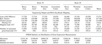
High-throughput paired-ended sequencing using Illumina technology was performed to generate gene-expression profiles of three cortical areas from two individual postmortem brains. Altogether seven samples (six cDNA libraries and one technical replicate) were sequenced in different lanes of the same Illumina Hi-Seq flowcell as 75-mer reads. The number of clusters of identical cDNA fragments generated during the amplification step of sequencing ranged from 133.5 to 186.8 million per lane. Between 100.9 million and 126.7 million (~75%) of these clusters passed quality filtering by Illumina (Table 1). The total number of reads produced for each sample ranged from approximately 73.5 million to 86.0 million; the yields in read count per lane were comparable. Of these reads, 73.5 million to 86.3 million (~70%) were mapped to the reference genome and exon junctions were annotated. The mapping statistics are comparable to those in published studies (Liu, Lin, Jiang, Wang, & Xing, Reference Liu, Lin, Jiang, Wang and Xing2011; Marioni et al., Reference Marioni, Mason, Mane, Stephens and Gilad2008; Wang et al., Reference Wang, Sandberg, Luo, Khrebtukova, Zhang and Mayr2008).
Sequencing output, RNA-Seq reads mapping to Human genome build 19, and statistics on the distribution of gene expression measurements across seven studied brain samples corresponding to the three cortical areas from two individuals, including the technical replicate

Note: FPKM, fragments per kilobase of exon per million mapped fragments.
aTechnical replicate.
bEstimated as the percentage of the total number of reads.
cEstimated as the percentage of 15,096 genes that have shown expression (FPKM = 0.001 and more) in at least one of the seven samples.
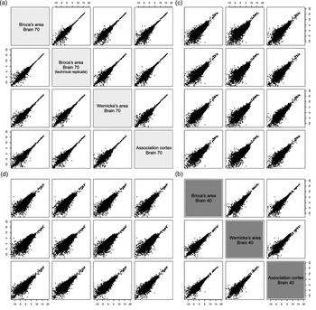
The comparison of technical replicates demonstrated good reproducibility of the RNA-Seq output (Table 1; Figure 1). The correlation coefficient of log2FPKM between replicates was extremely high (.99), and there were no statistically significant (p < .01) differences found in read counts between the two technical replicates.
Pairwise plots of transcript abundance levels (log2 of fragments per kilobase of exon per million mapped fragments) between seven sequenced complementary DNA samples including technical replicates. Each subplot shows the comparison of samples i versus j, each coordinate corresponds to a sample, and each dot corresponds to a gene/transcript. (a, b) Subplots represent the results of intraindividual comparisons between three cortical areas: Broca, Wernicke, and the association cortex. (c, d) Subplots represent interindividual comparisons of transcript abundance levels in the cortical areas.

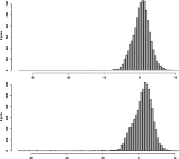
The number of RNA-Seq reads mapped per gene varied from 0 to tens of thousands (online-only supplemental Table S.1), which corresponds to the variation of expression levels among genes from 0 to 21,598 FPKM (Table 1). The median of the distribution of gene-expression levels varied among seven sequenced samples from 0.6058 to 1.3085 FKPM, indicating the dominance of low-expressed genes among the genes detected (Table 1 and Figure 2). Genes that showed FPKM ≥ 0.001 (corresponding to 1 or more fragments per 10 kb of transcript per 100 million mapped reads) in at least one sample were considered expressed. According to these criteria, 15,096 expressed genes were detected in the analyzed cortical areas (Table 1), which represents approximately 68% of the total number of genes (of ~22,000) contained in the Gencode (Harrow et al., Reference Harrow, Denoeud, Frankish, Reymond, Chen and Chrast2006; http://www.gencodegenes.org).
The distribution of expression levels (log2 fragments per kilobase of exon per million mapped fragments [FPKM]) of genes detected in Broca area of Brain 70 (the upper diagram) and the same area in Brain 40 (the lower diagram). Genes with FPKM of 0 are excluded. This plot shows that there are few genes that have an FPKM between 0 and 0.001; the latter corresponds to approximately –10 on the logarithmic scale. The threshold of 0.001 was used as a minimum FPKM for determining expressed genes.

Comparative analysis of gene-expression profiles across six cortical samples
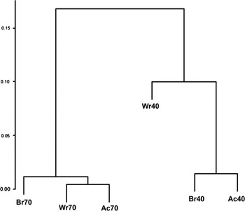
To identify the interarea and interindividual variability of gene expression, we performed pairwise comparisons of the expression profiles among three cortical areas across the two brains (Figure 1). The distributions indicate the prevalence of differences in the expression levels among low expressed genes. All plots representing the interindividual pairwise comparisons demonstrate more dispersed distributions compared to intraindividual comparisons. This indicates that more of the genes characterized by wide ranges of expression levels show more differential expression in inter- than intraindividual comparisons. Hierarchical clustering of the six samples, based on correlations between their gene-expression profiles, confirms this finding: the cortical areas from an individual brain cluster together in a single branch of the dendrogram, and the clusters for the two individuals are far removed from each other (Figure 3). Overall, these results indicate that the interindividual differences in gene expression are significantly greater than the intraindividual area differences in these samples.
Hierarchical clustering of cortical areas from the two individual brains on the basis of the correlations of their gene-expression profiles. The cortical areas are shown in the table as Broca area (Br), Wernicke area (Wr), and association cortex (Ac); the numbers 40 and 70 correspond to the two individuals. The dendrogram shows the significant predominance of interindividual differences in gene expression in the brain over differences between cortical areas within each individual's brain tissue.

Statistical analyses of gene-expression variations among the cortical areas and individuals listed in Table 1 indicate consistent differences in gene expression between these individuals across all three cortical areas. The variation of gene expression in all cortical areas of the older individual is characterized by lower medians (0.6058–0.6487) and higher standard deviations (185.63–188.16) than those found in the brain tissue from the individual 40 years of age (1.0206–1.3085 and 83.54–119.14, for the medians and standard deviations, respectively). This suggests lower expression levels but higher heterogeneity of gene expression in the brain of the older individual.
DEGs analysis
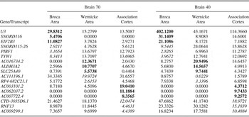
The analysis of DEGs between cortical areas within each individual showed negligible intraindividual difference between the gene-expression profiles: no genes showed statistically significant (p ≤ .05) differences in their expression levels. Despite the lack of statistical significance in the differential expression analysis, a set of transcripts (584 of 25,165 detected in the brain tissue) showed a concordance in their abundance levels in the studied areas of both individuals. In both individual brains, these genes showed 1.5-fold and more differences in expression levels in one particular cortical area compared to the others (online-only supplemental Table S.2). As an example, the top list of these genes, which have shown a higher dispersion of expression levels among cortical areas, is represented in Table 2. This observation indicates the potential of finding area specificity in gene expression, or an association between gene expression in distinct areas of the neocortex and the different functions they control in studies involving more cortical areas and more individual tissue samples to increase the power of analysis.
The top list of genes showing distinctively lower (italic) or higher (bold) expression levels (fragments per kilobase of exon per million mapped fragments) in particular cortical areas in both intraindividual comparisons

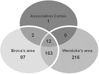
In contrast to the previous comparison, we identified significant interindividual differences in gene expression within each cortical area. Of 15,096 expressed genes, 491 (~3%) demonstrated differential expression between individual brain samples at p ≤ .05 (see online-only supplemental Table S.3 for the list of DEGs). Of these 491 genes, 177 were differentially expressed in at least two cortical areas (Figure 4). The number of DEGs between individuals varied in different cortical areas: only 15 DEGs were identified in the association cortex, whereas 274 and 391 DEGs were detected in the BA and Wernicke area, respectively (Figure 4). Of 491 DEGs, 133 genes (~27%) had significantly higher expression levels and 358 genes (~73%) significantly lower expression levels in the brain tissue from the older individual compared to those from the younger (online-only supplemental Table S.3). Again, this finding indicates the likelihood of a lower level of gene expression in the brain of the older individual.
A comparison among three cortical regions, in which 491 genes are detected as significantly (p ≤ .05) differentially expressed between two individual brains within the same areas: Broca, Wernicke, and the association cortex, respectively. Digits in circles represent both the number of differentially expressed genes detected in a particular cortical area and the number of overlapping genes across the areas.

Functional annotation of the DEGs
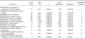
To identify the specificity of gene-expression differences found between individuals, we investigated the functional relationships between genes, which showed significant interindividual differences in their expression levels across all studied cortical areas. The set of 491 DEGs included 419 genes with known functions, 414 of which were annotated to different functional clusters using DAVID annotation tools (online-only supplemental Table S.4). Table 3 lists the GO terms that are significantly overrepresented among the genes differentially expressed between individual brain tissues. The analysis of the GO annotation suggests that these DEGs are especially enriched in genes involved in the regulation of the transmission of nerve impulses, nervous system development, and neurogenesis.
Summary of functional annotation of 491 genes differentially expressed between individual brains within the same cortical area

Note: The p values of the DAVID tool and the Benjamini corrections of the scores from the tool were used as inclusion criteria to trim the overrepresented term lists. Fold enrichment in each gene group compared to the base set of genes in the DAVID databases (19,235 genes). The trend of age-related change in the expression of genes belonging to a particular functional group is shown as ↑ and ↓, reflecting an increase and decrease of the expression levels, respectively. GO, gene ontology.
Functional annotation of genes downregulated and upregulated in the aged neocortex
To identify the direction of age-related changes in gene expression for the different functional groups of genes we performed separate functional annotations of genes that were found to be downregulated or upregulated in the brain tissue from the older individual. The lists of GO terms for genes that decreased and increased their expression in the aged neocortex are presented in online-only supplemental Table S.5 and Table S.6, respectively. Genes involved in the control of neuron development, the transmission of nerve impulse, including such functional groups as synaptic vesicles, ion transport, and channel complexes, are overrepresented in the list of 358 genes that showed significantly decreased levels of expression in the brain tissue from the individual 70 years of age (Table 3; online-only supplemental Table S.5). In addition, a number of genes known to be involved in the regulation of such higher cognitive functions as visual behavior and learning also showed decreased expression levels in the neocortex of the elderly individual.
The list of 133 genes significantly increased expression, or upregulated, in the brain tissue from the older individual is especially rich in protein-coding genes that are involved in divalent metal cation binding (Table 3; online-only supplemental Table S.6). This group is represented by four genes from the metallothionein superfamily: MT1E, MT1G, MT1F, and MT2A.
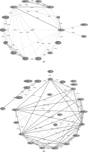
Two of the functional groups overrepresented in the list of DEGs (cellular chemical homeostasis and regulation of nervous system development) contain genes that have shown expressional changes in the brain tissue from the 70-year-old individual in both increased and decreased expression levels (Table 3). It is notable that the coexpression networks reconstructed for genes belonging to these two functional groups indicate high positive correlations within each of the sets of up- and downregulated genes, respectively, and high negative correlations between those subgroups of genes (Figure 5a, b). Moreover up- and downregulated genes have different functions in the regulation of cellular homeostasis and nervous system development. Thus, five of six genes (GLI2, HES1, ID4, PAX6, and SOX2) involved in the regulation of nervous system development and upregulated in the neocortex of the older individual, are factors of transcription regulation (Figure 5a). The genes involved in signal transduction pathway regulation (YWHAG and YWHAH), neuron regeneration and axonal myelinization (AMIGO1 and CD24), and genes coding receptors (KIT, GRIN1, CHRNB2) prevailed among genes belonging to the same functional group, but showing a decrease in the expression in the aged neocortex (Figure 5a).
Coexpression networks of genes involved in (a) the regulation of nervous system development and (b) cellular chemical homeostasis that have shown differential expression in the neocortex from the individual 70 years of age compared to that in the brain tissue from the individual 40 years of age. The network is constructed based on Pearson correlation coefficients between expression levels of these genes in the human neocortex (http://www.genenetwork.org). Interactions for an absolute correlation above .50 are shown; the correlation coefficients are represented. The positive correlations are shown as bold (r > .7) and normal (.5 < r < .7) lines; the negative correlations are shown as dashed lines. The nodes with symbols of genes upregulated in the brain tissue from the older individual are marked by a dark gray circle.

Similar to the previous finding within the functional group “cellular chemical homeostasis,” the list of genes that have shown significantly decreased expression in the older neocortex is especially populated by genes controlling the cellular signaling system, such as genes encoding for receptors (e.g., GPR21, CHRNB2, GRIN1, GPR6, MCH1R/SLC1, AVPR1A, and DRD1), signal transducers and neurotransmitters (e.g., GNG3, CD24, and TAC1; Figure 5b). Among the genes significantly downregulated, or showing decreased expression in the aged neocortex, there were genes involved in the electroneutral exchange of sodium and potassium ions, and potassium-chloride cotransport in mature neurons (SLC9A6 and SLC12A5). In contrast, a number of genes involved in bivalent ion (mainly Ca2+ cations) transport and homeostasis, and, as a result, the activation of signaling cascades, showed significantly increased expression in the aged cortex, such as FXYD1, GRIN2C, LPAR3, and NTSR2. Specifically, the FXYD domain containing ion transport regulator 1 (FXYD1) involved in chloride transport and calcium ion homeostasis; glutamate-activated NMDA-receptor (GRIN2C) triggers an influx of Ca2+ into the postsynapse; LPAR3 involved in releasing of calcium from the endoplasmatic reticulum; and neutrotensin receptor 2 (NTSPR2) indirectly activates a calcium second messenger system.
Taken together, these findings indicate that the main interindividual difference in gene expression in the neocortexes corresponds to the significant variability in activity of genes involved in the control of the cellular signaling system. This difference is specifically related to a decrease in the expression of genes coding receptors, signal transducers and neurotransmitters, as well as to significant changes in the expression of an orchestra of genes controlling ion channels, ion transport, and homeostasis in the neocortex from the aged brain.
Discussion
To investigate age-related changes in gene expression in the neocortex as whole and in its different functional areas, we performed RNA-Seq on the transcriptome in the prefrontal association cortex, which is involved in the regulation of a number of higher cognitive functions such as memory, thinking, and executive skills, and the two cortical areas controlling speech and language (BA and Wernicke area) of two postmortem female brains. The preliminary experiment involved brain tissue from two individuals of different ages (40 and 70 years of age), representing different adult development stages. Thus, at the middle adult stage (40 years of age) the brain has matured and transcriptional status is relatively stable (Lu et al., Reference Lu, Pan, Kao, Li, Kohane and Chan2004; Somel et al., Reference Somel, Franz, Yan, Lorenc, Guo and Giger2009); at the late adult stage (70 years of age), the changes in the morphophysiology (Dekaban & Sadowsky, Reference Dekaban and Sadowsky1978; Kabaso et al., Reference Kabaso, Coskren, Henry, Hof and Wearn2009; Masliah et al., Reference Masliah, Mallory, Hansen, DeTeresa and Terry1993; Terry et al., Reference Terry, DeTeresa and Hansen1987) and transcriptional status (Lu et al., Reference Lu, Pan, Kao, Li, Kohane and Chan2004; Somel et al., Reference Somel, Franz, Yan, Lorenc, Guo and Giger2009) of the brain related to normal aging are clearly detectable.
In our comparative study of the transcriptome we focused on the identification of differential expression patterns across distinct cortical regions and differently aged individuals, as well as on the analyses of sets of DEGs in terms of the functions and metabolic pathways those genes control. The main goal of the study, which must be considered preliminary due to the small number of subjects, was to trace the general directions of the comparative relationships between gene expression in the neocortex and higher cognitive functions and the concordance of their age-related dynamics.
The gene-expression profiling of the transcriptome in the neocortex based on RNA-Seq data has shown that at least 68% of known genes (~22,000 genes contained in the Gencode DB) are expressed in the brain. This finding is consistent with reported evidence on the high expression levels in the brain tissue in terms of number of expressed genes as 30%–50% of the genes contained in the human genome (Colantuoni et al., Reference Colantuoni, Purcell, Bouton and Pevsner2000; Myers et al., Reference Myers, Gibbs, Webster, Rohrer, Zhao and Marlowe2007). The statistical analysis of the distribution of gene-expression measurements (Table 1) showed the consistency in the expression level and the variability among all of the studied cortical regions within an individual brain.
Spatial variability of the gene expression in the neocortex (i.e., the differences between cortical areas within an individual brain) was found to be very low and not detectable at a high level of confidence. Thus, pairwise comparisons of the expression profiles among three cortical areas across the two individual brains showed negligible differences between the gene-expression profiles of distinct cortical areas within an individual brain: no genes showed statistically significant differences in their expression levels. This might be due to both the high similarity in expression profiles across distinct areas of the cerebral cortex and the presence of main gene-expression differences across cortical regions among less expressed genes, for which differences might be detectable and perhaps meaningful, but not significant statistically. The first interpretation is consistent with other studies showing low variability in gene expression among different sites of the cerebral cortex (Khaitovich et al., Reference Khaitovich, Muetzel, She, Lachmann, Hellmann and Dietzsch2004; Roth et al., Reference Roth, Hevezi, Lee, Willhite, Lechner and Foster2006). The second interpretation is consistent with the data presented in the previous section on the dominance of low-expressed genes in the transcriptome of the studied cortical tissues. All together these facts and the low power of the statistical apparatus for the reliable detection of differential expression of low-expressed genes constitute serious limitations in comparative studies of transcriptomes from tissues for which a priori low differences in gene expression are expected.
To determine the potential of identifying area specificity in gene expression we compared gene-expression variability between different cortical areas within each individual and across both individuals. This analysis showed that within each individual brain there are sets of genes that distinguish cortical areas from each other. The differences in the expression levels of these genes among cortical regions are not statistically significant but they are consistent across two individual comparisons (Table 2). Overall, these results of the comparative study of gene-expression variation among distinct cortical areas indicate the potential of finding region specificity in gene expression with increased power of analysis, including increased sample size or the involvement in the analysis of more cortical areas and more individual tissue samples.
The pairwise comparisons of global expression profiles using hierarchical cluster analyses of expression patterns across cortical areas and individuals showed that interindividual differences in gene expression are significantly greater than intraindividual differences between cortical areas. At the whole-genome level of expression, this difference is especially clear in the variation of gene-expression levels within each expression profile. Thus, all three cortical areas of the older individual brain were characterized by lower median and higher standard deviations of gene-expression levels compared to those estimated for the cortical areas of the younger individual brain. This indicates a decrease in global gene expression and an increase in the heterogeneity of expression in the aging neocortex. These trajectories of age-related changes in gene expression have been previously reported and have been explained as a possible outcome of accumulating stochastic changes in somatic cells, such as mutations, and cellular and DNA damage, especially in gene promoters (Lu et al., Reference Lu, Pan, Kao, Li, Kohane and Chan2004; Somel et al., Reference Somel, Khaitovich, Bahn, Paabo and Lachmann2006, Reference Somel, Guo, Fu, Yan, Hu and Xu2010). A systematic decrease in gene expression in the CNS has been reported as a transcriptional event associated with different mental disorders. Thus, significant downregulation of genes coding microRNA has been found in the brain tissue of the patients with schizophrenia and bipolar disorder (Moreau et al., Reference Moreau, Bruse, David-Rus, Buyske and Brzustowicz2011).
The analysis of differential expression patterns in distinct cortical areas between individual brains has shown that several hundred genes have significant interindividual difference in the expression level. Moreover, the variation in the expression differences makes the association cortex especially distant from the other two cortical areas. Thus, the number of DEGs found in this area is an order of magnitude lower than those found in Broca and Wernicke areas (15 DEGs vs. 275 and 391 DEGs, respectively). If confirmed by studying an extended sample of individuals, this finding might indicate the specificity and difference in the rate of age-related change in the transcriptome in different cortical sites of the brain, especially taking into account that heterogeneity has been reported in age-related morphological and molecular changes among different cortical regions (Hof & Morrison, Reference Hof and Morrison2004; Raz, Rodrigue, Head, Kennedy, & Acker, Reference Raz, Rodrigue, Head, Kennedy and Acker2004).
According to the functional annotation clustering of DEGs between individuals, the most crucial age-related changes in gene expression occur in metabolic pathways involved in the regulation of the transmission of nerve impulses, nervous system development, and neurogenesis. Once again, this finding is consistent with previously published data suggesting an age-related decrease in the expression of genes related to neurodevelopmental processes and signaling pathways (Somel et al., Reference Somel, Franz, Yan, Lorenc, Guo and Giger2009; Von Bernhardi, Tichauer, & Eugenin, Reference Von Bernhardi, Tichauer and Eugenín2010). We identified, as differentially expressed in the two brains, genes encoding for a number of receptors involved in the transmission of nerve impulses, among them dopamine receptor D1 (DRD1), glutamate receptor IN 1 (GRIN1), cholinergic receptor NB2 (CHRNB2), arginine vasopressin receptor 1A (AVPR1A), and a number of G protein-coupled receptors (such as GPR6, GPR21, and MCH1R), which play a key role in the plasticity of synapses and are believed to underlie memory and learning. Iterations in cellular signaling systems are known as a possible mechanism underlying atypical development. Thus, a study on the genomic etiology of autism spectrum disorder (ASD), based on the interaction networks of the genes implicated in ASD and expressed in the developing brain, has shown that the classical cytokine signaling system is a central pathway involved in ASD (Ziats & Rennert, Reference Ziats and Rennert2011).
Other changes in gene expression found in the aging neocortex are related to the cellular ionic homeostasis and transmembrane transport of ions. Thus, genes belonging to solute carrier families 9 and 12 (SLC9A6 and SLC12A5), which are involved in electrolytic exchange of sodium-potassium protons and potassium-chloride cotransport, respectively, were significantly downregulated in the neocortex tissue from the older individual brain. It has been reported that mutations in those genes, and insufficient quantities of their products, are associated with such outcomes as mental retardation, microcephaly, and cortical dysplasia, and might result in intellectual disability (see, e.g., Gilfillan et al., Reference Gilfillan, Selmer, Roxrud, Smith, Kyllerman and Eiklid2008; Munakata et al., Reference Munakata, Watanabe, Otsuki, Nakama, Arima and Itoh2007).
It is notable that one of the main ideas our findings suggest is that there are age-related gene-expression changes in the neocortex, mostly affecting the cellular signaling system than neuron morphogenesis. This observation is consistent with animal aging studies that reported, in the neocortex compared to the hippocampus, a predominance of downregulation of cell signaling pathways over downregulation of processes related to neurons in the neocortex compared to the hippocampus from the aging brain (Buechel et al., Reference Buechel, Popovic, Searcy, Porter, Thibault and Blalock2011). Altogether, these data may indicate the variability in age-related transcriptional changes across different structures of the brain, and, as a result, the variability in the risk of distinct functional declines related to normal aging process and pathological development of the CNS.
At the same time, a set of genes involved in the control of cellular ionic homeostasis significantly increase their expression in the neocortex from the 70-year-old individual compared to the middle-aged individual. Dominant among them are genes involved in the Ca2+ region: the release of Ca2+ from the endoplasmatic reticulum (LPAR3), its transport by means of the activation of the calcium second messenger system (NTSPR2), and its influx into the postsynaptic area through glutamate-operated receptor/channels, such as NMDA receptors (GRIN2C). The literature describing the aging-related changes in the calcium signaling system in neurons provides evidence that even the same types of Ca2+ channels appear to respond differently to aging. Thus, there was reported a decrease in the effectiveness of NMDA glutamate-operated receptors (Magnusson, Reference Magnusson1998) in contrast to other, AMPA, glutamate-operated receptors, which did not show any aging-related change in their activity (Toescu, Reference Toescu and Riddle2007). Differential response to aging has been shown for the low and the high voltage-activated Ca2+ channels in terms of their density and activation/inactivation (Toescu, Reference Toescu and Riddle2007). In general, our preliminary results allow the assumption that some important players involved in the calcium signaling system can be significantly upregulated at the expression level in the aging neocortex. In view of the general decrease in gene expression with aging, the increase in the expressional activity of some players involved in calcium homeostasis might be a compensatory mechanism for maintaining the active state of the calcium cascade, which is crucially associated with neuronal activation and nerve impulse transmission.
Several functional groups of genes that have shown a significant increase of their expression in the aging neocortex might be considered a confirmation of the possible activation of compensatory mechanisms at the transcriptional level responding to age-related changes. Among them, genes coding metallothionein (MT1E, MT1G, MT1F, and MT2A) and factors of the transcription regulation (GLI2, HES1, ID4, PAX6, and SOX2) were especially overrepresented in the list of genes upregulated in the aging cortex. Metallothionein is a stress-sensitive protein: its main function is the binding of various heavy metals related to the removal of free radicals. As a result, an increase in metallothionein abundance is associated with a number of biological consequences, specifically those associated with such positive outcomes as a decrease in apoptosis, oxidative damages, and neurodegeneration, and an increase in neuroprotection, cell growth, and regeneration (Swindell, Reference Swindell2011). It was recently reported that a decrease in the expression level of metallothionein genes, together with alterations in serotonergic and glucocorticoid signaling systems in the cortex, is associated with suicidal behaviors of patients with mood disorder (Sequeira et al., Reference Sequeira, Morgan, Walsh, Cartagena, Choudary and Li2012). Although the role of metallothionein in the aging process has not been specifically investigated, it has been reported that the protein is related to the basic mechanisms of aging (Swindell, Reference Swindell2011). In addition, there is evidence of an association between metallothionein's overexpression and longevity in mice (Yang et al., Reference Yang, Doser, Fang, Nunn, Janardhanan and Zhu2006), and between genotype variation of metallothionein and life span in humans (Cipriano et al., Reference Cipriano, Malavolta, Costarelli, Giacconi, Muti and Gasparini2006).
Another interesting finding is the overexpression of a number of transcriptional factors in the aging brain. Some of them are known as regulators of genes controlling CNS development, and are expressed in the developing brain (e.g., SOX2 and PAX6); other genes are involved in the response to DNA damage (e.g., HES1). The literature describing age-related changes in gene expression in brain tissue (Colantuoni et al., Reference Colantuoni, Lipska, Ye, Hyde, Tao and Leek2011; Kang et al., Reference Kang, Kawasawa, Cheng, Zhu, Xu and Li2011; Lu et al., Reference Lu, Pan, Kao, Li, Kohane and Chan2004; Somel et al., Reference Somel, Franz, Yan, Lorenc, Guo and Giger2009) provides similar findings on the positive correlation between age and the activity of genes involved in DNA repair and binding, and the regulation of transcription.
In general, the results of this study are consistent with published studies on aging-related dynamic changes in gene expression in the brain. Our results highlight the fact that, being in general stochastic, gene-expression changes have a systematic directed component: factors of the regulation of genome activity and genes coding stress-sensitive proteins can significantly increase their expression with aging. At the same time, a global decrease in genome activity at the transcriptional level, especially a significant decrease in the activity of genes regulating the neuron signaling, throughout normal aging might crucially affect the neocortex functioning and underlie age-related declines in intellectual functions.
Conclusion
The results of our preliminary study have provided some important findings on the structure and diversity of the transcriptome in the human brain. It has become obvious that interindividual diversity in gene expression in the neocortex is a substantial one order of magnitude higher than gene-expression variation between distinct cortical areas within individuals. This conclusion might be important for planning a comparative study of gene expression in cortical regions of the brain. Despite the limitations of the small sample size and, as a consequence, the lack of statistically significant results, our work has indicated the potential use of novel experimental technologies, high-throughput second-generation sequencing of the transcriptome, to detect region-specific expression profiles in distinct functional areas of the neocortex. The results of our investigation, even based on one pairwise comparison of transcriptomes from two individuals of different ages, have shown that most of the found interindividual gene-expression differences might be explained in terms of the age-related dynamics of the transcriptome in the brain. We demonstrated that most of the gene-expression changes in the aging neocortex are related to different cellular signal systems; this might significantly affect signal transduction in the neocortex and might be associated with declines in brain function.
Continued investigation in this field might contribute to a better understanding of the relationships between higher cognitive functions and the transcriptional status of the CNS, as well as the dynamics of these relationships during the life span, throughout different development stages. Such studies, focused on molecular mechanisms underlying normal development, might be especially informative for investigations of relationships between interruptions of these mechanisms and disorders in CNS development and functioning.
Supplementary Materials and Methods
The supplementary material referred to in this article can be found online at http://journals.cambridge.org/dpp