Introduction
One of the authors (K-L. v.B.) first noticed the new mineral in 2009 as tiny green spots on a few specimens offered by mineral dealer Donny Riekert (deceased) in the town of Kuruman, Northern Cape Province, Republic of South Africa. Thorough queries revealed that the material originates from the N'Chwaning III mine of the Kalahari Manganese Field. To date, only a very limited number of saccoite specimens have been verified.
Preliminary investigations by single-crystal X-ray diffraction methods in the years 2012 to 2014 (G.G.) suggested the presence of a hitherto unknown microporous mineral, however only further examinations in 2018 allowed the full characterisation as a new species. This work is part of a long-term study on the occurrence of rare minerals described for the Kalahari Manganese Field (Giester and Rieck, Reference Giester and Rieck1994, Reference Giester and Rieck1996; Rieck et al., Reference Rieck, Pristacz and Giester2015; Giester et al., Reference Giester, Lengauer, Pristacz, Rieck, Topa and von Bezing2016).
Mineral name and type material
The new mineral species is named for Guido Sacco (1900–1994) and his son Desmond Sacco (born 1942). They both played a pivotal role in the exploration and development of mining in the Postmasburg and Kalahari Manganese Field of the Northern Cape Province, Republic of South Africa, including the type locality. Desmond Sacco has accepted the proposal of the name ‘saccoite’. Mineral and name have been approved by the Commission on New Minerals, Nomenclature and Classification of the International Mineralogical Association under the number IMA2019-056 (Giester et al., Reference Giester, Lengauer, Topa, Gutzmer and Von Bezing2019). The holotype material is deposited in the mineral collection of the Natural History Museum Vienna, Austria, inventory number O1784.
Occurrence
The samples originate from the N'Chwaning III underground mine, Kalahari Manganese Field, Northern Cape Province, Republic of South Africa (27°7′50″S, 22°51′31″E). They were found in the Northwest section in the upper part of the ore body. Saccoite was noted as isolated sprays of light green needles, not longer than a millimetre in length, lining and filling minute vugs between interlocking sparry white calcite and baryte crystals in hydrothermally altered high-grade bixbyite–hematite ores. Baryte and calcite overgrow very coarse crystalline, euhedral bixbyite cubes with edge lengths reaching up to a centimetre. Locally, these bixbyite crystals are found to be covered by millimetre-sized light brown garnet crystals, most likely andradite. Further associated minerals are minor braunite, gypsum, chlorite, sturmanite and ettringite.
The coarse crystalline bixbyite–hematite assemblage is typical for so-called Wessels-type ores (Gutzmer and Beukes, Reference Gutzmer and Beukes1993, Reference Gutzmer and Beukes1996; Cairncross et al., Reference Cairncross, Beukes and Gutzmer1997). These ores formed by intensive, structurally-controlled hydrothermal alteration from carbonate-rich sedimentary manganese ores of the Palaeoproterozoic Hotazel Formation (Gutzmer and Beukes, Reference Gutzmer and Beukes1993). Hydrothermal fluid flow responsible for the alteration has been dated to ca. 1.01 Ga (Gnos et al., Reference Gnos, Armbruster and Villa2003) thus coinciding with the Namaqua–Natal Orogeny along the western edge of the Kalahari Craton (Cairncross et al., Reference Cairncross, Beukes and Gutzmer1997).
Appearance, physical and optical properties
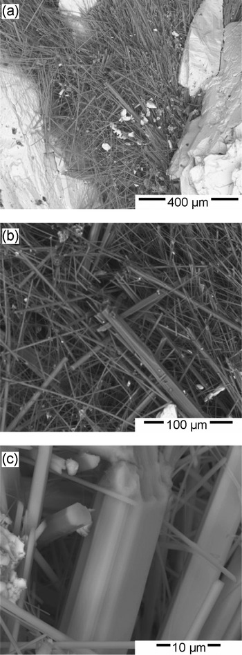
Small needles (see Fig. 1a–c) up to 1.5 mm along [001] and 10 μm in thickness are found in vugs between baryte and bixbyite crystals. Felted crystal masses to 5 mm fill small cavities. Dendritic crystals are developed occasionally on cleavage planes in baryte. Observed forms are prisms and pinacoid, no twinning but parallel intergrowth is abundant.
Secondary electron images of saccoite needles within a void between baryte crystals. Fragment from holotype specimen.

The mineral is brittle, neither cleavage nor preferred parting has been observed. The Mohs hardness and density could not be determined reliably due to the fragility of the needles, the calculated density is 2.73 g⋅cm–3 based on the cell volume refined from single-crystal X-ray diffraction data and empirical chemical formula.
Saccoite is transparent with pale to olive-green colour, and has white to light green streak and vitreous lustre. This colour is presumably caused by minor incorporation of Cu2+ into the structure. No luminescence was observed under either long-wave or short-wave ultraviolet irradiation. A single needle was mounted on a glass fibre and inspected under a Leitz Ortholux polarising microscope equipped with a Trelle micro-refractometer spindle stage using Cargille refractive index liquids (B series [n: 1.644–1.700, Δn 0.004) and M series (n: 1.71–1.80, Δn 0.01)]. The optical orientation of the indicatrix was determined by extinction curves applying the refractive index liquid (n = 1.70) and EXCALIBR software (Gunter et al., Reference Gunter, Bandli, Bloss, Evans, Su and Weaver2004). Saccoite was proven to be uniaxial (–) with a straight extinction. The refractive indices at 589(1) nm are ω = 1.705(5) and ɛ = 1.684(2), with birefringence –0.021. The pleochroism is distinct (bluish green // ω – yellow green // ɛ), the dispersion is weak, with r < v. Calculation of the equilibrium constant K c for the empirical formula using the Gladstone–Dale relationship (Gladstone and Dale, Reference Gladstone and Dale1863) with the values given by Mandarino (Reference Mandarino1976), combined with the optical data, resulted in a compatibility index for the empirical formula of –0.005, which is rated as superior (Mandarino, Reference Mandarino1981).
Chemical composition
Chemical analyses (11 spots) were carried out with an JEOL JXA-8500F EPMA system (WDS mode, 15 kV, 20 nA and 15 μm beam diameter). The H2O content could not be determined directly due to the scarcity and limited mass of pure material but is derived from the structure refinement. Analytical data are given in Table 1. The empirical composition calculated on the basis of Σ(Ca,Mn3+,Cu,Mg) = 4 is Ca2.06Mn3+1.78Cu0.10Mg0.07F0.97(OH)8.02(SO4)0.39; simplified Ca2(Mn3+,Cu2+)2F(OH)8⋅0.5(SO4). The ideal formula Ca2Mn3+2F(OH)8⋅0.5(SO4) requires CaO 28.53, Mn2O3 40.16, SO3 10.18, F 4.83, O≡F –2.03, H2O 18.33, total 100 wt.%.
Chemical composition (wt.%) of saccoite.

S.D. = standard deviation (2σ uncertainty); nd = not detected.
* H2O was calculated from stoichiometry.
Powder X-ray diffraction
Powder X-ray diffraction data (Table 2) were collected with a Gandolfi camera (114.59 mm diameter) utilising CuKα radiation. The refined unit-cell parameters (Holland and Redfern, Reference Holland and Redfern1997) are a = 12.834(3) Å, c = 5.622(2) Å and V = 926.0(4) Å3, in good agreement to the ones obtained from single-crystal X-ray work. The strongest lines in the powder X-ray pattern [d in Å (I/I100) (hkl)] are: 9.0735 (35) (110), 4.5370 (95) (220), 4.0644 (20) (310), 3.0105 (100) (321), 2.8117 (20) (002), 2.7242 (75) (411), 1.9755 (35) (611) and 1.8142 (20) (550).
Powder X-ray diffraction data (d in Å) for saccoite.

Note: Bragg peaks with relative intensities ≥20% are marked in bold.
Single-crystal X-ray diffraction data and crystal-structure refinements
Needles of saccoite with homogeneous extinction, mounted on glass capillaries with laboratory grease, were selected for single-crystal X-ray data collections. The samples were studied at room temperature on a Bruker APEXII diffractometer equipped with a charge-coupled device (CCD) area detector and an Incoatec Microfocus Source IμS (30 W, multilayer mirror and MoKα). Several sets of phi- and omega-scans with 1.5° scan width were collected at a crystal–detector distance of 40 mm up to 70°2θ full sphere. The absorption was corrected by the evaluation of partial multi-scans. Reflection data were processed using the Bruker APEX3 software suite (Bruker, Reference Bruker2020). The crystal structure was solved by direct methods in space group P4/ncc and refined by full-matrix least-squares techniques with the Shelx software (Sheldrick, Reference Sheldrick2015, Reference Sheldrick2018). Selected crystal parameters and a summary of data collection and structure refinements are given in Table 3. Saccoite is characterised by channels along [001], which house disordered and only partially occupied groups, especially SO42–, which could be only crudely localised. Final atom positions and important interatomic distances and angles are listed in Tables 4 and 5, respectively. The crystallographic information file has been deposited with the Principal Editor of Mineralogical Magazine and is available as Supplementary material (see below).
Crystal data and details of the single-crystal intensity measurement and structure refinement for saccoite.

* R 1= Σ F o – F c / Σ F o; wR 2 = [Σw(F o2 – F c2)2 / Σw(F o2)2]½.
** w = 1 / [σ2(F o2) + (a×P)2+ b×P]; P = {[max of (0 or F o2)] + 2F c2} / 3.
Atom coordinates and displacement parameters (Å2) for saccoite.

* Mn = (Mn0.89, Cu0.05, Mg0.03 and Ca0.03) based on EPMA.
** The disordered atoms within the channel, S and O3, were refined with positions half occupied.
Selected interatomic distances (d, Å) and bond valence strengths in valence units (vu) for saccoite.

Raman spectroscopy
Raman spectra of saccoite were obtained at room temperature by mean of a dispersive Horiba LabRAM Evolution spectrometer. This system was equipped with an Olympus BX41 series optical microscope and a Peltier-cooled CCD detector. The focal length is 800 mm. A diffraction grating with 1800 grooves per mm was used to disperse the scattered light. Raman spectra were excited by a 532 nm frequency-doubled Nd:YAG laser. The laser power (measured behind the objective) was 1.2 mW. Note that preliminary tests with 12 mW laser power resulted in local sample disintegration, which is assigned to significant warming in the focal-spot area due to too intense absorption of laser light. An Olympus 50× objective (numerical aperture 0.55) was used to focus the laser beam onto the sample surface and to collect the scattered light. The spectral resolution was ~1 cm−1. Wavenumber calibration was done using the Rayleigh line and Kr-lamp emissions, resulting in a wavenumber accuracy better than 0.5 cm−1. For further analytical details see Nasdala et al. (Reference Nasdala, Akhmadaliev, Artac, Chanmuang, Habler and Lenz2018).
The Raman spectrum of saccoite is shown in Fig. 2. It is dominated by a pair of intense bands at 535 and 565 cm−1, and a narrow, intense band at 986 cm–1. Band assignments of the former remain unclear whereas the latter is interpreted as ν1(SO4) mode (i.e. symmetric stretching vibrations of sulfate groups), hence supporting the presence of SO4 groups as derived from results of chemical analyses. The hydroxyl-stretching range shows a distinct band at 3602 cm–1. This band has a shoulder at ca. 3556 cm–1 and clear asymmetry towards the low-energy side (3500–3600 cm–1), suggesting that there exist (lower amounts of) additional, non-equivalent OH sites in the structure.
Raman spectrum (532 nm excitation) of saccoite, shown in comparison with the – somehow apparently similar – spectrum of kleinite.

To the best of our knowledge, no related Raman spectrum exists, which seems to correspond well to the fact that there is no other mineral whose structure is related closely to that of saccoite. In database searches, however, analysts may be puzzled by the apparent similarity of the Raman spectrum of saccoite with that of kleinite, Hg2N(Cl,SO4)⋅n(H2O) (Sachs, Reference Sachs1905; Giester et al., Reference Giester, Mikenda and Pertlik1996). There is no structural similarity between kleinite and saccoite, except both minerals are characterised by SO4 groups in channels. As the Raman spectrum of kleinite has not yet been described in the literature and is only contained in databases, we provide it here as a reference in comparison with the saccoite spectrum. The reference spectrum was obtained from kleinite from the Terlingua District, Brewster Co., Texas, USA (sample #H3845 in the mineral collection of the Natural History Museum, Vienna); the identity of this sample was confirmed using single-crystal X-ray diffraction. Note that in spite of similar double bands at around 550 cm–1, distinguishing the Raman spectra of saccoite and kleinite is straightforward, based on differences in their low-energy (<400 cm–1) and OH-stretching ranges.
Structure description and discussion
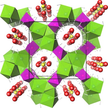
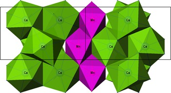
The crystal structure of saccoite features a heteropolyhedral framework (see Fig. 3) composed of edge- and corner sharing CaF2(OH)6 and M(OH)6 polyhedra (M = Mn3+, Cu2+, Mg2+ and Ca2+) thus establishing large, eight-membered channels along [001]. The CaF2(OH)6 polyhedron can be fairly well described as a tetragonal antiprism with Ca–F distances of 2.3 Å, the Ca–OH distances are in the range of 2.44–2.58 Å. These CaF2(OH)6 antiprisms share common edges among each other, arranged in columns along [001]. The F atom, tetrahedrally coordinated to calcium ions, is the central element of these columns. The M(OH)6 octahedron is strongly 4+2 Jahn-Teller distorted (4 × ~1.92 Å, 2 × 2.27 Å). M(OH)6 groups are linked to each other via common edges O2–O1 and O1–O1 to form zig-zag chains along [001] and further share edges with CaF2(OH)6 antiprisms (Fig. 4). A one-dimensional, non-interconnecting microporous framework results with 4.43 to 5.33 Å as a maximum diameter of a ‘sphere’ that can be accommodated in the channel (Foster et al., Reference Foster, Rivin, Treacy and Delgado Friedrichs2006), estimated on the basis of (O1–O1) and (O2–O2) separation distances, respectively. Bond valence calculations (Table 5, Brese and O'Keeffe, Reference Brese and O'Keeffe1991) yield bond strengths of 2.02 and 3.06 valence units (vu) for the Ca and Mn sites, respectively. The hydrogen atoms of the OH groups formed by O1 and O2 point into the channel, which houses disordered and only partially occupied molecular groups, especially SO42–. Hydrogen bonds probably occur towards the O3 oxygen atom of the anionic groups within the channel, ranging from 2.7 to 3.0 Å.
Perspective view of the structure of saccoite projected along [001]. CaF2(OH)6 antiprisms are illustrated in green, M(OH)6 octahedra in magenta and oxygen atoms of disordered sulfate groups in red. All structure drawings were done with ATOMS software (Dowty, Reference Dowty2016)

Structure detail of saccoite projected along [110]. CaF2(OH)6 antiprisms are illustrated in green, M(OH)6 octahedra in magenta. Hydrogen atoms are omitted.

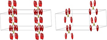
The extra-framework content of the channel is strongly disordered and only partially occupied. Due to space group P4/ncc (number 130, origin choice 2) of the [Ca2Mn3+2F(OH)8] framework host, the channel is symmetry constrained and arranged around a 4-fold axis, which does not allow a tetrahedral SO4 coordination. Attempts failed to describe the extra-framework content with ordered sulfate groups reliably in subgroups of lower symmetry. Data collections of a second crystal as well as a low-temperature measurement revealed no relevant changes of the model although a slight lattice distortion to orthorhombic metrics at 100 K cannot be excluded. As the distance c/2 of ~2.8 Å is far too short for neighbouring S atoms belonging to sulfate groups, one can assume the channel to be filled by only 50%, which is in accordance with chemistry and balance of charges required to compensate for the framework [Ca2Mn3+2F(OH)8]1+. Minor substitution of Mn3+ by divalent ions Cu2+, Mg2+ and Ca2+ would – as indicated by the chemical analysis – further lead to a reduction of necessary anionic sulfate groups from 0.5 to ~0.4 per formula unit. Because of the observed strong disorder, and combined with atomic displacement parameters exhibiting strongly elongated ellipsoids (see Fig. 5) along [001], the formal S–O distances (1.8 Å) of such a simplified model are much too high. Therefore, the hydrogen bonding system can only be estimated. The strongest residual electron density peaks (~1.7 and 1.3 e – Å–3) are located on the 4-fold axis within the channel.
Selected detail of the channel filling in saccoite. Thermal ellipsoids (50% probability) for disordered atoms of the sulfate group are shown. Left: half-occupied disordered SO4 groups: Right: Arbitrary channel filling disregarding space group symmetry.

From the chemical point of view, saccoite may be classified as 7.BC.65 in the Strunz Mineralogical Tables (Strunz and Nickel, Reference Strunz and Nickel2001), i.e. sulfates with additional anions and without H2O, with chemical similarities to despujolsite. However, the observed chemistry as an anhydrous Ca, Mn, F bearing sulfate is presently unique. According to its main one-dimensional structural motive saccoite can also be classified as a heterometallic, heteropolyhedral microporous mineral.
Acknowledgements
The authors are grateful to Uwe Kolitsch (Natural History Museum, Vienna) for providing a kleinite reference sample (inventory number H3845) for Raman analysis. Helpful comments of three anonymous reviewers and the Structural Editor Peter Leverett are kindly acknowledged.
Supplementary material
To view supplementary material for this article, please visit https://doi.org/10.1180/mgm.2022.60
Competing interests
The authors declare none.