In fetuses, neonates and children who have died suddenly, or following an illness, or following an intervention, a post-mortem study is desirable.1 Perinatal post-mortem can demonstrate new diagnoses in up to three-quarters of cases, and confirm the diagnosis in almost all the remainder,2 with similar figures seen for post-mortem studies of the heart alone.3
Over the last decade, there has been a continuing decline in the number of fetal and pediatric post-mortem studies performed in the United Kingdom.1, 4, 5 Some parents have always been reluctant to consent to conventional histopathological post-mortem assessment, but this has been further exacerbated by recent publicity about retention of organs following post-mortem examinations in the United Kingdom.6
A non-invasive method of accurately assessing the detailed anatomy of the heart and great vessels would therefore be useful. It could provide information for diagnosis and clinical audit and create a permanent electronic record of the findings, whilst allying parental concern with regard to retention of organs or conventional invasive post-mortems.
Magnetic resonance imaging is well suited as a non-invasive modality for post-mortem assessment.7 Magnetic resonance imaging of the excised brain, spine, and heart has been successful performed. Preliminary studies of whole-body post-mortem magnetic resonance imaging in fetuses and infants have also been reported.8–10 In all these studies, imaging of the central nervous system proved most accurate, whilst cardiac imaging proved more difficult.
In this case report, we demonstrate the usefulness of post-mortem magnetic resonance imaging in the assessment of the cardiovascular system.
Case report
A female infant was born with interruption of the aorta distal to the left subclavian artery, and a perimembranous ventricular septal defect. Initial surgery was performed through a left thoracotomy, with repair of the interrupted aorta by division of the left subclavian artery, anastomosis of the subclavian flap to the descending aorta, and placement of a band round the pulmonary artery. Subsequent anastomotic stricture of the aortic repair required balloon dilation at 4 months of age.
At 9 months of age, the ventricular septal defect was closed, the band removed from the pulmonary trunk, and a patch placed to augment the descending aortic anastomosis, all procedures being performed through a midline sternotomy. After a period of 39 days following the second operation, the patient presented to her local hospital with haemoptysis and circulatory shock. Insertion of a chest drain during resuscitation revealed presence of blood in the left hemithorax. The patient died during transfer to a cardiac surgical centre.
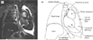
Conventional histopathological autopsy was declined, although the parents gave informed consent for post-mortem magnetic resonance imaging. Magnetic resonance imaging was performed using a 1.5 Tesla scanner (Symphony, Seimens Medical Solutions, Erlangen, Germany). Standard T1- and T2-weighted turbo spin echo images, inversion recovery T1- and T2-weighted turbo spin echo images, T2* gradient echo images, and three-dimensional gradient echo images were all acquired. The time required for imaging was 60 minutes. The images revealed a large false aneurysm of the descending aorta, with distal rupture, mediastinal haemorrhage, and a large left haemothorax (Figs 1 and 2).

Figure 1. (a) Coronal inversion recovery T2-weighted image (repeat time – 8500 ms, echo time – 114 ms, inversion time – 150 ms, flip angle – 150°, averages – 2, slice thickness – 3 mm, field of view – 200 × 200 mm, matrix – 224 × 320, resolution = 0.9 × 0.6 × 3 mm), with schematic representation (b). Altered blood (heterogeneous signal) is seen within the false aneurysm, which lies just lateral to the site of aortic surgery in the descending aorta, and posterior to the left main bronchus (not seen on this image slice). The heterogeneous signal suggests blood of varying ages – acute haemorrhage to clot. The left lung is collapsed towards the midline. There is a rim of high signal peripherally in the left hemi-thorax, which may represent signal from either very acute haemorrhage or serous fluid. Beneath this, there is an area of intermediate signal, consistent with recent haemorrhage or retracted clot within the left hemi-thorax.

Figure 2. (a) Coronal inversion recovery T2-weighted image (image parameters as in Figure 1), just posterior to image in Figure 1, and (b), coronal fat saturated T1-weighted gradient echo image (repeat time – 4.4 ms, echo time – 2.3 ms, flip angle – 12°, slice thickness-2 mm, field of view – 151 × 220 mm, matrix – 123 × 256, resolution = 1.2 × 0.9 × 2 mm) in the same plane. Note that the inferior-lateral wall of the false aneurysm is deficient, and is presumably the site of aneurysmal rupture (white arrow).
Discussion
With this case report, we have demonstrated that magnetic resonance imaging can be used to assess a subject in whom the pre-mortem diagnosis, though suspected, was not confirmed. The post-mortem magnetic resonance images were diagnostic, and enabled the clinician caring for the subject to explain the cause of death to the parents, and complete the death certificate with confidence. Furthermore, magnetic resonance imaging proved an acceptable form of post- mortem assessment for the parents, after request for traditional autopsy had been declined.
Magnetic resonance imaging post-mortem may be helpful in the assessment of children with congenital cardiovascular disease. There remains a need, however, for validation of magnetic resonance against conventional histopathology, still considered the “gold-standard” for autopsy. Such studies are now under way.7
Acknowledgement
Andrew Taylor is funded by the Higher Education Funding Council for England.