Introduction
Paragangliomas are rare tumours of the head and neck, and comprise approximately 0.6 per cent of all neoplasms in this area.Reference Suárez, Sevilla and Llorente1 They are neuroectodermal tumours originating in the paraganglia, a collection of small neuroendocrine organs. Although paragangliomas of the middle ear are common, only four cases have been reported in the external auditory canal.Reference Singh, Hanna and Dinnen2–Reference Hyams, Batsakis and Michaels4
In this paper, we present the first published report of a painful paraganglioma in the external auditory canal. The clinical diagnosis was delayed by this atypical presentation of a rare tumour in an unusual anatomical location, which masqueraded as non-resolving otitis externa.
Case report
A 49-year-old African woman was referred to our urgent referral clinic with suspected otitis externa. She had presented with a three-week history of left-sided otalgia, hearing loss and tinnitus.
Physical examination revealed a firm swelling arising from the superior portion of the left external auditory canal, covered by skin and completely obscuring the tympanic membrane. A full otorhinolaryngological examination was otherwise unremarkable.
Pure tone audiometry showed a conductive hearing loss of 25 dB.
The patient was managed with antibiotic eardrops and the insertion of an otowick for a few weeks, with no improvement.
Attempted aspiration of the swelling resulted in brisk bleeding, which was controlled by packing.
Computed tomography (CT) of the temporal bones revealed a non-specific, soft tissue mass in the left external auditory canal, with a thickened tympanic membrane and no erosion of the underlying bony canal.
A biopsy was performed under general anaesthesia. Once again, there was localised haemorrhage. The mass was completely excised via an endaural approach. The tympanic membrane was normal. However, the mass had slightly eroded the auditory canal anterosuperiorly, so a bony canalplasty was performed to facilitate complete removal. The ear canal was then lined with a split skin graft.
At one year post-operative follow up, the patient had a widely patent external auditory canal with a normal hearing threshold, and was clinically free of recurrence.
Histopathology
Macroscopically, the tumour measured 25 × 20 × 7 mm and lay beneath the epidermis.
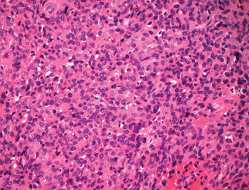
Histological examination showed a proliferation of cells with inconspicuous cytoplasm and uniform, dense nuclei, which were intimately associated with blood vessels (Figure 1). No mitotic activity or necrosis was identified.
Photomicrograph of the tumour showing moderate nuclear pleomorphism and small stromal blood vessels. There is no clear evidence of peripheral sustentacular cells. (H&E; ×400)

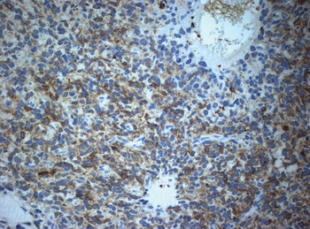
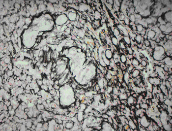
Immunohistochemical analysis of the surgical specimen revealed patchy positivity for neuron-specific enolase (NSE) (there had been strong positivity in the earlier biopsy) (Figure 2) and for cluster of differentiation 56 protein. The proliferation fraction, as identified by Ki67, was low. Reticulin staining showed a nesting pattern (Figure 3).
Photomicrograph of the tumour prepared with neuron-specific enolase stain. (×400)

Photomicrograph of the tumour prepared with reticulin stain, showing a discernable nesting pattern. (×400)

After seeking further expert opinion, a diagnosis of paraganglioma of the jugulotympanic system was made, with no evidence of a round cell tumour or primary bone sarcoma (suspected previously).
Discussion
Paragangliomas are neoplasms of neuroectodermal origin. They are embryologically derived from the neural crest cells, which originate in the paraganglia, a collection of small neuroendocrine organs. Paragangliomas are usually divided into two categories: those developing in the head and neck region and those arising elsewhere. The adrenal medulla is the most common site.Reference Suárez, Sevilla and Llorente1 Paragangliomas have been found accompanying Arnold's nerve, deriving innervation from the vagus nerve and blood supply from the inferior tympanic branch of the ascending pharyngeal artery.Reference Singh, Hanna and Dinnen2, Reference Gulya5
Normal paraganglia play an important role in homeostasis, with most in the head and neck acting as chemoreceptors. Paragangliomas comprise approximately 0.6 per cent of all neoplasms in this area and hence are very rare. The presented patient's external auditory canal tumour was postulated to have arisen from paraganglionic tissue associated with Arnold's nerve.Reference Singh, Hanna and Dinnen2 In a previous review of tumours of the middle ear and the surrounding tissue, paragangliomas of the external auditory canal represented 2 per cent of all such lesions; subsequently, only two more cases have been described in the English language literature.Reference Singh, Hanna and Dinnen2–Reference Hyams, Batsakis and Michaels4 Neither of these cases presented with pain, in contrast to our patient.
Pain has previously been described in cases of type II laryngeal paraganglioma.Reference Ali, Aird and Bihari6 These tumours are exquisitely painful and characteristically inconspicuous, eluding diagnosis. Eighty-five per cent present with metastasis. The pain has been ascribed to the secretion of a pain-inducing substance close to sensory nerve endings. However, there has been no previous report of a painful aural paraganglioma. We hypothesise that, in the presented case, the auricular branch of the vagus nerve, which relays sensory input from the postero-superior part of the external auditory canal, conducted the nociceptive stimuli. Fortunately, there was no clinical or histological evidence of malignancy in this case, as suggested by the low proliferation fraction.
The diagnosis of paraganglioma is made from the observed histology. In the presented patient, the brisk bleeding after aspiration raised the possibility of a vascular tumour. In addition, the tumour lacked some of the characteristic features of paragangliomas occurring at other sites, making diagnosis more difficult. The typical nesting pattern was focal and only appreciated with silver staining. This is postulated to be a problem with paragangliomas occurring in the external auditory canal, for which crush artefact can be very marked. There were no demonstrable S100 sustentacular cells, which does not preclude the diagnosis but makes it more challenging. The marked delicate vascularity of the tumour contributed to the diagnosis. In this patient, the use of special stains was invaluable to the diagnosis.
Imaging may be helpful in the diagnosis of paragangliomas. Computed tomography scanning, although useful for delineating the adjacent bony detail, does not assist the diagnosis.Reference Suárez, Sevilla and Llorente1 Magnetic resonance imaging (MRI) was used in a similar case to successfully demonstrate the extent of involvement of the tympanic membrane.Reference Skinner, Curran, Barnes and Timon3 In our case, the tympanic membrane thickening seen on CT was due to a piece of otowick lodged behind the mass; this was retrieved during examination under anaesthesia, which also revealed the tympanic membrane to be macroscopically free of tumour. Hence, an MRI was deemed unnecessary in our case.
• Unusual external ear tumours may masquerade as atypical patterns of common diseases
• Early intervention, with imaging and biopsy, is needed
• Diagnosis may be difficult; additional stains and awareness of atypical histopathology may be required
• Junior doctors should seek early advice for non-resolving otitis externa; neoplastic disease is not uncommon
The other cited advantage of imaging in such cases is to detect synchronous tumours in familial cases.Reference Skinner, Curran, Barnes and Timon3 Most synchronous tumours are discovered incidentally.Reference Spector, Ciralsky, Maisel and Ogura7 Such tumours are commoner in patients with a heredofamilial pattern (who account for up to 40 per cent of cases), with 7 per cent occuring in non-familial cases.Reference Hodge, Byers and Peters8 Thus, there is an argument for imaging patients with paraganglioma in order to detect synchronous tumours.
Paragangliomas tend to be locally destructive. However, in our own and previous cases, the tumour was confined to the bony canal without invasion of the surrounding tissue. This can be explained by the fact that most paragangliomas grow slowly at a median rate of 1 mm per year, with a median tumour doubling time of 4.2 years.Reference Suárez, Sevilla and Llorente1 Their growth in the relatively confined, superficial position of the external auditory canal leads to disability in the form of hearing loss, which favours early diagnosis.