Introduction
More than 700 million grazing animals and over 180 million people are at risk of fasciolosis (Liu et al., Reference Liu, Gasser, Young, Song, Ai and Zhu2014). Bovine fasciolosis has a global geographical distribution. Its prevalence ranges from 1.15 to 80% (Ibarra et al., Reference Ibarra, Montenegro, Vera, Boulard, Quiroz, Flores and Ochoa1998; Phiri et al., Reference Phiri, Phiri, Sikasunge and Monrad2005; Pfukenyi et al., Reference Pfukenyi, Mukaratirwa, Willingham and Monrad2006; Cabán-Hernández et al., Reference Cabán-Hernández, Gaudier, Ruiz-Jiménez and Espino2014; Charlier et al., Reference Charlier, Vercruysse, Morgan, Dijk and Williams2014; Liu et al., Reference Liu, Gasser, Young, Song, Ai and Zhu2014; Mufti et al., Reference Mufti, Afshan, Khan, Zafar, Rizvi, Nazir and Qayyum2014). Fasciolosis is a major cause of huge economic losses in the livestock industry. It reduces animal productivity, including decreased growth rate, meat and milk production, fertility, feed efficiency and draught power (Asrat, Reference Asrat2004).
Control of bovine fasciolosis is hampered by limitations of currently available anthelmintics, and the fact that there is no effective vaccine against fasciolosis. Most anthelmintics have a low efficacy against the immature stages of Fasciola, and there is evidence for the development of drug resistance in Pakistan (Hamad et al., Reference Hamad, Iqbal, Sindhu and Muhammad2013, Reference Hamad, Iqbal, Sindhu, Abbas, Khan, Muhammad and Epperson2014). This makes research into the prediction of risk periods, exploration of disease epidemiology, and thus the optimum application of available drugs, more pertinent.
Using conventional methods, the disease can only be diagnosed at week 14 post-infection, by which time major hepatic damage due to the migration of flukes has already occurred. Furthermore, this method does not work if the parasitic burden is low. Early diagnosis of the disease is essential to control and treat the infection. To overcome the deficiencies of diagnosis, various enzyme-linked immunosorbent assays (ELISAs) have been developed, which aim at detecting anti-fasciola antibodies in serum or milk, and antigens in serum or faeces. The excretory–secretory (ES) antigens of Fasciola (F.) gigantica, or their partially purified components, are the most common source of antigens used in ELISA. Antibodies to these antigens can be detected as early as 2 weeks, and their concentrations can be determined between 8 and10 weeks post-infection (Yadav et al., Reference Yadav, Saini, Raina, Nambi, Jadav and Sriveny2005; Awad et al., Reference Awad, Ibrahim and Salib2009; Varghese et al., Reference Varghese, Raina, Nagar, Garg, Banerjee, Maharana and Kollannur2012). An alternative to antibody detection is the direct measurement of F. gigantica antigens that are shed into the serum or faeces of infected animals or humans (Valero et al., Reference Valeroa, Ubeira, Khoubbane, Artigas, Muino, Mezo, Pérez-Crespo, Periagoa and Mas-Coma2009). A monoclonal antibody has been produced that can detect F. gigantica antigen in the serum and faeces of infected humans (Demerdash et al., Reference Demerdash, Diab, Aly, Mohamed, Mahmoud, Zoheiry, Mansour, Attia and El-Bassiouny2011) and cattle (Estuningsih et al., Reference Estuningsih, Spithill, Raadsma, Law, Adiwinata, Meeusen and Piedrafita2009). Antibody detection assays are most commonly used due to the relative simplicity of the assays, and early seroconversion (usually 1–2 weeks), high sensitivity and easy usage (Jalali et al., Reference Jalali, Zadeh, Abadi and Yamchi2012).
The sensitivity and specificity of a coproantigen-based ELISA (Mezo et al., Reference Mezo, González-Warleta, Carro and Ubeira2004) has been investigated by experimental infection (Cornelissen et al., Reference Cornelissen, Gaasenbeek, Borgsteede, Holland, Harmsen and Boersma2001), or by testing it on two distinct populations: a positive population selected from an enzootic area and a negative population from a fluke-free area (Ibarra et al., Reference Ibarra, Montenegro, Vera, Boulard, Quiroz, Flores and Ochoa1998; Mezo et al., Reference Mezo, González-Warleta, Carro and Ubeira2004; Salimi-Bejestani et al., Reference Salimi-Bejestani, McGarry, Felstead, Ortiz, Akca and Williams2005). However, the sensitivity and specificity of diagnostic tests vary by population (Greiner & Gardner, Reference Greiner and Gardner2000), and thus results cannot always be extrapolated to the area and field situation where the test will be applied. Hence, in various countries scientists have developed ELISA using the local antigens. ELISAs based on ES antigens have been developed in many countries, including Bolivia (Hillyer et al., Reference Hillyer, de Galanes, Buchon and Bjorland1996), Mexico (Ibarraa et al., Reference Ibarra, Montenegro, Vera, Boulard, Quiroz, Flores and Ochoa1998), Vietnam (Anderson et al., Reference Anderson, Luong, Vo, Bui, Smooker and Spithill1999), Peru (Ortiz et al., Reference Ortiz, Claxton, Clarkson, McGarry and Williams2000), Belgium (Charlier et al., Reference Charlier, Meulemeester, Claerebout, Williams and Vercruysse2008) and Iran (Jalali et al., Reference Jalali, Zadeh, Abadi and Yamchi2012). However, no such effort has ever been made in Pakistan. Enhanced epidemiological understanding of disease and early detection of fasciolosis with a highly sensitive assay will improve the health status of livestock, and thus result in the alleviation of poverty of livestock farmers.
The present objectives include the development and standardization of ELISA using ES antigens of F. gigantica from infected bovines, the validation of indigenous and commercial ELISAs under field conditions, and an estimation of economic losses due to bovine fasciolosis from the Sargodha district, Pakistan.
Materials and methods
Collection of adult F. gigantica and blood samples from slaughterhouses
The livers of cattle and buffaloes from slaughterhouses of Sargodha district (an administrative division of a province, Punjab, of Pakistan) were cut into 10-mm-thick pieces and thoroughly examined for the presence of any Fasciola. Only mature F. gigantica were isolated after identification. Whole blood samples of the same animals were collected and centrifuged at 2500 rpm for 15 min. Serum was aspirated carefully and stored at − 20°C after taking aliquots of 0.5 ml in serum cups.
Coprological and immunological protocols
The presence of Fasciola eggs in faecal samples was evaluated using a sedimentation–flotation technique. Briefly, a mixture of 4 g of faeces in 200 ml water was filtered three times through a tea sieve and allowed to stand for 30 min, after which the sediment was collected in a test tube and centrifuged at 2000 rpm for 3 min. After centrifugation, the sediment was suspended in zinc chloride and centrifuged at 1000 rpm for 3 min. A meniscus was prepared by adding zinc chloride solution, over which a coverslip was placed. Finally, the coverslip was examined microscopically at 100 × magnification (Charlier et al., Reference Charlier, Meulemeester, Claerebout, Williams and Vercruysse2008). Identification of eggs and flukes was carried out using standard parasitological keys, as described by Soulsby (Reference Soulsby2006).
The method described previously by Anderson et al. (Reference Anderson, Luong, Vo, Bui, Smooker and Spithill1999) was followed for the preparation of ES antigens from adult F. gigantica. Briefly, adult parasites (in batches of about 10), were added to 25 ml of phosphate-buffered saline (PBS) at 37°C, washed three times in PBS for 20 min and then incubated in fresh PBS for 6–8 h at 37°C. After incubation, the fluid containing the ES antigens was filtered through a 0.22-μm filter and stored at − 20°C, until required. The protein concentration of this solution was estimated spectrophotometrically at 280 nm and was found to be 2.8 mg/ml.
Optimum concentrations of antigen and antibodies were determined by checkerboard titration. Antigen dilution at column 2 (1/200) and serum dilution at row B (1/100) produced maximum OD at higher dilution. First of all, the antigen at a 1/200 dilution was coated on a 96-well ELISA plate and incubated overnight at 4°C. The wells were then washed five times with PBS containing 0.05% Tween and incubated with 50 μl of 5% skimmed milk in PBS for 1 h at 37°C. The plate was washed five times, 50 μl of 1/100 dilution of serum were added to each well and incubated for l h at 37°C. After another wash, 50 μl of a 1/10,000 dilution of rabbit anti-bovine IgG antibodies conjugated to horseradish peroxidase (HRP) were added to each well, and the plate was incubated for 1 h at 37°C. After incubation, 50 μl of the substrate, tetramethyl benzidine, was dispensed into each well, incubated for 15 min at 37°C and the optical density (OD) measured at 450 nm using an ELISA reader. Comparative sensitivity and specificity were calculated using the following formulae:
 $$\begin{eqnarray} Sensitivity = \frac {Positive}{Positive + False\ Positive}\times 100 \end{eqnarray}$$
 $$\begin{eqnarray} Sensitivity = \frac {Negative}{Negative + False\ negative}\times 100 \end{eqnarray}$$
Liver infection served as the gold standard for true positive and true negative.
Field sampling of bovines
To validate the sensitivity and specificity of the newly standardized ELISA under field conditions, a survey of bovines of Sargodha district was performed. Sample size was calculated using simple random sampling as described by Thrusfield (Reference Thrusfield2008). Taking an expected prevalence of 25.46% (Khan et al., Reference Khan, Sajid, Khan, Iqbal and Iqbal2009) and 5% precision level, sample size was calculated as 300 each for cattle and buffalo. The number of animals to be sampled from each of the six sub-districts was determined by a proportional allocation method (Lauridsen, Reference Lauridsen2005).
After determining the sample size and selecting the sampling units, we visited the locations fortnightly to collect faecal and blood samples. A total of 5580 faecal (l0 g) and 600 blood (10 ml) samples were collected from selected animals and transported to the laboratory for coprological examination and ELISA, respectively. Information regarding age, sex, types of feeding systems and watering systems were collected through a questionnaire that used close-ended questions of dichotomous and multiple choice types. Samples were collected from animals of four age groups: ≤ 2 years, >2–4 years, >4–6 years and >6 years. Information regarding the types of feeding systems (grazing or stall feeding) and watering systems (river water, pond water or tap water) used in the study area were recorded.
Coprological and immunological protocols
Faecal samples collected during the field survey of Sargodha district were examined for the presence of any Fasciola ova through coprological examination, as described above. Serum was separated from blood samples and tested for the presence of anti-fasciola antibodies using both indigenous and commercial ELISA (DRG International, Springfield Township, New Jersey, USA). For the indigenous ELISA, the protocol described above was followed, while the manufacturer's instructions were followed for the commercial ELISA kit.
Estimation of economic losses
Economic losses caused by the condemnation of infected livers due to bovine fasciolosis in slaughterhouses of the six sub-districts of the Sargodha district (viz. Sargodha, Bhalwal, Shahpur, Kot Momin, Sahiwal and Silanwali) were calculated by the formula described by Tsegaye et al. (Reference Tsegaye, Begna and Mulugeta2011), considering the overall prevalence of the disease and the price of liver in the Sargodha market:
 $$\begin{eqnarray} LC = CSR\times LCo\times P \end{eqnarray}$$
where LC = losses due to liver condemnation, CSR = average number of slaughtered bovines, LCo = average cost of one liver, P = prevalence of fasciolosis.
Data analysis
Results of ELISA tests were analysed for sensitivity, specificity, positive predictive value, negative predictive value and screening comparative diagnostics using Winepiscope, by undertaking coprological examination as the standard test. Both results were compared using the chi-square test. Results of the coprological survey were analysed by chi-square test and multiple logistic regression analysis. Paired characteristics were analysed by odds ratio. All procedures were carried out using the SAS statistical software package at 95% confidence level (SAS, 1998).
Results
The mean OD value of the positive group (0.6891 ± 0.1371) was significantly higher (χ2= 29.761) than that of the negative group (0.0712 ± 0.0403), with a cut-off value of 0.1922.
Evaluation of indigenous ELISA
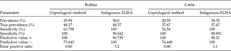
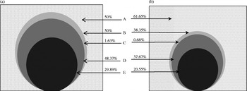
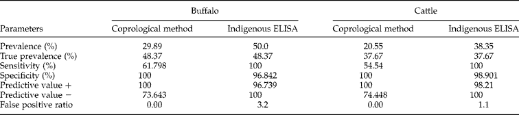
Evaluation parameters of indigenous ELISA and coprological examination were calculated. The sensitivity of ELISA was found to be 100% in both buffaloes and cattle (table 1) compared with coprological examination (buffalo, 61.79%; cattle, 54.54%). Indigenous ELISA was also found to be highly specific, with values of 96.84 and 98.90% in buffaloes and cattle, respectively. The standard test in this study was post-mortem liver examination. The false positive ratio of indigenous ELISA was 3.2 in the buffalo population, which fell to 1.1 in the cattle population (table 1). All animals that yielded Fasciola ova in coprological examinations were positive by ELISA. Also, all animals positive in liver examination yielded positive results in ELISA. Three buffaloes were found to be false positives while only one cattle serum was wrongly detected as positive (fig. 1).
Evaluation parameters of indigenous ELISA and coprological examination, using liver examination of buffalo and cattle as standard; ELISA appears to be more sensitive in both host types (P=0.000) and coprological examination is more specific in buffalo (P=0.083) and cattle (P=0.317).
Comparative positivity rates in the diagnosis of bovine fasciolosis based on liver, coprological and ELISA tests in (a) buffaloes and (b) cattle. (A) Healthy animals using all tests; (B) using ELISA; (C) false positivity; (D) using liver; (E) using the coprological test.

Serological and coprological evaluation
During the coprological evaluation of bovines of Sargodha district, Fasciola ova were identified in 1962 animals out of the 5580 cattle and buffaloes examined (35.16%). The buffalo population had a significantly higher prevalence (χ2= 5.8399; P value = 0.0157; OR = 0.563) compared with cattle. Prevalence of F. gigantica was significantly higher than that of F. hepatica (χ2= 70.6325; P value = 0.0001) between the two species (table 2).
The prevalence (%) of fasciolosis in buffalo and cattle from Sargodha district using coprological examination; N= number of animals examined.

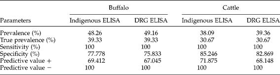
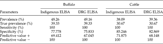
Both the indigenous ELISA and DRG ELISA detected a higher percentage of anti-fasciola antibodies in buffaloes (48.26% and 49.16% respectively) compared with coprological examination (true prevalence = 39.33%). Similar results were recorded in cattle (table 3), where we found higher sero-prevalence in the indigenous ELISA (38.09%) and DRG kit ELISA (39.36%) compared with coprological examination (30.67%). Both tests were 100% sensitive when compared statistically using Winepiscope, although indigenous ELISA was more specific than DRG ELISA for comparative diagnostics screening (table 3).
Evaluation of indigenous ELISA and commercial ELISA (DRG) tests using coprological examination; both ELISA tests are equally sensitive, with the indigenous ELISA being more specific.
Risk factors and estimates of economic losses
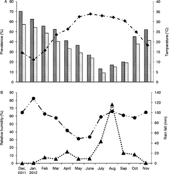
The month of the year was associated with risk of infection in both buffaloes (χ2= 46.888; P value < 0.0001) and cattle (χ2= 38.671; P value < 0.0001). The highest prevalence was found in December, while the lowest prevalence was found in July, suggesting a clear association between levels of infection and meteorological parameters (fig. 2). The lowest prevalence was recorded during the month of July when the temperature was high and relative humidity was low. On the other hand, disease prevalence increased after a peak in rainfall (August) and continued to increase post monsoons.
The relationship between seroprevalence (%) of fasciolosis in buffalo (dark grey bars) and cattle (light grey bars) relative to (A) temperature (- ·♦- ·) and (B) humidity (…●…) and rainfall (…▲…) in the district of Sargodha from December 2011 to November 2012.

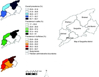
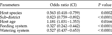
The prevalence of bovine fasciolosis was found to be significantly different between all sub-districts of Sargodha (OR = 0.823; χ2= 78.8745; P value < 0.0001). A relatively higher prevalence was found in bovines of Bhalwal, Sahiwal and Shahpur sub-districts compared with Sargodha, Kot Momin and Silanwali (fig. 3). When the effect of sub-districts was studied using the confounding variables age, sex and month, significant differences of prevalence among the six sub-districts were found.
The prevalence of bovine fasciolosis in six tehsils within Sargodha district.

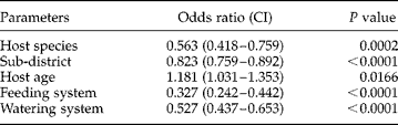
The risk of fasciolosis was significantly associated (OR = 1.181; χ2= 105.6757; P value < 0.0001) with age (table 4). Bovines ≤ 2 years old had the lowest prevalence of fasciolosis, whereas animals between the ages of >2 and 4 years had the highest prevalence of the disease. With increasing age, the chances of disease occurrence decreased. A P value >0.05 indicated that there was no significant difference in the risk of disease between sexes. Slightly higher prevalence was recorded in males compared with females (table 2).
Multivariate logistic regression analysis of bovine fasciolosis in the Sargodha district; CI=95% confidence intervals.
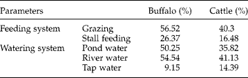
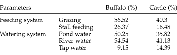
Feeding and watering systems were significantly associated with the risk of infection. Significantly higher prevalence of disease was recorded in grazing groups (χ2= 61.3443; P value < 0.0001) compared with stall-fed ones (OR = 0.327) in both cattle and buffaloes (table 5). Analysing data related to the watering system revealed that animals drinking pond or river water had a higher chance of contracting the disease compared with animals fed tap water (table 5) (OR = 0.527).
The prevalence (%) of fasciolosis in buffalo and cattle relative to feeding and watering systems.
The economic losses, due to the condemnation of bovine fasciolosis livers infected in Sargodha district, were estimated as US$35,697 (PKR 3,141,360), with a disease prevalence of 43.63%. The average cost of one bovine liver was PKR 800 during the study period. Since, on average, half a liver per infected bovine was condemned, the value of LCo was considered to be PKR 400.
Discussion
For a number of years, the diagnosis of fasciolosis was based on the microscopic detection of Fasciola ova in faeces. However, this method is not very sensitive because Fasciola ova do not appear in faeces until the fluke has become an adult and reached the biliary system of the liver. Following infection, Fasciola take 10–14 weeks to mature and release ova into faeces (Anderson et al., Reference Anderson, Luong, Vo, Bui, Smooker and Spithill1999), by which time hepatic damage has already occurred. Therefore, early detection of infection is necessary to prevent disease progression and avoid losses caused by bovine fasciolosis. To overcome the shortcomings of faecal examination, serological detection of antibodies by ELISA was adopted as an alternative approach. Excretory–secretory product was the most commonly used antigen in serological detection of bovine fasciolosis (Santiago & Hillyer, Reference Santiago and Hillyer1988; Fagbemi & Guobadia, Reference Fagbemi and Guobadia1995).
In the present study, ES antigens of F. gigantica were used to detect bovine fasciolosis. However, results can be extrapolated in general as bovine fasciolosis, due to the fact that in the study area F. hepatica infection is limited to 3%, as reported by Khan et al. (Reference Khan, Sajid, Khan, Iqbal and Iqbal2009). The results of indigenous ELISA were compared with those of coprological examination in the first experiment. Lower prevalence was recorded by coprological examination when compared with serological testing. Khan et al. (Reference Khan, Sajid, Khan, Iqbal and Iqbal2009) and Maqbool et al. (Reference Maqbool, Hayat, Akhtar and Hashmi2002) recorded lower prevalence in the same population compared with the sero-prevalence observed in the present study. The higher infection rate found during coprological examinations in this and previous studies (Maqbool et al., Reference Maqbool, Hayat, Akhtar and Hashmi2002; Khan et al., Reference Khan, Sajid, Khan, Iqbal and Iqbal2009) indicate a failure of strategies to control bovine fasciolosis in the study area. This may be due to the development of drug resistance against commonly available fasciolicides. The lower sensitivity of coprological examination may also be due to the fact that Fasciola ova do not appear in the faeces of a number of animals that have flukes in their liver. Absence of ova in faeces of infected animals may be due to mature flukes only intermittently shedding ova, low parasitic burden and/or the presence of immature flukes. These results are in accordance with those of Anderson et al. (Reference Anderson, Luong, Vo, Bui, Smooker and Spithill1999). The low sensitivity of coprological examination could be due to the presence of a large number of animals with low levels of infection, in the sampled group, and sampling animals that have recovered from infection (i.e. with no flukes present), but in which anti-fasciola antibodies are still circulating. However, sensitivity could be improved by increasing sample size. Previously, it was reported that sensitivity of faecal examination improved from 69% to 90% by three serial faecal examinations of the same samples (Rapsch et al., Reference Rapsch, Schweizer, Grimm, Kohler, Bauer, Deplazes, Braun and Torgerson2006) – a labour-intensive process. Detection of fasciolosis is hampered due to the low sensitivity of methods, which cannot detect small numbers of ova in large volumes of ruminant faeces. This is clearly illustrated in the present study where the predictive value for a positive egg count was 100%, whereas the predictive value for a negative count was only 73.64% and 74.45% in buffalo and cattle, respectively. This low value was largely due to the zero egg counts in faeces of animals with flukes in their liver.
An essential characteristic of ELISA-based assays is high sensitivity. Welch et al. (Reference Welch, Smith, Malone, Holmes and Geagan1987) recorded that 26% of animals shedding Fasciola ova were ELISA-negative, and Hillyer et al. (Reference Hillyer, de Galanes, Buchon and Bjorland1996) found seven animals as ELISA negative, which were positive in faecal examination. However, this was not the case in the present study. Indigenous ELISA was 100% sensitive. In all cattle and buffalo examined, none of the animals that yielded Fasciola ova during coprological examination were negative in ELISA. All the animals that were positive in liver examination yielded positive results by ELISA. With the help of indigenous ELISA, it was possible to detect anti-fasciola antibodies in sera of 34 buffaloes and 24 cattle, whereas no Fasciola ova were identified during coprological examination of faeces of these animals. However, it is possible that infection in these animals may have been mild and the ova count was too low to be detected by the employed method, or at the time of examination the flukes had not matured, and hence not released ova. A major disadvantage of the indirect serological test is that the presence of antibodies is not indicative of active infection, as it has been demonstrated that antibody titres could remain high in hosts after they are treated (Ibarra et al., Reference Ibarra, Montenegro, Vera, Boulard, Quiroz, Flores and Ochoa1998). In the present study, three buffaloes were false positive, while only one cattle serum was wrongly detected as positive. This may be due to low infection, which could have been missed during liver examination, or due to common antigenic epitopes with other helminths (Fagbemi & Obarisiagbon, Reference Fagbemi and Obarisiagbon1991). Recent administration of fasciolicide may have eliminated flukes from bile ducts, but the antibodies persist in the serum for 2–7 months (Santiago & Hillyer, Reference Santiago and Hillyer1988). Information regarding fasciolicide treatment and previous history of animals examined in this study was not collected. Therefore, positive and negative likelihood ratios are calculated without any knowledge of the history of the animal. However, these may increase or decrease if this information is used in conjunction with the test result. In conclusion, in the present study an indirect ELISA, which has high sensitivity and specificity for bovine fascioliasis, was developed and standardized.
However, antibody-based detection assays have some limitations. They also detect antibodies of previous infections, but cannot detect antibodies after flukes have reached bile ducts as excretory–secretory products are not available to the immune system of host, but are excreted with bile through faeces. Yet antibody detection assays are most commonly used due to high sensitivity and easy usage. Epidemiologists have now turned their attention to direct detection of antigen in serum and faeces to differentiate between active and previous infections. Antigen detection-based assays can detect infection earlier than antibody detection assays as antigens are available earlier than antibodies in serum. The specificity of these assays is 100% as purified/recombinant antigens do not cross-react with other antigens.
To validate the sensitivity and specificity of the standardized test under field conditions, a survey of bovines of Sargodha district was performed. This is the first survey of bovine fasciolosis in Pakistan using a highly sensitive diagnostic technique rather than just conventional microscopy, which does not give the real picture of disease burden. Prevalence values (48.26% and 49.16%) recorded in this survey were higher compared with previous studies, i.e. 25.59% (Maqbool et al., Reference Maqbool, Hayat, Akhtar and Hashmi2002) and 25.46% (Khan et al., Reference Khan, Sajid, Khan, Iqbal and Iqbal2009). This is due to the fact that the more sensitive ELISA technique was employed, rather than simple microscopy used in previous studies. The higher prevalence of fasciolosis in buffalo as compared with cattle may be attributed to their usual habit of living in muddy and marshy areas, which are favourable for the growth of the intermediate host snail of F. gigantica.
Soulsby (Reference Soulsby2006) stated that the two most important factors that influence the prevalence of fasciolosis are temperature and moisture, as these affect the hatching of eggs of Fasciola. In the present study, elevated prevalence of fasciolosis was observed after peak rainfall in the month of August as relative humidity had increased. Cruz-Mendoza et al. (Reference Cruz-Mendoza, Ibarra-Velarde, Quintero-Martínez, Naranjo-García, Lecumberri-López and Correa2005) observed an increase in population of infected and non-infected snails after a rise in humidity, and some weeks later an elevated prevalence of fasciolosis in cattle.
The relatively higher prevalence of fasciolosis found in bovines of Bhalwal, Sahiwal and Shahpur sub-districts, may be due to the rivers Jhelum and Chenab, which border these sub-districts. Alves et al. (Reference Alves, Carneiro, Martins, Bernardo, Donatele, Pereira, Almeida, Avelar and Leão2011) studied the prevalence of F. hepatica in ten municipalities in Brazil, and determined an association between the presence of flooded areas in municipalities and prevalence of fasciolosis. The results of the present study are in agreement with those of Khan et al. (Reference Khan, Sajid, Khan, Iqbal and Iqbal2009) who have recorded the highest prevalence post monsoons. Prevalence of fasciolosis was recorded to be highest in the age group >2 to 4 years, after which prevalence decreases gradually with advancing age. The lowest prevalence in animals of < 2 years age may be attributed to the fact that their chances infection were smaller due to shorter exposure time compared with older animals. The association of disease with age, found in the present study, was in agreement with the results of Anderson et al. (Reference Anderson, Luong, Vo, Bui, Smooker and Spithill1999), Fatima et al. (Reference Fatima, Chishti, Ahmad and Lone2012) and Pfukenyi et al. (Reference Pfukenyi, Mukaratirwa, Willingham and Monrad2006). However, Khan et al. (Reference Khan, Sajid, Khan, Iqbal and Iqbal2009) found no association between age of the animal and prevalence of disease. The decrease in prevalence at ages >4 years is due to the fact that cattle develop resistance to challenge infections. Hillyer et al. (Reference Hillyer, de Galanes, Buchon and Bjorland1996) stated that adult cattle are more resistant to reinfection or they may self-cure. There are some reports that resistance is not totally immunologically based. Rather, it is suggested that hepatic fibrosis resulting from primary infection may be the reason for resistance to reinfection (Mulcahy et al., Reference Mulcahy, Joyce, Dalton and Dalton1999). Both Ross (Reference Ross1967) and Boray (Reference Boray and Soulsby1967) suggested that hepatic fibrosis resulting from the primary infection may be an important factor contributing to resistance to challenge infections. Doyle (Reference Doyle1971) also pointed out that the duration of the primary infection, and hence the extent of liver fibrosis, correlated with the level of resistance.
Both sexes were found to be equally susceptible in the present study. These results are in agreement with those of Maqbool et al. (Reference Maqbool, Hayat, Akhtar and Hashmi2002). Although Phiri et al. (Reference Phiri, Phiri, Sikasunge and Monrad2005) found significantly higher prevalence in females compared with males, Khan et al. (Reference Khan, Sajid, Khan, Iqbal and Iqbal2009) did not find any statistical association between sex and disease.
The economic losses, due to the condemnation of bovine fasciolosis livers infected in Sargodha district, were estimated to be US$35,697 (PKR 3,141,360). Fasciolosis is known to cause significant economic loss in animal productivity: an estimated 9% reduction in weight gain in growing cattle, 10% reduction in milk yield, an extension of the service period by 13 days and an increment of 0.75 services per conception (Charlier et al., Reference Charlier, Vercruysse, Morgan, Dijk and Williams2014). A benefit–cost ratio of 3.9 is gained after treating cattle with some fasciolicide, while investigating increase in milk quantity and quality (Khan et al., Reference Khan, Sajid, Khan, Iqbal and Iqbal2009).
In conclusion, we have developed a sensitive and specific diagnostic ELISA for F. gigantica infection in buffalo and cattle that has been fully validated using sera from populations of known infection status.
Conflict of interest
None.