Introduction
Superior semi-circular canal dehiscence syndrome was first described in the late 1990s by Lloyd Minor and colleagues.Reference Minor, Solomon, Zinreich and Zee1,Reference Kutz, Phillips and Erskine2 Individuals with this condition often describe vestibular symptoms elicited by loud sounds, as well as other pressure-induced symptoms, including sneezing, coughing and straining. Individuals often also experience autophony, hyperacusis, cognitive dysfunction, spatial disorientation, anxiety and migraine headaches. Abnormal audiovestibular findings include an increased air–bone gap and/or suprathreshold bone conduction values with significantly lower cervical vestibular-evoked myogenic potential thresholds.Reference Crane, Carey, Minor, Brackmann, Shelton and Arriaga3
The aetiology of superior semi-circular canal dehiscence syndrome is not fully understood, and debate continues regarding whether this condition is developmental or acquired.Reference Davey, Kelly-Morland, Phillips, Nunney and Pawaroo4 The diagnosis of superior semi-circular canal dehiscence syndrome requires a careful and detailed history and examination, as well as detailed audiometry, high-resolution computed tomography and cervical vestibular-evoked myogenic potentials testing.
Case report
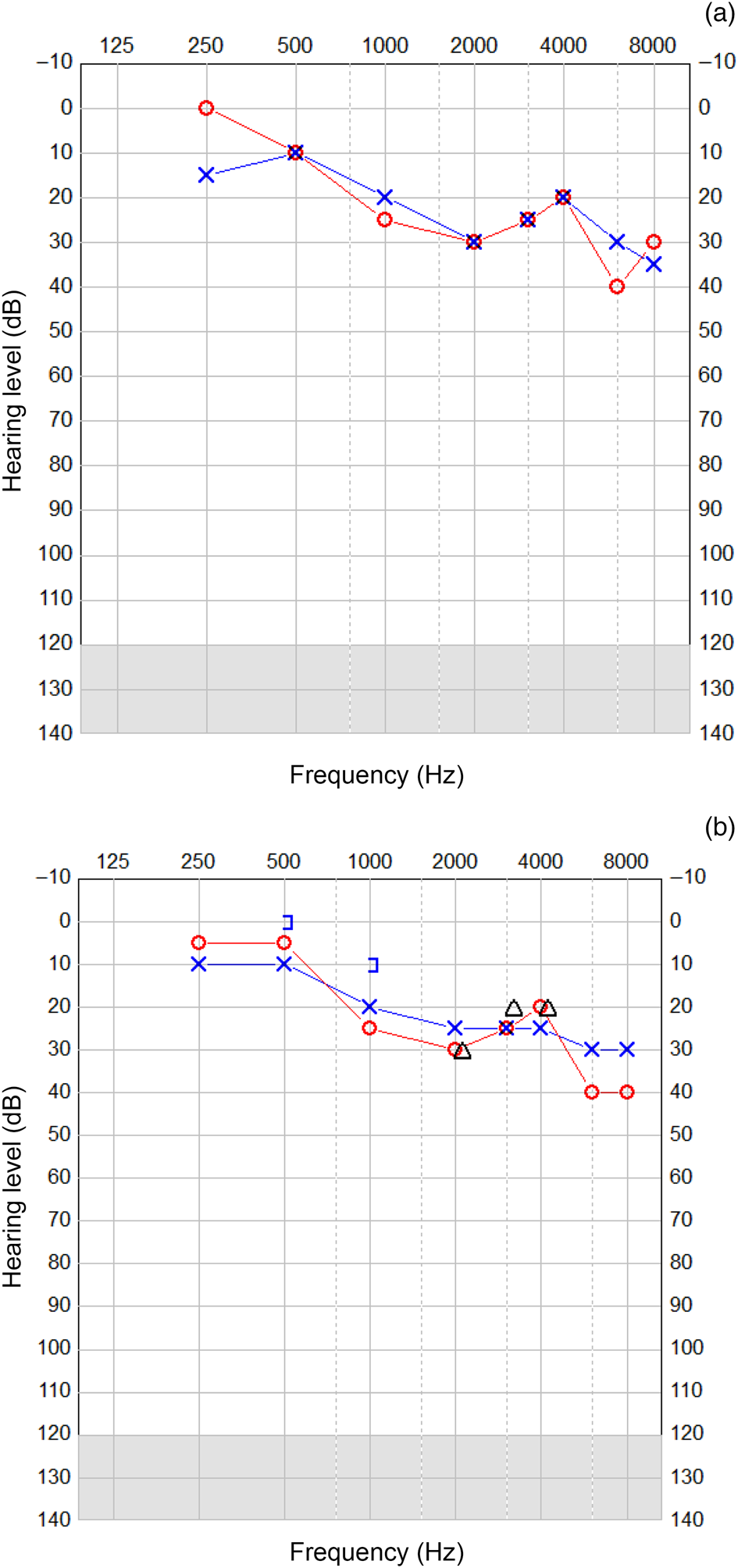
A 60-year-old lady presented to the Otolaryngology Department at the Norfolk and Norwich University Hospital with a 7-year history of progressive pulsatile tinnitus, imbalance, autophony and the perception of hearing her eyes move. Her symptoms in her own words were ‘driving her mad’. Her hearing was normal at presentation (Figure 1a).
Audiometry at presentation (a), and post-operatively (b). ○ = Air conduction right ear; × = air conduction left ear; ] = masked bone conduction left ear; Δ = unmasked bone conduction

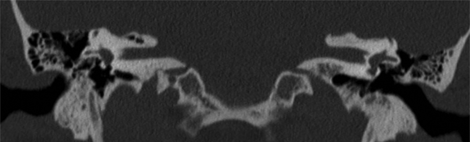
The patient's symptoms were investigated using pure tone audiometry (Figure 1a), computed tomography imaging of the temporal bones (Figure 2), and vestibular testing that included the provision of cervical vestibular-evoked myogenic potentials. The results of these investigations supported a clinical diagnosis of left-sided superior semi-circular canal dehiscence syndrome.
Coronal computed tomography scan of the temporal bones along the long axis of the left superior semi-circular canal, showing dehiscence of the superior semi-circular canal for the left temporal bone. The bone is thin over the right semi-circular canal but intact.

The diagnosis and associated controversies were fully explained to the patient, together with the relative merits and risks of contemporary treatments for superior semi-circular canal dehiscence syndrome. This discussion included the option of no intervention.
The senior author fully disclosed his proposed procedure to the hospital's ‘new procedures committee’. Permission was granted in view of the nuances of his proposed procedure in the context of similar procedures currently undertaken for other neurotological conditions.
Surgical procedure
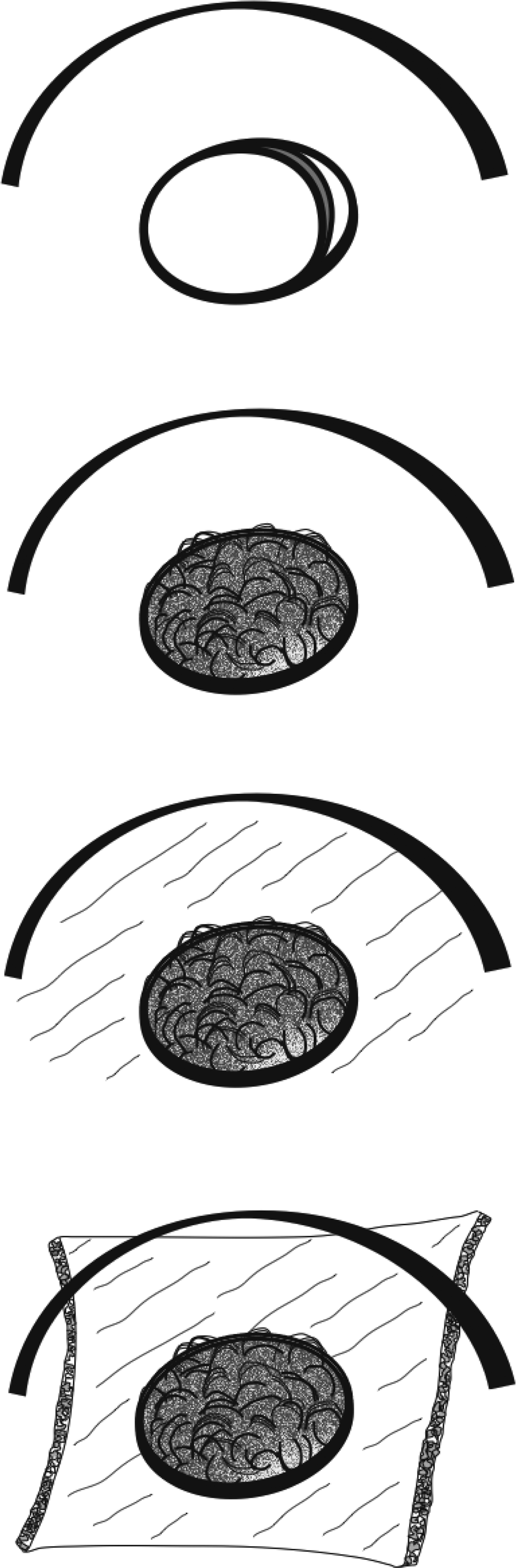
Under general anaesthesia, a permeatal tympanotomy was performed. The round window niche was identified and then filled with fat harvested from the patient's ear lobule. The bony meatus leading to the round window was ‘scratched up’ with a curved needle and then the tragal perichondrium was placed as a double layer to secure the fat within the round window niche. The surgical steps involved are illustrated in Figure 3.
The surgical steps involved in reinforcing the round window: fat is placed within the round window niche and onto the round window membrane; the round window niche is ‘scratched up’; and, finally, a double layer of perichondrium is applied to secure the reinforcement.

Outcome
The patient was reviewed eight weeks after surgery. The patient reported the surgery as being a ‘resounding success’. When asked to quantify the degree of symptom improvement, she stated that her synchronous pulsatile tinnitus had entirely disappeared. In addition, her balance was 75 per cent better, and the symptoms of autophony and perception of hearing her eyes moving within her head improved by 80 per cent. Otoscopy revealed a healthy and intact ear drum, and hearing was unchanged (Figure 1b).
Discussion
A variety of techniques have been described for the surgical management of superior semi-circular canal dehiscence syndrome. Historically, the ‘gold standard’ technique involved a middle fossa approach to the superior semi-circular canal; however, newer techniques involve a transmastoid approach. Once the dehiscence has been identified, rectification of the pathological defect may be achieved by either plugging or resurfacing the superior semi-circular canal.
• Superior semi-circular canal dehiscence syndrome is characterised by vestibular symptoms elicited by loud sounds, and other pressure-induced symptoms
• Symptoms occur due to the presence of a ‘third window’ created by the superior semi-circular canal dehiscence
• This paper describes a permeatal procedure with a round window niche reinforced with fat and a double layer of perichondrium
• The case supports ‘soft reinforcement’ as a simple and effective technique to treat superior canal dehiscence syndrome symptoms
Over the last decade, a number of minimally invasive remedies have been proposed that involve obliterating or reinforcing the round window.Reference Silverstein and Vas Ess5–Reference Succar, Manickam, Wing, Walter, Greene and Azeredo8 More recently, the literature has become polarised regarding the effectiveness of round window surgery for individuals with superior semi-circular canal dehiscence syndrome.Reference Silverstein and Vas Ess5–Reference Succar, Manickam, Wing, Walter, Greene and Azeredo8 Whilst many experts support round window surgery as a first-line treatment to avoid more invasive surgical options, some experts advise against this.Reference Succar, Manickam, Wing, Walter, Greene and Azeredo8
It is difficult to draw many conclusions from an isolated single case report. However, when considered in the context of previous work and the fact that our patient reported this procedure to have been ‘life-changing’, this report adds to the mounting literature that supports the use of minimally invasive treatments for superior semi-circular canal dehiscence syndrome.
Our particular surgical technique may be partly responsible for the success of the procedure. Whilst many techniques have been described, we have determined the fundamental principle of employing a ‘soft reinforcement’ technique to be of great importance. We harvested fat to provide a soft material to reinforce the round window membrane, rather than cartilage to provide a stiff obliteration of the round window niche. The additional benefit of applying a double layer of perichondrium to an appropriately prepared site adds to the robustness of the reinforcement. Other advantages of this technique are its amenability to being performed under local anaesthesia, its use of autologous materials, and the fact that it would be simple to reverse.
Conclusion
The case provides support for the use of ‘soft reinforcement’ as a simple and effective technique to treat superior canal dehiscence syndrome symptoms.
Competing interests
None declared