Introduction
Subjective memory complaints increase with age, with nearly half of adults over the age of 65 years reporting memory problems (Bassett and Folstein, Reference Bassett and Folstein1993). Previous studies have found that standardized measures of self-perceived changes in memory and cognition relate significantly to performance on objective neuropsychological tests (Bassett and Folstein, Reference Bassett and Folstein1993; Jonker et al., Reference Jonker, Geerlings and Schmand2000; Amariglio et al., Reference Amariglio, Townsend, Grodstein, Sperling and Rentz2011). While several studies have demonstrated that self-awareness of memory change predicts future risk of cognitive decline, including development of Alzheimer's disease (AD) and related dementias (Jonker et al., Reference Jonker, Geerlings and Schmand2000; Jessen et al., Reference Jessen2010), other studies have not (Blazer et al., Reference Blazer, Hays, Fillenbaum and Gold1997; Jorm et al., Reference Jorm, Christensen, Korten, Henderson, Jacomb and Mackinnon1997; Minett et al., Reference Minett, Dean, Firbank, English and O'Brien2005). Inconsistencies in this relationship can be ascribed to differing study methodologies, design, or control of covariates known to be associated with cognitive function such as age, education, and depression (Blazer et al., Reference Blazer, Hays, Fillenbaum and Gold1997; Jonker et al., Reference Jonker, Geerlings and Schmand2000; Merema et al., Reference Merema, Speelman, Kaczmarek and Foster2011).
Prior research has indicated varying degrees of relationship between brain imaging measures and subjective memory complaints. Structural magnetic resonance imaging (MRI) studies have found greater atrophy in memory-relevant temporal lobe structures in older adults with subjective memory complaints (Striepens et al., Reference Striepens2010). A study using positron emission tomography (PET) after infusion of 2-[18F]fluoro-2-deoxy-D-glucose (FDG) in subjects without dementia found decreased frontal lobe glucose metabolism in older adults relying more heavily on memory aids (Small et al., Reference Small1994). Self-perceived memory loss was likewise found to be associated with global cerebral metabolic decline (Ercoli et al., Reference Ercoli2006). In contrast, previous analysis of data from the Australian Imaging, Biomarkers, and Lifestyle (AIBL) study of aging comparing the dichotomous presence or absence of subjective memory complaints (% complainers) to (11)C-Pittsburgh Compound B (PiB) PET positivity or negativity (PiB-PET cutoff level 1.5) did not show any relationship between subjective memory complaints and brain beta-amyloid (Rowe et al., Reference Rowe2010; Pike et al., Reference Pike2011).
Previously, we have reported that performance on objective memory tests, including the Wechsler Memory Scale Verbal Paired Associations task and the Buschke–Fuld Selective Reminding Test, correlate with the levels of amyloid plaques and tau neurofibrillary tangles in vivo as measured by PET scans using 2-(1-{6-[(2-[F-18]fluoroethyl)(methyl)amino]-2-naphthyl}ethylidene)malononitrile (FDDNP) (Small et al., Reference Small2006). Others have shown that the Memory Functioning Questionnaire (MFQ), a standardized instrument measuring self-appraisal of memory ability with high internal consistency, has moderate concurrent validity with objective memory performance measures, such as immediate and delayed list recall and recognition (Gilewski et al., Reference Gilewski, Zelinski and Schaie1990; Zelinski et al., Reference Zelinski, Gilewski and Anthony-Bergstone1990).
In this study, we report the relationship between types and degree of subjective memory complaints, as measured by the MFQ factor scores (Gilewski et al., Reference Gilewski, Zelinski and Schaie1990), and load of amyloid senile plaques and tau neurofibrillary tangles in non-depressed older adults without dementia using FDDNP-PET scans. We focused our investigation on a sample of older adults with subjective memory complaints, and either normal cognition or mild cognitive impairment, but not dementia (Small et al., Reference Small2006; Ercoli et al., Reference Ercoli2009), to determine whether specific types or degrees of subjective memory complaints reflect an increased burden of plaque and tangle pathology in memory-relevant brain regions.
Methods
Subjects
A total of 57 subjects without dementia who completed the MFQ and underwent FDDNP-PET scans were drawn from a larger, longitudinal study of predictors of cognitive decline, detailed elsewhere (Small et al., Reference Small2006; Ercoli et al., Reference Ercoli2009). Briefly, volunteers from the community were recruited through advertisements of a study of mild memory impairment, media coverage of the study, and referrals by physicians and families. Members of the research staff screened potential volunteers via telephone interviews. Subjects received neurological and psychiatric evaluations and routine screening laboratory tests to rule out treatable causes of cognitive impairment or potential cognitive confounding factors (e.g. severe sensory deficits or medication interactions). Most of the subjects (N = 54) underwent MRI; the remaining subjects who could not tolerate MRI (because of claustrophobia or metal in the body) underwent computed tomography (CT). All subjects were proficient in English and underwent comprehensive clinical and cognitive assessment to characterize cognitive status at the time of baseline assessment.
A neuropsychological test battery was administered to quantify cognitive performance and confirm the diagnostic category of each study subject (normal aging, mild cognitive impairment (MCI), or dementia). Subjects with dementia were excluded while subjects without dementia were classified as normal aging or MCI using standard criteria (Smith et al., Reference Smith, Petersen, Parisi and Ivnik1996). The neuropsychological test battery was administered to assess five cognitive domains: (1) memory, including the Wechsler Memory Scale Third Edition (WMS-III), Logical Memory (Delayed score), the Buschke Selective Reminding (Total score), and the Rey–Osterrieth Complex Figure Recall (3-min delayed recall score); (2) language, including the Boston Naming Test and Letter (FAS) and category (Animal Naming Test) fluency; (3) attention and information-processing speed, including Trailmaking A, Stroop Color Naming (Kaplan version), and the Wechsler Adult Intelligence Scale Third Edition (WAIS-III) Digit Symbol; (4) executive functioning, including Trailmaking B, and Stroop Interference (Kaplan version); and (5) visuospatial ability, including WAIS-III Block Design, and the Rey–Osterrieth Complex Figure Copy. We used standard diagnostic criteria for MCI (i.e. memory impairment without other cognitive impairments), which include (1) patient awareness of a memory problem, preferably confirmed by another person who knows the patient; (2) memory impairment detected with standard assessment tests; and (3) ability to perform normal daily activities (Smith et al., Reference Smith, Petersen, Parisi and Ivnik1996). Subjects were categorized as normal if they had neither objective deficits on neuropsychological test measures after correction for age and education nor functional deficits in daily functioning and did not meet the MCI criteria. The Hamilton Rating Scales for both Depression and Anxiety were administered to assess mood and anxiety, respectively. For the current analysis, subjects meeting criteria for dementia, depression, or anxiety disorders were excluded.
Degrees of subjective memory complaints were reported by subjects via mailed survey by using the MFQ, a widely used instrument developed to evaluate self-perception of everyday memory functioning (Gilewski et al., Reference Gilewski, Zelinski and Schaie1990; Zelinski et al., Reference Zelinski, Gilewski and Anthony-Bergstone1990). The MFQ consists of 64 items rated on a seven-point scale, and provides four unit-weight factor scores measuring Factor 1, frequency of forgetting (including ratings of how often forgetting occurs in 28 specific situations and five ratings of general memory performance); Factor 2, seriousness of forgetting (memory failure ratings from 18 different situations); Factor 3, retrospective functioning (changes in current memory ability relative to five time points earlier in life); and Factor 4, mnemonics usage (frequency of mnemonics usage in eight specific situations). Higher scores indicate higher (more favorable) levels of perceived memory functioning, i.e. fewer forgetting incidents and less frequent use of mnemonics. Factor structure is stable across age groups and internal consistency is high, with Cronbach's α values for its four-factor scores ranging from 0.83 to 0.94 (Zelinski et al., Reference Zelinski, Gilewski and Anthony-Bergstone1990). Self-reported health ratings and education contribute to the variance of some of the factors, but together these variables account for only 9% of variance of any factor (Gilewski et al., Reference Gilewski, Zelinski and Schaie1990). The MFQ shows moderate concurrent validity with objective memory performance (Zelinski et al., Reference Zelinski, Gilewski and Anthony-Bergstone1990).
Neuroimaging
FDDNP was prepared at very high specific activities (>37 GBq/mol) (Liu et al., Reference Liu2007). All scans were performed with the ECAT HR or EXACT HR+ tomograph (Siemens-CTI, Knoxville, TN) with subjects supine and the imaging plane parallel to the orbitomeatal line. A bolus of FDDNP (320–550 MBq) was injected via an indwelling venous catheter, and consecutive dynamic PET scans were performed for 2 hours. Scans were decay-corrected and reconstructed using filtered back-projection (Hann filter, 5.5-mm FWHM) with scatter and measured attenuation correction. The resulting images contained 47 contiguous slices with plane separation of 3.37 mm (ECAT HR) or 63 contiguous slices with plane separation of 2.42 mm (EXACT HR+). Non-parametric Wilcoxon two-sample tests within MCI and cognitively normal groups separately found no significant differences in regional FDDNP signals between the two PET scanners (p-values ranging from 0.18 to 0.84).
The FDDNP-PET binding levels were quantified as described by Small et al. (Reference Small2006). Briefly, we performed the Logan graphical analysis (Logan et al., Reference Logan, Fowler, Volkow, Wang, Ding and Alexoff1996) with cerebellum as the reference region for time points between 30 and 125 minutes. The slope of the linear portion of the Logan plot is the relative distribution volume (DVR), which is equal to the distribution volume of the tracer in a region of interest (ROI) divided by that in the reference region. We generated DVR parametric images and analyzed them using gray matter ROIs drawn manually on the FDDNP-PET image obtained in first 5 minutes after injection (the perfusion image). This image shows the perfusion pattern and has sufficient anatomical information to identify the cerebellum and cerebellar gray matter. ROIs were drawn bilaterally on the medial temporal (containing limbic regions, including hippocampus, parahippocampal, and entorhinal areas), lateral temporal, posterior cingulate, parietal, frontal, and cerebellar regions, as described previously (Kepe et al., Reference Kepe2006). Each cerebral region DVR or binding value was expressed as an average of left and right regions. Rules for ROI drawing were based on the atlas by Talairach and Tournoux (Reference Talairach and Tournoux1988), which we used as a visual guide for identifying the important gyral and sulcal landmarks needed in delineating the ROI. An individual blind to clinical assessments performed the ROI determinations; previous inter-rater reliability studies have confirmed high consistency and reliability using this method (Small et al., Reference Small, Stern, Mandelkern, Fairbanks, Min and Guze1992).
Among the 54 subjects who underwent MRI, anatomical brain MRI scans were obtained using either a 1.5 T (N = 9) or 3 T (N = 45) magnet (General Electric-Signa, Milwaukee) scanner. Fifty-four transverse planes were collected throughout the brain, superior to the cerebellum, using a double-echo and fast-spin echo series with a 24-cm field of view and 256 × 256 matrix with 3 mm/0 gap (repetition time = 6000 [3 T] and 2000 [1.5 T]; echo time = 17/85 [3 T] and 30/90 [1.5 T]).
Statistical analysis
Data were screened for outliers and normality assumptions. The MFQ factor scores that were more than 2.5 SD from the mean of the sample were removed (3 scores for MFQ1 and 2 scores for MFQ3). As stated previously, the MFQ was self-administered via mailed survey, thus the scales of some subjects had incomplete items. For the subjects who completed less than 80% within each factor of the questionnaire, item-wise deletion was performed (4 for MFQ1, 3 for MFQ2, and 2 for MFQ3). For the remaining subjects with missing data, the MFQ factor scores were imputed as follows: the observed score was multiplied by the ratio of the total possible score of all items to total score for the answered items within each factor. In this way, 8 subjects' MFQ1 scores, 10 MFQ2 scores, 1 MFQ3 score, and 3 MFQ4 scores were imputed. This method of imputation is valid if the probability that an item is missing can be related to the values of observed items alone, not the missing items, i.e. the data are missing at random. We examined missingness by normal versus MCI groups. For all four factors, missingness was not significantly different between groups: MFQ1 χ2 (1) = 0.3, p = 0.6; MFQ2 χ2 (1) = 2.9, p = 0.1; MFQ3 χ2 (1) = 0.1, p = 0.7; MFQ4 χ2 (1) = 1.6, p = 0.2. Further, all analyses were repeated using only the subjects with complete data to determine whether the imputation procedure had any effect on the findings.
Pearson correlation coefficients were used to examine associations between the MFQ factors and FDDNP-PET binding levels. We first assessed whether the MFQ factors and FDDNP-PET binding levels were associated with age and education. MFQ1 was found to be related to age (Pearson correlation coefficient, r = −0.33, p = 0.02); thus, we controlled for age in all the analyses involving MFQ1. Education was not associated with either MFQ factors or FDDNP-PET binding levels. In order to limit the number of tests, we then analyzed the relationship between the MFQ factors and the global (overall average) FDDNP-PET binding levels. Only those factors that were found to have a significant association globally were further analyzed to determine region-specific associations. We also determined if MCI and normal aging subjects, as well as subjects with and without one or more copies of the apolipoprotein E-4 (APOE-4) allele, differed in their MFQ-FDDNP-PET associations by estimating ANCOVAs with the MFQ factor scores as dependent variables, and FDDNP-PET binding levels, cognitive status (MCI vs. normal aging), APOE-4 status, and the interaction of cognitive status/APOE-4 status and FDDNP-PET binding levels as predictors.
In addition, for MFQ1, which has 32 items, we conducted an item analysis to determine if a subset of the items performs as well as the total. To identify the subset, we calculated Cronbach's α and retained only those items that had a correlation coefficient of at least 0.75 with the total score. We then obtained MFQ1Sub as the sum of the retained items in MFQ1 and repeated the above analyses examining the relationship between FDDNP-PET binding levels and MFQ1Sub. All tests were two-tailed and a significance level of 0.05 was used for all inferences.
Results
Demographic and clinical characteristics of the sample were as follows. The mean age of all the subjects was 66.3 ± 10.6 (SD) years and men comprised 49% of the patient sample. Most subjects were Caucasian (91%), and subjects had an average of 17 ± 3 (SD) years of formal education. Positive family history of AD was reported in 66.7% of the sample, and 53% of the subjects were carriers of at least one copy of the APOE-4 allele. Average score on the Mini-Mental State Examination was 28.6 ± 1.4 (SD). Subjects meeting the criteria for MCI based on their performance on neuropsychological testing comprised 56% (N = 32 of 57 subjects; amnestic MCI = 17, non-amnestic MCI = 5, multiple domain MCI = 10) of the study sample. As this study involved pre-screening to rule out subjects with clinically significant anxiety or depression, subjects did not display significant levels of anxiety (the mean Hamilton Rating Scale for Anxiety scores = 4.5 ± 4.2 SD) or depression (the mean Hamilton Rating Scale for Depression scores 3.4 ± 2.8).
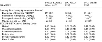
The mean scores for each of the MFQ factors and the global plus regional FDDNP-PET binding levels are shown for all subjects in Table 1. After controlling for age, ANCOVA performed for all subjects did not reveal significant interaction terms, indicating that the two cognitive subgroups of subjects (normal control and MCI) and subjects with and without the APOE-4 allele exhibited similar MFQ-FDDNP-PET associations. MFQ1 for all subjects was significantly associated with global FDDNP-PET binding levels (r = −0.33, p = 0.02; Figure 1a). The remaining MFQ factors were not significantly related to FDDNP-PET binding levels. Further analysis of the MFQ1 scores with measured FDDNP-PET levels revealed significant correlations in the following regions of interest: medial temporal (r = −0.29, p = 0.05), parietal (r = −0.30, p = 0.03), and frontal (r = −0.35, p = 0.01) regions.
Test scores and plaque and tangle loads

Figures indicate mean with SD.
FDDNP-PET = 2-(1-{6-[(2-[F-18]fluoroethyl)(methyl)amino]-2-naphthyl}ethylidene)malononitrile positron emission tomography; MCI = Mild Cognitive Impairment; NC = Normal Control.
a For the Memory Functioning Questionnaire (MFQ), higher scores indicate fewer self-reported memory problems and less use of mnemonics. Norms for people aged 60–69 and 70–79 years: frequency of forgetting = 152 (28) and 149 (29); seriousness of forgetting = 86 (20) and 84 (21); retrospective functioning = 18 (6) and 18 (6); and mnemonics usage = 31 (9) and 30 (10).
b MFQ1 mean values are significantly different for NC and MCI groups: t (48) = 2.5, p = 0.02.
c FDDNP-PET cortical binding levels are different for NC and MCI groups. Global: t (55) = 3.7, p = 0.0005; medial temporal lobe: t (55) = 4.4, p < 0.0001; lateral temporal lobe: t (55) = 3.1, p = 0.0004; parietal: t (55) = 3.1, p = 0.003.
Plot of global [F-18]FDDNP binding with MFQ1: (a) frequency of forgetting, and (b) MFQ1Sub, the sum of a subset of five items from MFQ1.

Analyses using only data from complete cases (N = 36) yielded similar findings to those from the entire sample. After controlling for age, MFQ1 was significantly associated with global FDDNP-PET binding levels (r = −0.43, p = 0.01). The remaining MFQ factors were not significantly related to FDDNP-PET binding levels. Further analysis of the MFQ1 scores with measured FDDNP-PET levels in complete cases revealed significant correlations in parietal (r = −0.46, p = 0.005), and frontal (r = −0.44, p = 0.01) regions. The association in medial temporal lobe did not reach significance (r = −0.30, p = 0.08).
Item analysis of the MFQ1 factor indicated that five of the 32 items had a correlation coefficient greater than 0.75 with the total score and, in addition, worsened Cronbach's α when removed. Therefore, these items were retained and are presented in Table 2. The sum of these five items, MFQ1Sub, had a mean of 24.0 ± 6.0 SD. After controlling for age, MFQ1Sub was significantly associated with global FDDNP-PET binding levels (r = −0.52, p = 0.0002) (Figure 1b). Regional analysis of the MFQ1Sub scores with FDDNP-PET levels showed significant correlations: medial temporal (r = −0.38, p = 0.01), parietal (r = −0.43, p = 0.002), posterior cingulate (r = −0.30, p = 0.04), and frontal (r = −0.51, p = 0.0002) regions. As with MFQ1, normal aging and MCI subjects did not differ in their FDDNP-PET associations with MFQ1Sub.
Five questions reproducing results from item analysis of the MFQ frequency of forgetting factor*

* Item analysis of the MFQ Frequency of Forgetting factor revealed that five questions reproduce the results obtained from the full 32-item scale when relating MFQ to FDDNP-PET binding.
Discussion
This study reports that the degree of self-perceived memory loss correlates significantly with FDDNP-PET binding levels in healthy middle-aged and older adults without dementia. In particular, our results show that the MFQ frequency of forgetting factor significantly relates to FDDNP-PET binding levels both globally and in medial temporal, parietal, and frontal lobe regions of interest. Item analysis of the frequency of forgetting factor enabled consolidation of the scale from 32 questions down to five questions, and confirmed the above associations, with the additional significant correlation of frequency of forgetting to posterior cingulate FDDNP-PET binding levels. These findings complement previous work (Small et al., Reference Small2006), which has shown that the global and regional FDDNP-PET uptake levels differentiate controls and MCI subjects, and also yield a significant relationship with objective cognitive testing performance (Ercoli et al., Reference Ercoli2009).
In contrast to findings from the AIBL study of aging (Rowe et al., Reference Rowe2010; Pike et al., Reference Pike2011), which used the amyloid-specific PET ligand PiB, this study demonstrates a significant relationship between the degree of subjective memory complaints and an in vivo neuroimaging marker of amyloid plaque and tau neurofibrillary tangle levels, namely FDDNP. Another small pilot study of PiB-PET found that only one of the five older adults in the study with subjective cognitive impairment had elevated PiB-PET levels (Rodda et al., Reference Rodda, Okello, Edison, Dannhauser, Brooks and Walker2010). The approach used in these prior studies focused on the dichotomous presence or absence of subjective memory complaints (% complainers) compared with PiB-PET positivity or negativity (PiB cutoff level: 1.5). This differs from the current approach, which examined these variables in a continuous fashion, relating degrees of subjective memory complaints to FDDNP-PET binding levels. Even though FDDNP-PET signals tend to be lower than PiB, as is common to many 18-F PET tracers, assessing subjective memory complaints on a continuum may have compensated for this property of FDDNP-PET.
These results may also differ because of the ability of FDDNP-PET to detect both amyloid plaque and tau tangle levels, in comparison with the sole detection of amyloid plaques by PiB-PET. In comparison with amyloid plaques, the tau tangle pathology is known to increase progressively throughout various brain regions as the patient progresses in severity from preclinical to clinical stages of dementia (Braak and Braak, Reference Braak and Braak1991; Delacourte et al., Reference Delacourte1999). As such, memory self-appraisal measures may be among the earliest measures that detect these underlying tau-mediated neuropathological changes that are occurring, in addition to any contribution from progression of amyloid pathology. While no autopsy studies have examined the relationship between subjective memory changes and brain pathology, the current results suggest that subjective memory complaints may reflect the accumulation of neurodegenerative changes, in particular tau pathology more so than amyloid pathology, prior to emergence of objective cognitive changes.
In the present study, the MFQ frequency of forgetting factor was related to FDDNP-PET binding in memory-relevant brain regions. This factor score assesses commonly observed recent memory problems in older adults by querying how often subjects have problems remembering details of things like names, conversations, recent events, and reading materials (novels and magazines/newspapers). Such tasks are known to be dependent on intact function and connectivity between the medial temporal lobe and frontal cortex. The current results demonstrating increased FDDNP binding values in these brain regions in subjects with a greater degree of self-reported memory difficulties support the hypothesis that subjective memory complaints represent worsening underlying brain pathology (Jonker et al., Reference Jonker, Geerlings and Schmand2000; Jessen et al., Reference Jessen2010; Merema et al., Reference Merema, Speelman, Kaczmarek and Foster2011) and not just subjective anxiety over normal age-related changes in memory function (Blazer et al., Reference Blazer, Hays, Fillenbaum and Gold1997; Jorm et al., Reference Jorm, Christensen, Korten, Henderson, Jacomb and Mackinnon1997; Minett et al., Reference Minett, Dean, Firbank, English and O'Brien2005).
The current results complement earlier work demonstrating a relationship between subjective memory complaints and biomarkers of brain function such as FDG-PET (Small et al., Reference Small1994, Reference Small, Komo, Rue, Kaplan and Mandelkern1995; Ercoli et al., Reference Ercoli2006). In particular, a prior FDG-PET analysis by our group in a similar population of older adults without dementia found a significant relationship between global cerebral glucose hypometabolism and reported frequency of forgetting regardless of APOE-4 genetic risk for AD (Ercoli et al., Reference Ercoli2006). Moreover, in that study, the factor score for mnemonics use significantly correlated with metabolic decline in the temporal regions in APOE-4 carriers but not in non-carriers. The degree of mnemonic use had previously been related to frontal but not temporal or parietal hypometabolism in subjects (Small et al., Reference Small1994) without dementia. While our current study found a trend for a relationship between the degree of mnemonic use and global FDDNP-PET binding levels (p = 0.07), this relationship was not significant and, thus was not explored further in our subregion analysis. Although FDG-PET and FDDNP-PET results can overlap and show inverse correlations (Small et al., Reference Small2006), these two probes measure different targets – FDG reflects neuronal function, whereas FDDNP targets pathological structures – so that findings from these two probes would be expected to vary.
The current results highlight the potential clinical importance of asking about memory symptoms in older adults. While our initial analysis found a significant relationship between FDDNP-PET binding and the 32-question frequency of forgetting factor score of the MFQ, our item analysis found that as few as five questions about recent memory relating to common, everyday situations may reflect the underlying brain pathology. Such questions may be useful for clinicians who evaluate patients presenting with subjective memory complaints. In addition, an interesting follow-up study would be to observe whether or not the severity of these initial subjective memory complaints and associated elevations in FDDNP uptake are predictive of future clinical disease progression.
Reisberg et al. (Reference Reisberg2008) recently introduced the concept that memory changes noticed by otherwise cognitively intact older adults represent a mild and long-lasting pre-MCI stage of age-related cognitive decline known as subjective cognitive impairment (SCI), which has been estimated to last for an average of 15 years prior to the development of MCI. In support of this notion, a greater degree of MRI atrophy has been observed in the temporal lobe of older adults without dementia reporting subjective memory changes when compared with those who do not (Saykin et al., Reference Saykin2006; Striepens et al., Reference Striepens2010). In this proposed mildest stage of memory loss, patients and/or their family members report subjective changes in memory function; however, the performance on neuropsychological testing and daily functioning remains intact. When taken in the context of the previously published findings relating the degree of objective memory performance to both FDDNP-PET binding levels (Small et al., Reference Small2006) and MFQ-measured subjective memory complaints (Zelinski et al., Reference Zelinski, Gilewski and Anthony-Bergstone1990), these results suggest that subjective memory complaints may be a guide to the fairly new and compelling concept of preclinical AD (Sperling et al., Reference Sperling2011). The current results in non-depressed middle-aged and older adults without dementia suggest that the degree of neuropathology underlying this subtle degree of memory loss may be assessed in clinical settings by simplified bedside versions of memory self-appraisal tests such as the MFQ (see Table 2). This may be of particular importance since amyloid imaging is unlikely to be practical for large populations of cognitively healthy older adults.
An important limitation of our data set is that it is a relatively small non-representative sample of convenience. The findings need to be replicated in a larger sample, which could address the question of whether subjective complaints are more sensitive to imaging findings than objective measures. In addition, several subjects had missing data values. However, findings from analyses, including only those subjects with complete data, were similar to those with the larger sample. Further, we limited the potential effect of missing data values by only including subjects who had returned mailed questionnaires with at least 80% of each section completed. Our method of imputation does assume that the missing data are similar to the complete data, i.e. the probability of missingness depends on the complete data but not the missing data, an assumption that may not be true.
In conclusion, these findings suggest that self-awareness of memory loss may reflect the extent of cerebral amyloid and tau brain pathology in people without dementia. Such self-perceived memory abilities might predict and track neuropathological changes in the brain years before symptoms of dementia are present. Thus, subjective memory complaints may be an important indicator that signals the need for further clinical assessment and monitoring.
Conflict of interest declaration
The University of California, Los Angeles, owns a US patent (6274119) entitled “Methods for Labeling Beta-Amyloid Plaques and Neurofibrillary Tangles,” which uses the approach outlined in this paper. Drs. Small, Huang, Cole, and Barrio who are among the inventors, have received royalties, and may receive royalties on future sales. Dr. Small reports having served as a consultant and/or having received lecture fees from Dakim, Eisai, Forest, Medivation, Novartis, and Pfizer. He also reports having received stock options from Dakim. Dr. Lavretsky reports having received lecture fees from Eisai, Janssen, and Pfizer, and a grant from Forest. Dr. Huang reports having received lecture fees from GlaxoSmithKline. Dr. Barrio reports having served as a consultant and having received lecture fees from Nihon Medi-Physics Co., Bristol-Meyer Squibb, PETNet Pharmaceuticals, and Siemens. Drs. Merrill, Siddarth, Ercoli, Burggren, Kepe, Miller, Kim, Huang, and Bookheimer and Nathan Saito have no financial conflicts of interest.
Description of authors' roles
David A. Merrill designed the study, supervised data collection and statistical analysis, and wrote the paper. Prabha Siddarth helped design the study, was primarily responsible for the statistical design of the study and for carrying out the statistical analysis, and helped write the paper. Nathan Y. Saito helped with data collection and analysis. Linda M. Ercoli helped design the study, supervised the neuropsychological data collection, and helped write the paper. Alison C. Burggren helped design the study and write the paper. Vladimir Kepe helped collect and analyze the neuroimaging data and write the paper. Helen Lavretsky helped recruit and consent subjects, collect data, and write the paper. Karen J. Miller and Jeanne Kim helped collect and analyze neuropsychological data. S. C. Huang helped supervise collection and analysis of the neuroimaging data and write the paper. Susan Y. Bookheimer, Jorge R. Barrio, and Gary W. Small helped design the study, supervise collection and analysis of the data, and write the paper.
Acknowledgments
The authors thank Anel Dzmura and Colin Shinn for help in subject recruitment, data management, and study coordination, and Gerald Timbol and Anasheh Halabi for help in image processing. Drs. Siddarth and Kepe had full access to all of the data in the study and take responsibility for the integrity of the data and the accuracy of data analysis.
This study was funded by NIH grants P01-AG025831, AG13308, P50 AG 16570, MH/AG58156, MH52453, AG10123, M01-RR00865, the Department of Energy (DOE contract DE-FC03-87-ER60615); General Clinical Research Centers Program; the Fran and Ray Stark Foundation Fund for Alzheimer's Disease Research; the Ahmanson Foundation; the Larry L. Hillblom Foundation; the Lovelace Foundation; the Sence Foundation; the McMahan Foundation; the Judith Olenick Elgart Fund for Research on Brain Aging; the Elizabeth and Thomas Plott Endowment in Gerontology; the UCLA Claude Pepper Older Americans Independence Center funded by the National Institute on Aging (5P30AG028748); and AFAR, the John A. Hartford Foundation and the Centers of Excellence National Program.