In some species of Bryoria (Parmeliaceae), including Bryoria bicolor (Ehrh.) Brodo & D. Hawksw. and B. fuscescens (Gyeln.) Brodo & D. Hawksw., thallus reactions with spot tests are patchy, and phrases such as “Pd+ bright red at least in parts” have been used in descriptions for many years (e.g. Hawksworth Reference Hawksworth1972). It has recently been discovered that the extracellular lichen substances (‘extrolites’) in individual thalli of some species in the genus are more varied than hitherto assumed (Hawksworth et al. Reference Hawksworth, Crespo, Rico and Ruibal2011; Myllys et al. Reference Myllys, Velmala, Holien, Halonen, Wang and Goward2011). We hypothesized that this variation in extrolites and the patchiness phenomenon could be due to one or more of several factors, including: 1) chemosyndromic variation; 2) variations in concentration and the sensitivity of detection methods; 3) differences between basal, median and apical regions of the thallus; or 4) the localization of particular compounds in particular anatomical features, such as pseudocyphellae and soralia. As the localization of compounds is known to occur in at least one species of the genus, viz. the yellow pigment vulpinic acid in the soralia of Bryoria fremontii (Brodo & Hawksworth Reference Brodo and Hawksworth1977), we decided to explore the last possibility first.
Long-wave ultra-violet (UV) fluorescence (at 350 nm) has proved a valuable tool in the separation of thalli of similar crustose and macrolichens, and is also used routinely in the examination of thin-layer chromatographic (TLC) plates; xanthones fluoresce shades of yellow, orange and red, while depsides and depsidones generally fluoresce blue to white or shades of grey, although atranorin gives a yellow hue (Orange Reference Orange, James and White2010). In addition, fluorescence microscopy has been used to explore the location of lichen products in sections of a range of macrolichens (Kauppi & Verseghy-Patay Reference Kauppi and Verseghy-Patay1990). We therefore decided to explore whether fluorescence microscopy could be used to determine the localization of extrolites in whole lichen thalli, as a supplement to reagent tests, especially as material subjected to Pd reactions has to be discarded. In addition, we wished to determine whether fluorescence would disclose sites: 1) not revealed by reagent tests, that is sites where compounds were present in concentrations too low to yield visible spot test reactions; and 2) which fluoresced in different colours, suggesting the presence of more than one compound.
For this pilot investigation, we used specimens from populations of Byoria sect. Implexae (Myllys et al. Reference Myllys, Velmala, Holien, Halonen, Wang and Goward2011) collected in several European countries, but especially in the central mountains of Spain (deposited in MAF-Lich). We examined transverse and longitudinal sections cut by hand, and also thallus portions, mounted in water. Spot tests and TLC were performed using standard methods and solvents A, B, C, and G (Orange et al. Reference Orange, James and White2010). For auto-fluorescence we used a Nikon microscope: D-LF epi-fluorescence module coupled to an Eclipse-80i, with bright field and DIC, and connected to a DS-Fi1 camera and DS-C2 control unit. Two filter blocks were used: Nikon UV-2A (Ex 330–380 nm, DM 400, BA 420) and B-2A (Ex 450–490 nm, DM 505, BA 520).
The most striking fluorescence was the red under the UV-2A and B-2A blocks, attributable to the chlorophyll of the included Trebouxia cells. This fluorescence is widely used in plant and lichen physiology as an indicator of the condition of chloroplasts and algal cells (Maxwell & Johnson Reference Maxwell and Johnson2000; Jensen Reference Jensen, Kranner, Beckett and Varma2002; Jensen & Kricke Reference Jensen, Kricke, Nimis, Scheidegger and Wolseley2002). Red fluorescence, therefore, disappears in old collections (e.g. MAF-Lich. 584 from Madrid collected in 1973, MAF-Lich. 586 from Navarra collected in 1984, MAF-Lich. 4248 from British Columbia collected in 1994). We also found that if fresh thalli were exposed to the UV sources for just 15 min, the red fluorescence becomes white, indicating chlorophyll damage. Furthermore, some regions of thallus branches were whitish blue in the algal layer instead of red, which suggests algae in some parts of thalli may be dying. Interestingly, in low-magnification bright-field microscopy, the same algae are not significantly damaged.
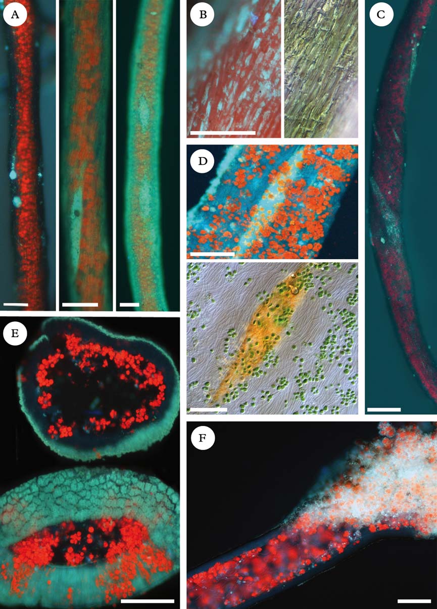
A bluish green fluorescence under UV-2A was evident in the granular outer layer (Fig. 1B). This consisted of small granules when sparse, but a cracked film when abundant. This variation in the surface features of Byoria sect. Implexae specimens is evident in scanning electron micrographs (SEM; Hawksworth Reference Hawksworth1969: figs 1a–d, 2a–c). The granules are extracellular and develop at a short distance behind the growing tips, and can be removed by treatment with KOH (K) and the lipase/protease enzymes in biological washing powders (Greenhalgh & Whitfield Reference Greenhalgh and Whitfield1987). Rikkinen (Reference Rikkinen1995) postulated that these granules might have a light-scattering function. Bryoria fuscescens can have an almost black to an almost white cortex. Under fluorescence microscopy, we found that in dark thalli this substance was hardly evident (Fig. 1A left), while in whitish thalli the granules covered the entire cortex which fluoresced bluish green (Fig. 1A right). Our material of Bryoria capillaris (Ach.) Brodo & D. Hawksw., with a whitish grey thallus, always fluoresced intensely bluish green in the granular outer layer (Fig. 1E). This granular fluorescent substance was more abundant in older parts of the thalli, and rarer or absent in the tips, an observation consistent with those of Greenhalgh & Whitfield (Reference Greenhalgh and Whitfield1987).
Bryoria auto-fluorescence, using UV-2A cube (except in B right and D down) and DIC. Red fluorescent colours are produced by Trebouxia chlorophyll. A, Bryoria fuscescens, granular outer layer: left, black specimen (Spain, Canary Islands, Gran Canaria, MAF-Lich. 18859); centre, brown specimen (Spain, Asturias, Caso, MAF-Lich. 18860); right, pale grey specimen (Spain, Canary Islands, Tenerife, MAF-Lich. 18861). Fusiform brightest areas correspond to an inconspicuous pseudocyphella and circular bright areas to an incipient soralium; B, B. fuscescens, dark olive specimen granular outer layer with (left) and without fluorescence (Spain, Madrid, MAF-Lich. 18862); C, B. fuscescens, dark grey lacinia with bluish fluorescent pseudocyphellae (Spain, Segovia, MAF-Lich. 18863); D, B. fuscescens (Spain, Madrid, MAF-Lich. 18862), dark olive specimen with inconspicuous pseudocyphellae, containing norstictic acid as unique lichen compound (TLC): up, with epi-fluorescence; down, acicular crystals after adding K, without fluorescence; E, B. capillaris, cross-sections of a pale grey specimen, showing the granular outer layer (Spain, Segovia, MAF-Lich. 18865); F, B. fuscescens, longitudinal lacinia section of a dark grey specimen with brightly fluorescent soralium (Spain, Segovia, MAF-Lich. 18864). Scales=100 µm.
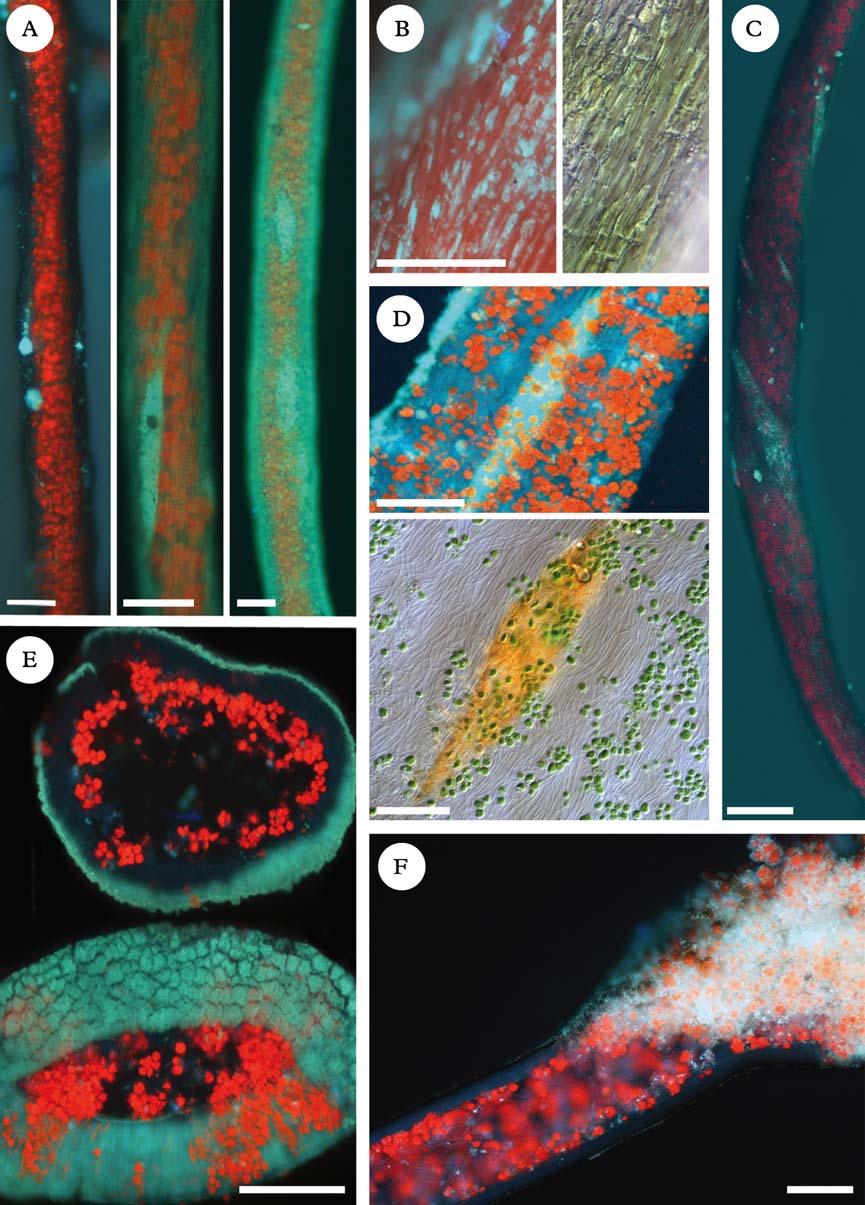
Protocetraric/fumarprotocetraric, norstictic/connorstictic, and psoromic acids gave variations of a whitish blue to greenish blue sequence of fluorescence colours, not clearly differentiated. However, the autofluorescence did serve to demonstrate extrolite distribution in the thallus using three consecutive portions of one branch of a specimen with only one TLC-detected extrolite: 1) TLC, to identify the unique lichen substance; 2) autofluorescence (UV-2A) before and after a K spot test; and 3) extrolite removal with an acetone bath (2 h at 20°C), then autofluoresence (UV-2A) before and after a K spot test. An example is specimens of Bryoria fuscescens with norstictic acid confined to soralia or pseudocyphellae with whitish blue fluorescence (Fig. 1D top). Adding K after fluorescence observation, the typical red acicular crystals formed in these laciniae areas, while other parts of the thallus gave no reaction (Fig. 1D bottom). After removing norstictic acid with acetone and adding K, the red crystals of the reaction were not observed by bright-field microscopy, indicating the acid had been removed. However, when that sample was examined under fluorescence, a less intense whitish blue colour remained, suggesting that either a little of the acid remained or there was another fluorescent substance present not soluble in acetone. Pseudocyphellae and soralia fluoresced the brightest (Fig. 1C & F). Further tests using the described combined method showed that this fluorescence was not exclusively attributable to substances detectable by TLC. This phenomenon was not restricted to Bryoria sect. Implexae. We also studied specimens of Bryoria bicolor, which accumulate fumarprotocetraric acid in the thallus but not in the pseudocyphellae (Pd−). Under the fluorescence microscope, the pseudocyphellae fluoresced brightly with the same colour as those of B. fuscescens. This suggests that this localized fluorescence in the absence of the acids is attributable to different substances. We speculated if hydrophobins could be the cause, peptide-containing proteins that self-assemble on the surfaces of hyphae, but these are not expected to fluoresce, unless labelled (Wang et al. Reference Wang, de Vocht, de Jong, Poolman and Robillard2002). The nature of this fluorescent material remains obscure and merits detailed study.
In summary, this preliminary investigation into the possibilities of the use of fluorescence microscopy for the localization of extrolites in Bryoria thalli, indicates that it has the potential to precisely demonstrate heterogeneous deposition sites. However, at least with the method and UV-blocks used and the compounds accumulated in Bryoria, the colours produced were not sufficiently distinctive to enable particular lichen products to be differentiated by eye, though it is probable that they could be separated spectrophotometrically. Furthermore, the method evidently has to be used with caution as we discovered areas of localized fluorescence where lichen acids were absent or had been eluted, notably granules on the surface, soralia, and pseudocyphellae.
This contribution was prepared with support from the Spanish Ministerio de Economía y Competitividad project CGL2011-25003.