Introduction
The anatomy of the temporal bone is extremely complicated, and most medical students have difficulty in understanding its three-dimensional nature. The skills required for ear surgery can best be achieved by practising dissection of human temporal bone specimens. However, not all trainees will have this opportunity because of the scarcity of real, cadaveric temporal bones.
Previously, we have created a three-dimensional temporal bone model using the selective laser sintering method, and we have examined this model's validity as a training and educational aid.Reference Suzuki, Ogawa, Kawano, Hagiwara, Yamaguchi and Ono1, Reference Suzuki, Ogawa, Hagiwara, Yamaguchi and Ono2 We have also reported the usefulness of such a model for presurgical planning and simulation.Reference Suzuki, Hagiwara, Kawaguchi and Ono3
In this study, we replicated the bony structures together with the soft tissue, to establish a more realistic model, and we used this model for surgical planning and navigation for a case of extensive cholesteatoma.
Materials and methods
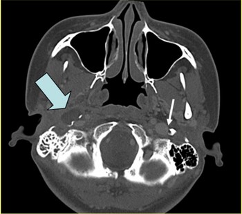
Raw data from computed tomography (CT) scanning of the head were used. Axial CT images were taken using an Asteion multidectector-row CT (MDCT) system (Toshiba, Tokyo, Japan), with a 0.5 mm slice width, 2.5 helical pitch and 0.1 mm image production interval. Data were transferred into the digital imaging and communications in medicine (DICOM) system. The intensity value for extracting the bone shadow was determined based upon these image slices. Three-dimensional data thus derived were converted into standard triangulated language (STL) computer files. In order to create a soft tissue model, the CT intensities of the vessels and the cholesteatoma were determined separately and converted into standard triangulated language (STL) files (Figure 1).
Axial computed tomography scan showing a parapharyngeal lesion (slightly low density; large arrow) and a vessel (higher intensity; small arrow).

Powder substrate (polyamide nylon with added glass beads) was laser sintered according to the STL data, after the target shadow had been extracted. The sintered layers were accumulated, with a 0.1 mm pitch, until the final model was formed. Details of the laser sintering method have been reported elsewhere.Reference Suzuki, Ogawa, Kawano, Hagiwara, Yamaguchi and Ono1
Case presentation
Clinical course
Our patient was a 31-year-old woman whose chief complaint was right-sided otorrhoea.
The patient had first visited our clinic at the age of eight years (in 1985), because of otorrhoea in the right ear. She had been diagnosed with congenital cholesteatoma developing in the meso- and hypotympanum. The right ear had been found to have an average conductive hearing loss of 50 dB. The cholesteatoma had been removed via a postauricular incision. The post-operative course had been uneventful, and the patient had been followed until 11 years of age.
In 1995, aged 21 years, the patient had reattended our clinic because of right-sided otalgia and otorrhoea. Recurrent cholesteatoma had been found in the hypotympanum, and she had undergone surgery.
Two years later (1997), the patient had undergone a third surgical procedure for her cholesteatoma, which had been found to extend further medially into the neck. Since the patient had not wanted radical surgery, she had been followed up for occasional removal of cholesteatoma debris. At this stage, her right ear hearing had deteriorated to 70 dB on average.
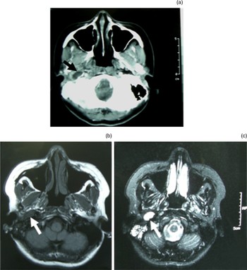
At the age of 30 years (2007), the patient complained of pain in the right lateral neck. A CT scan showed a round, low density lesion in the right parapharyngeal space (Figure 2). The patient was admitted to our hospital for administration of antibiotics. She soon began to develop vertiginous attacks, occurring several times a month. This vertigo was diagnosed to be due to right vestibular disorder. The patient hoped that eradication of the cholesteatoma would resolve her vertigo and otorrhoea, and so surgery was scheduled.
Computed tomography (CT) and magnetic resonance imaging (MRI) findings. (a) Axial CT scan showing low intensity, round lesion in the parapharyngeal space with ring enhancement (arrow). (b) T1-weighted MRI showing low intensity lesion (arrow). (c) T2-weighted MRI showing high intensity lesion (arrow).

Models
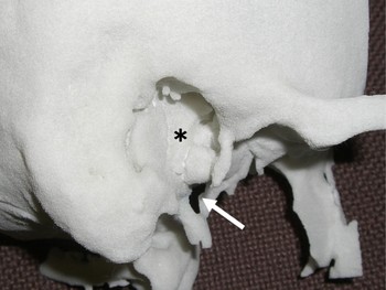
Prior to surgery, laser-sintered models of the patient's ear structures were created. The first model was created solely from bone intensity CT data. This model showed a bony defect of the right styloid process sheath (also termed vagina processus styloidei) (Figure 3).
The three-dimensional, laser-sintered bone model, showing the previously shaved tympanic cavity (*) and a bony defect in the styloid process sheath (arrow).

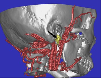
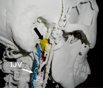
Figure 4 shows a three-dimensional CT image created from the combined STL files for bone, vessels and cholesteatoma. The laser-sintered model created from these combined data showed the cholesteatoma and the surrounding major vessels, including the internal jugular vein (IJV) and internal carotid artery (ICA) (Figure 5).
Three-dimensional computed tomography image derived from combined STL data for bone, cholesteatoma (arrow) and vessels.

Three-dimensional, laser-sintered model created from combined STL data for bone, cholesteatoma (arrow) and vessels. The cholesteatoma has been coloured yellow and the internal jugular vein (IJV) blue.

Surgery
Under general anaesthesia, a postauricular incision was extended caudally by 2 cm.
The granulation in the tympanic cavity was curetted. The mastoid air cells were removed to expose the vertical segment of the facial nerve. Mastoid tip bone was shaved to expose the main trunk of the facial nerve at the styloid process. The cholesteatoma matrix, extending from the hypotympanum through to the styloid process sheath, was dissected and removed.
At this point, the surgeon manually examined the disinfected plastic model to identify the bony defect of the styloid process sheath.
Dissection of the cholesteatoma was continued further, into the parapharyngeal space. The cholesteatoma matrix in the parapharyngeal space was removed, as was the connective tissue adherent to the ICA. Gentamicin was injected into the lateral semicircular canal and oval window, to achieve a chemical labyrinthectomy. Abdominal fat was harvested to obliterate the parapharyngeal space and the mastoid cavity. The external ear canal skin was sutured at the entrance of the canal.
The surgeon found the three-dimensional model useful for peri-operative surgical navigation, and for identification of the cholesteatoma extension from the styloid process sheath through to the ICA.
After surgery, the patient suffered mild dizziness and paralytic nystagmus for one week, which then cleared. Otherwise, she recovered well, without any facial palsy or mastication problems. The right hearing dropped to the scale-out level.
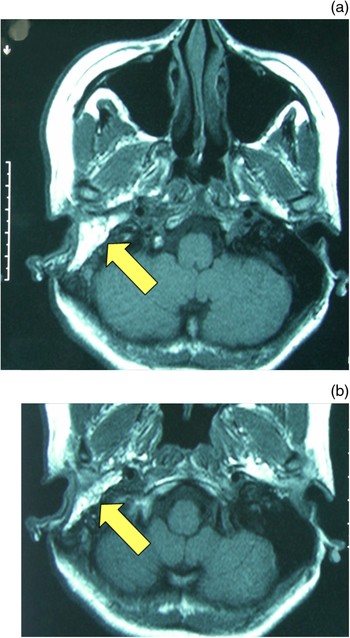
At the time of writing, the patient had remained free of otorrhoea and dizziness up until now for 18 months. A post-operative MRI scan showed a fat tissue shadow packing the operated space from the mastoid cavity through to the parapharyngeal space (Figure 6).
Post-operative, axial MRI scans, showing the parapharyngeal space filled with fatty tissue (arrows).

Discussion
Surgical procedures involving the temporal bone are a common part of otorhinolaryngological practice. However, acquisition of the requisite surgical skills is not easy, due to the complex anatomy of this region. In patients with lesions involving these complex structures, surgery becomes even harder. Given these difficulties, a three-dimensional model of the temporal bone may serve as a useful tool for education and surgical navigation.
Rapid prototyping can be used to create three-dimensional models from plastic liquid or powder material. This technique has been extensively used for designing and manufacturing prototypes in the fields of engineering and medicine.Reference Lightman, Cohen and Jacobs4–Reference Begall and Vorwerk6 There are two types of rapid prototyping: stereolithography and selective laser sintering. The former involves polymerisation and solidification of liquid resin, while the latter uses laser to fuse layers of plastic powder.
We have already reported the usefulness of a temporal bone model, prototyped by selective laser sintering, for medical education and surgical planning.Reference Suzuki, Ogawa, Kawano, Hagiwara, Yamaguchi and Ono1–Reference Suzuki, Hagiwara, Kawaguchi and Ono3 In this study, we attempted to replicate not only the bony structures but also the vessels and the cholesteatoma (which extended into the parapharyngeal space, close to the internal carotid artery (ICA)).
• The skills of middle-ear surgery are best attained by practising dissection of the human temporal bone
• This paper describes the creation of a three-dimensional plastic model using a selective laser sintering method, and examines the validity of such a model as a surgical aid and a training and educational tool
There are two methods of replicating bone and soft tissue in such prototyped models. The first is to determine the CT intensity of bone and soft tissue on the enhanced CT data. This allows replication of bone and enhanced tissue, such as vessels and malignant or vascular tumours. The second method is to determine the CT intensity of bone and soft tissue separately, on non-enhanced CT images, and then to combine the STL data into a single STL file, as shown in Figure 3. In the present case, the intensities of the cholesteatoma and the vessels were well distinguished from regular CT images (Figure 1), thus allowing replication of these structures.
The three-dimensional model clearly showed the bony defect of the styloid process sheath, which could not be easily judged on a conventional CT image. Surgery could then safely be conducted, using this bone defect as a landmark guiding the surgeon toward the parapharyngeal space. The plastic model also allowed three-dimensional visualisation of the cholesteatoma and the surrounding major vessels, including the IJV and ICA. Due to these advantages, surgery was easily performed via a minimal skin incision. The three-dimensional model was also useful to facilitate presurgical explanation to the patient and her family, and to aid the consent process. Image-guided navigation has been extensively utilised in surgery for lesions involving complex structures, and its usefulness is widely reported.Reference Selesnick and Kacker7 From the standpoints of economy and convenience, a rapid prototype model may serve as a good alternative.
Conclusion
Computed tomography data were used to create a three-dimensional, laser-sintered, plastic model of the ear structures, including bones, vessels and the cholesteatoma, and this model was used to aid surgery for recurrent cholesteatoma. Surgery was safely performed, providing evidence of the validity of such a three-dimensional model for presurgical planning and navigation.
Acknowledgements
The authors appreciate Mr. H. Ono for his technical suggestion.