Introduction
Neurocysticercosis, caused by the metacestode stage of the Taenia solium tapeworm, is a serious health problem in many developing countries (Sciutto et al., Reference Sciutto, Fragoso, Fleury, Laclette, Sotelo, Aluja, Vargas and Larralde2000; Flisser et al., Reference Flisser, Sarti, Lightowlers and Schantz2003). This pathology represents the major brain disorder in the Third World and affects around 50 million people worldwide, being currently considered an emergent disease in developed countries (White, Reference White2005). Taenia solium also infects pigs, its intermediate host, leading to major economic loss and transmission to humans (Nash et al., Reference Nash, Singh, White, Rajshekhar, Loeb, Proano, Takayanagui, Gonzalez, Butman, De Giorgio, Del Brutto, Delgado-Escueta, Evans, Gilman, Martinez, Medina, Pretell, Teale and Garcia2006). Humans are the sole final hosts of T. solium, developing a tapeworm infection following the ingestion of raw or undercooked contaminated pork meat. Taenia solium eggs are passed with the stools and contaminate the environment (Sciutto et al., Reference Sciutto, Fragoso, Fleury, Laclette, Sotelo, Aluja, Vargas and Larralde2000). When T. solium eggs are ingested by pigs or humans, larvae may develop throughout the body. In the human, most clinical symptoms become apparent when cysticerci are established in the central nervous system (White, Reference White2005). On the other hand, due to the intrinsic difficulties of working with the natural hosts (pigs and humans) of T. solium, the high costs of keeping sufficient pigs, plus slowness in data retrieval, we have repeatedly used the experimental approach of murine cysticercosis to gain knowledge on the complex host (H)–parasite (P) relationship in cysticercosis (Larralde et al., Reference Larralde, Sotelo, Montoya, Palencia, Padilla, Govezensky, Diaz and Sciutto1990, Reference Larralde, Morales, Terrazas, Govezensky and Romano1995; Morales-Montor & Larralde, Reference Morales-Montor and Larralde2005). Murine intraperitoneal cysticercosis is caused by the taenid Taenia crassiceps and the model has been used to explore the physiological host factors associated with porcine cysticercosis, and to some degree, with human neurocysticercosis (Larralde et al., Reference Larralde, Sotelo, Montoya, Palencia, Padilla, Govezensky, Diaz and Sciutto1990). Several features of T. solium cysticercosis have been found by extrapolation from experimental murine cysticercosis (Larralde et al., Reference Larralde, Sotelo, Montoya, Palencia, Padilla, Govezensky, Diaz and Sciutto1990).
Taenia solium and T. crassiceps are highly evolved parasites, which have developed diverse mechanisms of survival within the host that facilitate their establishment. Interestingly, both parasites exploit the host endocrine system, which has been found to facilitate their establishment, growth and reproduction (Damian, Reference Damian1997; Escobedo et al., Reference Escobedo, Roberts, Carrero and Morales-Montor2005). This exploitation mechanism provides parasites with a dual benefit: first, it provides them with amino acids for metabolism, and second it prevents surface-bound antibodies from interfering with cytotoxic cells that interact with the parasite (Locksley, Reference Locksley1997; Shibayama et al., Reference Shibayama, Serrano-Luna, Rojas-Hernández, Campos-Rodríguez and Tsutsumi2003). In this sense, many species of taennids have evolved structures similar to the protein hormone receptors expressed in upper vertebrates, with binding properties and terminal effects akin to the hormonal metabolites synthesized by the host (Maswoswe et al., Reference Maswoswe, Peters and Warhurst1985; Unnasch et al., Reference Unnasch, Bradley, Beauchamp, Tuan and Kennedy1999). Such cross-regulation between host and parasite has been described in at least eight parasitic infections caused by helminth parasites (Escobedo et al., Reference Escobedo, Roberts, Carrero and Morales-Montor2005), such as schistosomiasis (Remoue et al., Reference Remoue, To Van, Schacht, Picquet, Garraud, Vercruysse, Ly, Capron and Riveau2001), filariasis (Sani et al., Reference Sani, Vaid, Comley and Montgomery1985), porcine cysticercosis caused by T. solium and murine cysticercosis by T. crassiceps (Morales et al., Reference Morales, Velasco, Tovar, Fragoso, Fleury, Beltran, Villalobos, Aluja, Rodarte, Sciutto and Larralde2002; Escobedo et al., Reference Escobedo, Larralde, Chavarria, Cerbon and Morales-Montor2004), among others.
In order to understand how the host's endocrine system can, under certain circumstances, favour the establishment of T. solium and T. crassiceps in their natural hosts, we have explored the in vitro effects of steroid hormones, which play an important role during infection (Morales-Montor et al., Reference Morales-Montor, Mohamed, Ghaleb, Baig, Hallal-Calleros and Damian2001; Escobedo et al., Reference Escobedo, Larralde, Chavarria, Cerbon and Morales-Montor2004). Also, protein hormones, which are part of one of the most ubiquitous hormonal systems throughout the evolutionary scale, have been investigated, such as the insulin–insulin receptor system. This is the major topic of the present work and may be involved in the complex host–parasite relationship, due to its importance in glucose-dependent energy metabolism (Hay et al., Reference Hay, Sparks, Gilbert, Battaglia and Meschia1984; Zenobi et al., Reference Zenobi, Graf, Ursprung and Froesch1992; Pende et al., Reference Pende, Kozma, Jaquet, Oorschot, Burcelin, Le Marchand-Brustel, Klumperman, Thorens and Thomas2000; Doehner et al., Reference Doehner, Gathercole, Cicoira, Krack, Coats, Camici and Anker2008; Ravier et al., Reference Ravier, Nenquin, Miki, Seino and Henquin2008; Takazawa et al., Reference Takazawa, Noguchi, Hosooka, Yoshioka, Tobimatsu and Kasuga2008) and its role as activator of the second messenger machinery (including growth factor receptor binding protein-2 (Grb2), Sos, Raf-1, MEK, extracellular regulated kinases (ERK)) that promotes differentiation, growth and proliferation processes in a very wide range of cell types, such as liver, muscle and stem cells (Wang et al., Reference Wang, Corbett, Marshall and McDaniel1993; Philippou et al., Reference Philippou, Halapas, Maridaki and Koutsilieris2007; Alonso-Magdalena et al., Reference Alonso-Magdalena, Ropero, Carrera, Cederroth, Baquié, Gauthier, Nef, Stefani and Nadal2008; Breuhahn & Schirmacher, Reference Breuhahn and Schirmacher2008; Dyachok et al., Reference Dyachok, Idevall-Hagren, Sågetorp, Tian, Wuttke, Arrieumerlou, Akusjärvi, Gylfe and Tengholm2008; Johnson & Alejandro, Reference Johnson and Alejandro2008; Kim et al., Reference Kim, Abdellatif, Koul and Crystal2008; Narbonne & Roy, Reference Narbonne and Roy2008; Rhoads et al., Reference Rhoads, Meyer, Kolath, Lamberson and Lucy2008).
Moreover, insulin-related peptides are a group of hormonal growth factors involved in the regulation of key processes such as metabolism, growth control, reproduction and ageing (van der Geer et al., Reference van der Geer, Hunter and Lindberg1994; White & Kahn, Reference White and Kahn1994; Hubbard & Till, Reference Hubbard and Till2000; Gems & Partridge, Reference Gems and Partridge2001). Insulin binds to specific insulin receptors (IR), located at the surface of target cells in many organisms of different phyla (van der Geer et al., Reference van der Geer, Hunter and Lindberg1994; Roovers et al., Reference Roovers, Vincent, van Kesteren, Geraerts, Planta, Vreugdenhil and van Heerikhuizen1995; Ruan et al., Reference Ruan, Chen, Cao and Garofalo1995; Kimura et al., Reference Kimura, Tissenbaum, Liu and Ruvkun1997; Hubbard & Till, Reference Hubbard and Till2000; Konrad et al., Reference Konrad, Kroner, Spiliotis, Zavala-Góngora and Brehm2003). Due to the ubiquitous presence of insulin-like signalling pathways in metazoans, a similar system could also be used in helminth infections (Beall & Pearce, Reference Beall and Pearce2002). Nevertheless, particularly in taenids, very little information has been generated regarding the role of insulin and its signalling pathways, although its widespread importance in metazoans suggests that insulin/IR-mediated signalling may control parasite development and possibly several aspects of the host–parasite relationship.
The present in vitro experiments were designed to explore, with a variety of approaches, whether insulin has a direct effect upon T. crassiceps and T. solium cysticercus reproduction, differentiation and infectivity. In addition, we searched for preliminary evidence of the possible molecular mechanisms involved, which could provide a keystone in the design of a drug that would exclusively bind to the parasite insulin receptor, blocking the insulin effect on the pathogen and all signal transduction pathways mediated by this growth factor.
Materials and methods
Harvesting and preparing T. crassiceps and T. solium cysticerci for experimentation
A new stock of T. crassiceps cysticerci (ORF-Kuhn2 strain) (Culbreth et al., Reference Culbreth, Esch and Kuhn1972; Freeman, Reference Freeman1985) was donated to our laboratory by Dr R. Kuhn in 2000 and kept by serial intraperitoneal passage in BALB/cAnN female mice approximately every 4 months. Cysticerci for each experimental session were obtained from intraperitoneally infected female mice and placed in tubes containing sterile phosphate-buffered saline (PBS) (1 × ) supplemented with 100 U/ml of antibiotics–fungizone (Gibco, Grand Island, New York, USA) (Esch & Smith, Reference Esch and Smith1976). The tubes were centrifuged for 10 min at 1500 rpm at 4°C and the supernatant was discarded. The packed cysticerci were incubated in Dulbecco's modified Eagle's (DMEM) serum-free medium (Gibco BRL, Rockville, Maryland, USA). They were then washed by centrifuging three times for 10 min at 1500 rpm. After the final wash, the numbers of viable cysticerci (complete, translucent and motile cystic structures) were counted under a binocular microscope. Ten viable non-budding cysticerci of approximately 2 mm in diameter were then selected and dispensed into each well of 24-well culture plates (Falcon, BectonDickinson Labware, Franklin Lakes, New Jersey, USA) in 1 ml DMEM serum-free medium and incubated at 37°C under 5% CO2.
Taenia solium cysticerci were dissected from the muscle of infected male pigs in independent experiments. Pigs were humanely sacrificed by using head electroshock and posterior bleeding. The method was previously evaluated by the University Animal Care and Use Committee and governmental agencies to ensure compliance with federal regulations and guidelines. The fibrous capsule surrounding each parasite was carefully separated under a dissection microscope. Once dissected, cysticerci were placed in tubes containing sterile PBS (1 × ) supplemented with 100 U/ml of antibiotics–fungizone. The tubes were centrifuged for 10 min, at 800 g at 4°C and the supernatant was discarded. The packed cysticerci were incubated in DMEM serum-free medium. They were then centrifuged three times for 10 min at 800 g for washing. After the final wash, the numbers of viable cysticerci (complete and translucent cystic structures) were counted using a binocular microscope. Five viable cysticerci were then selected and dispensed into each well of six-well culture plates (Falcon, BectonDickinson Labware) in 3 ml DMEM-free serum medium and incubated at 37°C under 5% CO2.
For both species of parasites, a sufficient number of culture wells was prepared to accommodate the complete experimental design for evaluation of in vitro treatment effects of insulin on the cysticerci. Cultures were revised daily and their medium completely replaced when they turned yellowish.
Insulin concentration–response-time curves
Culture-grade insulin was obtained from Sigma (recombinant human insulin, Sigma-Aldrich, Inc., St. Louis, Missouri, USA). For in vitro tests, water-soluble insulin was dissolved in DMEM serum-free culture medium to the desired stock concentration, and sterilized by passage through a 0.2 mm millipore filter. The experiments employed the parasite-loaded wells as follows: six wells were used as untreated controls, six wells were supplemented with the same medium in which hormone was previously dissolved. Series of six wells were separately treated with 0.5, 1.0, 1.5 and 2.0 units (IU) of insulin. Hormone concentrations used in this study were under physiological and pharmacological conditions, as previously reported (Escobedo et al., Reference Escobedo, Larralde, Chavarria, Cerbon and Morales-Montor2004), and designed in order to approximate the levels in humans, gestant pigs and mice (Morales et al., Reference Morales, Velasco, Tovar, Fragoso, Fleury, Beltran, Villalobos, Aluja, Rodarte, Sciutto and Larralde2002). In hormone time–response curves, the number of T. crassiceps buds and differentiated T. solium scolexes as a function of days in culture were assessed as the response variable. From the dose–response curves of each hormone, we selected an optimal dose for insulin (1 IU), to be used in time–response curves.
Visual assessment, reproduction and viability of T. crassiceps and T. solium
Parasite reproduction was assessed as the total number of buds that were attached to the ten cysticerci in each well. Bud count was performed daily under an inverted light microscope. Cysticerci viability was determined daily in the wells using an inverted microscope (Olympus, MO21, Tokyo, Japan) at 4 × and 10 × magnification. Injury to cysticerci was recognized microscopically by progressive internal disorganization, development of whitish opaque areas on the parasite's tegument and loss of motility. Dead cysticerci were immobile, opaque and structurally disorganized. The T. crassiceps viability was measured as the percentage motility of cysts, while for T. solium viability was the percentage of evagination in the presence of pig bile (4%).
Insulin effect on viability and scolex evagination of cultured T. solium cysticerci
Cysticercus viability, scolex evagination and scolex growth of the T. solium cysticerci were determined daily for all wells, using an inverted microscope (Olympus, MO21, Tokyo, Japan) at 4 × and 10 × magnification. Injury to cysticerci was recognized microscopically by progressive internal disorganization and development of whitish opaque areas on the parasite's tegument. Once evaginated, dead parasites were immobile, opaque and structurally disorganized.
Measurement of bud diameter in T. crassiceps cysticerci
At every tested concentration of insulin, bud diameter was measured by means of a millimetre rule under an inverted microscope (4 × and 10 × magnification). Only buds on cysticerci were considered and these results are presented as the mean ± SD of bud diameter.
Infectivity assay of pre-treated T. crassiceps cysticerci
Six-week-old BALB/cAnN female mice were used in this study as recipients of insulin pre-treated cysticerci. Mice were fed with Purina Diet 5015 and water ad libitum. Larvae for intraperitoneal experimental infection were the ten in vitro cultured cysticerci after their pre-treatment for 5 days with 1 IU of insulin. Each recipient mouse was injected i.p. with the insulin pre-treated cysticerci using a 0.8 gauge needle. Recipient female mice were sacrificed after 8 weeks of infection by cervical dislocation after etherization, and their parasite load estimated by inverted microscope-aided visual counting of the total number of cysticerci collected from the infected mice after thorough peritoneal washes with 1 × PBS. The infectivity rate was measured as the total sum of the parasite load divided by the total number of cysts and buds that were injected into each mouse.
Insulin receptor gene amplification
Total RNA was isolated from insulin-treated T. solium and T. crassiceps cysticerci by the method based on guanidine isothiocyanate/phenol/chloroform extraction using Trizol reagent (Invitrogen, Carlsbad, California). In brief, cysticerci were disrupted in Trizol reagent (1 ml/0.1 g tissue) and 0.2 ml of chloroform was added per 1 ml of Trizol. The aqueous phase was recovered after 10 min of centrifugation at 14,000 g. RNA was precipitated with isopropyl alcohol, washed with 75% ethanol and redissolved in RNAse-free water. RNA concentration was determined by absorbance at 260 nm and its purity was verified after electrophoresis on 1.0% denaturing agarose gel in the presence of 2.2 m formaldehyde. Total RNA from hormone-treated cysticerci was reverse-transcribed, followed by specific polymerase chain reaction (PCR) amplification of insulin receptor, together with β-actin (control gene), as previously described (Escobedo et al., Reference Escobedo, Larralde, Chavarria, Cerbon and Morales-Montor2004). Primers were designed based on the sequence of previously cloned Echinococcus multilocularis (a related cestode) gene (Konrad et al., Reference Konrad, Kroner, Spiliotis, Zavala-Góngora and Brehm2003). Primer sequences were as follows:
Insulin receptor:
sense: 5′-TGTGCTACGTGCGGACTGTGA
antisense: 5′-CCCTGATGGATAGCGAACTGG
β-actin:
sense: 5′-GGGTCAGAAGGATTCCTATG
antisense: 5′-GGTCTCAAACATGATCTGGG.
Obtained PCR products were visualized by staining with ethidium bromide. In all cases, a single band was detected, corresponding to the expected size of the gene (640 bp). The expression of the parasite insulin receptors is numerically presented as the ratio of the optical densitometry (OD) of each studied gene relative to the expression in the same preparation of the β-actin gene, a constitutively expressed gene used as internal control for differences in the amplification procedure between experiments and in staining of the different gels. It is important to underline that in order to demonstrate that cysticerci were not contaminated by host-cell DNA or RNA, we performed the same reverse transcriptase (RT)-PCR using cDNA from T. crassiceps, T. solium, mouse and pig spleen to amplify the interferon-γ (IFN-γ) and variable domain region (VDR) genes, which are expressed with high specificity only in the mammalian hosts, since both molecules take part in physiological homeostatic regulation as important components of the immune response (IFN-γ) and the variable region of the IgG immunoglobulin. Sequences of primers were as follows:
IFN-γ:
sense: 5′-CAGAGCTGTCTCTAAACCGT-3′
antisense: 5′-AGGGTGTCTGAAGGAGGGGA-3′
VDR:
sense: 5′-CTGAATTCCA TGAAACACCTGTGGTTCTT-3′
antisense: 5′-GACTCTAGA GGGCTCACACTCA CCTCCCCT-3′.
Experimental design and statistical analysis
Hormone dose–response-time curves were estimated in ten independent experiments, each performed with five T. solium cysticerci and ten T. crassiceps cysticerci, freshly extracted from different infected donor pigs and mice, and cultured in the presence of insulin at four different concentrations, each replicated in six different wells. The response variable used in statistical analysis was the sum of buds (T. crassiceps) and the evaginated scolexes (T. solium) in the six wells of each experiment. The used hormone (one level), the dose of used hormone (four levels), the time of exposure (ten levels) and the number of experiments (ten levels) were the independent variables. All experiments to evaluate the effect of the optimal dose of insulin on the parasite proliferation indexes were performed five times. The data of the ten replicates of each experiment were pooled and expressed as their average. The data were analysed using analysis of variance (ANOVA) with hormone treatment (four levels) and number of experiments (ten levels) as independent variables and, as dependent variables: average of sum of buds, the total number of developed cysticerci in recipient female mice of pre-treated cysticerci for the infectivity assay. If significant differences between treatments were found by ANOVA, differences between the group means were assessed within each experiment by means of a Tukey test, using the residual variance estimated by ANOVA to test for significance. Differences were considered significant when P < 0.05.
Results
Dose–response curves
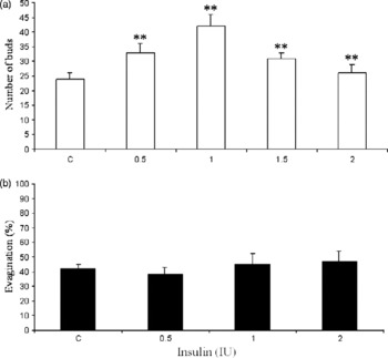
Different concentrations of insulin in the culture media were studied to explore a probable dose-dependent response pattern of T. crassiceps and T. solium. In T. crassiceps, the number of buds clearly increased as the insulin concentration rose, reaching a maximum value at 1 IU of this hormone (fig. 1a). Above this concentration, the effect of in vitro insulin progressively diminished until it reached similar values to the control groups (fig. 1a), showing a typical dose-dependent pattern of T. crassiceps cysticercus reproduction. However, T. solium cysticerci did not show a response to insulin, even at the highest concentration, because no significant difference in the scolex evagination percentage was observed between insulin-treated parasites and control cysticerci (fig. 1b).
Insulin effect upon Taenia crassiceps and Taenia solium. Concentration–response curves. Insulin clearly augmented the number of buds in T. crassiceps even at the lowest concentration (a). On the contrary, insulin had no significant effect upon scolex evagination of T. solium cysticerci (b). C, control cysticerci treated with vehicle. IU, International Units. **P < 0.05.

Time–response curves
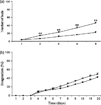
When T. crassiceps cysticerci were exposed to 1 IU of insulin in vitro, the hormone markedly increased parasite bud production (fig. 2a). In the insulin-treated group, the number of buds in relation to control values increased progressively, up to twice their initial number after 5 days in culture (fig. 2a). On the contrary, insulin treatment did not modify the evagination of the T. solium cysticerci after 20 days of in vitro exposure, as shown in the dose–response curves (fig. 2b). In consequence, no significant effect was observed upon scolex length in insulin-treated parasites (data not shown), emphasizing the dichotomistic effect of this hormone on T. crassiceps and T. solium. Interestingly, T. solium insulin-treated cysticerci remained alive throughout the time of in vitro culture, which supports the fact that insulin treatment had no negative effects on the viability of cultured parasites.
Insulin effect upon Taenia crassiceps and Taenia solium. Time–response curves. In vitro effect of insulin (1 IU) on T. crassiceps was maintained throughout (a). On the other hand, T. solium cysticerci were not affected by insulin treatment, either at optimal concentration of the hormone or throughout the time (b). ○, Insulin-treated cysticerci; ●, control cysticerci treated with vehicle. **P < 0.05.

Viability curves
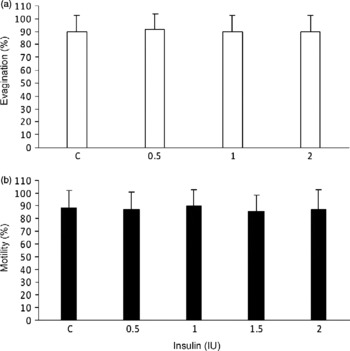
Figure 3 shows the viability assays of both parasites during experimental treatments. In T. crassiceps (fig. 3a) viability was recorded at every dose used, and compared against untreated cysticerci (control). Our results showed that insulin treatment had no negative effects on the viability of cultured parasites since, even at the higher dose used, viability remained unaltered. At the initial period of culture and at the end (5 days) viability was the same. The same result was obtained in T. solium evagination (fig. 3b), since insulin-treated cysticerci remained alive throughout the time of in vitro culture, which supports the fact that insulin treatment had no negative effects on the viability of cultured parasites.
Viability curves of Taenia crassiceps (a) and Taenia solium (b). Insulin did not modify the viability, measured as motility (T. crassiceps) or evagination (T. solium), between control and insulin-treated parasites. C, control cysticerci treated with vehicle; IU, International Units.

Morphological studies
Insulin-treated T. crassiceps cysticerci were translucent, mobile and surrounded by an increasing number of buds, also motile and rather large. Many multilobulated giant buds were frequently observed on insulin-treated parasites, in contrast with the aspect of untreated cysticerci. In contrast, insulin treatment did not produce any visible morphological alterations in T. solium cysticerci, which remained healthy, showing no progressive internal disorganization or development of opaque whitish masses in the tegument, including no loss of scolex motility (not shown).
Bud diameter
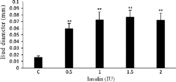
In fig. 4, it is very clear that insulin treatment increases the average bud diameter of T. crassiceps, which reached its maximum size at the concentration of 1 IU (86 ± 9.49 μm), fivefold larger than in untreated cysticerci. After that concentration, bud diameter reached a plateau and no bigger buds were recorded. Control bud diameter remained unaltered during all time of experimentation.
Bud diameter of Taenia crassiceps cysticerci. Insulin clearly increased fivefold the average diameter of buds in treated cysticerci. C, control cysticerci treated with vehicle; IU, International Units **P < 0.05.

Infectivity assays
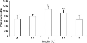
Figure 5 shows the parasite burdens obtained from female mice after 8 weeks of infection. Before being inoculated, cysticerci were exposed in vitro to optimal doses of insulin during 5 days in culture. Interestingly, mice inoculated with parasites previously exposed to 1 and 1.5 IU of insulin, showed a significant increase in parasite load (0.7- and 0.3-fold, respectively), compared to the recipient hosts infected with cysticerci pre-treated with 0.5 and 2.0 IU of insulin, which showed parasite numbers similar to that in controls (fig. 5). This finding indicates that insulin promoted parasite infectivity and establishment when cysticerci were exposed to this hormone prior to their inoculation into recipient female mice.
Infectivity of insulin pre-treated cysticerci. Infected females with Taenia crassiceps cysticerci pre-treated with 1 and 1.5 IU of insulin had significantly increased parasite loads in the peritoneal cavity, compared to control groups. C, control cysticerci treated with vehicle. IU, International Units. **P < 0.05.

Insulin receptor detection in T. crassiceps and T. solium cysticerci
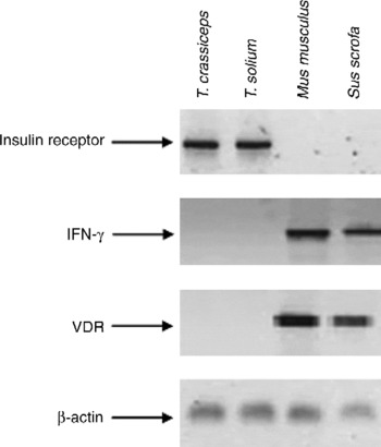
Amplification by RT-PCR of the insulin receptor and β-actin gene from T. crassiceps and T. solium is shown in fig. 6. Specific fragments that corresponded in size (624 bp) to the insulin receptor from other cestodes (Konrad et al., Reference Konrad, Kroner, Spiliotis, Zavala-Góngora and Brehm2003) were obtained from both T. crassiceps and T. solium. On the other hand, to dismiss the possibility that the obtained fragments of T. crassiceps and T. solium were a contamination product from host DNA or RNA (Mus musculus and Sus scrofa), we performed the same RT-PCR to amplify the IFN-γ and VDR genes, which are molecules synthesized exclusively in mammalian species, as they are important factors of the immune response and MHC-mediated recognition. As expected, both IFN-γ and VDR were well amplified for mouse and pig tissues (spleen) but not for T. crassiceps and T. solium (fig. 6). Using the same RNA samples, β-actin was detected in both cestodes and mammals (fig. 6), a finding that suggests insulin receptor amplification in T. crassiceps and T. solium was parasite-specific and not a cross-amplification from mouse and pig cells.
Insulin receptor amplification from Taenia crassiceps and Taenia solium cysticerci. Corresponding bands to insulin receptor (624 bp) were specifically detected in the helminth parasites T. crassiceps and T. solium, but not in mouse (Mus musculus) or pig (Sus scrofa). Concomitantly, IFN-γ and VDR genes were not amplified in either helminth, but they were clearly detected in host tissues (spleen), which in this case completely excludes the possibility of contamination between host and parasite cells.

Discussion
The relationship between parasites and their hosts implies close biochemical co-evolution and communication between their complex physiological and metabolic systems (Escobedo et al., Reference Escobedo, Roberts, Carrero and Morales-Montor2005; Morales-Montor & Larralde, Reference Morales-Montor and Larralde2005).
This work describes the effect of insulin on T. solium cysticerci in vitro evagination and T. crassiceps in vitro reproduction, in order to imitate the physiological conditions to which parasites can be exposed in their native hosts. Interestingly, our findings showed that insulin has a dichotomistic effect on the helminth cestodes T. crassiceps and T. solium. In fact, insulin directly promotes proliferation, viability and infectivity of the T. crassiceps cysticercus. In contrast, T. solium cysticerci were not affected by insulin treatment at any tested dose (fig. 1). In T. crassiceps, insulin effects depended on concentration and exposure time to the hormone. The effect of insulin on their reproduction and viability began at 24 h of culture but were maximal between experimental and control groups at 5 days of culture (fig. 2a). In T. solium, neither concentration nor exposure time showed significant effects upon scolex evagination of the treated parasites (fig. 2b), which is consistent with the initial findings that insulin has differential actions on T. crassiceps and T. solium.
Concomitantly, differences were not only related to the parasite's reproduction or differentiation, but also to their viability and morphological characteristics. In fact, T. crassiceps cysticerci exposed to insulin appeared larger than untreated parasites. Interestingly, their buds seemed larger (fourfold) than in control parasites, and also more spherical and motile (fig. 4). In addition, viability of T. crassiceps was not affected by insulin treatment, because parasites appeared motile on the culture plate, showing no damage, injury or disorganization of the tegument or vesicular liquid. On the contrary, T. solium cysticerci showed no response to in vitro insulin treatment; and the parasite's morphological appearance, viability and evagination percentage was equal in experimental and control groups (fig. 3b).
Since insulin showed significant effects only on T. crassiceps reproduction, growth and viability, we evaluated its influence on parasite infectivity (measured as the capability of cysticerci to establish in an immunocompetent host), which is probably the most important characteristic for parasite survival. Interestingly, we found that insulin treatment directly increased infectivity of the T. crassiceps cysticercus (fig. 5), maybe because this growth factor activates the signalling pathways related to the establishment of parasitic cells on the host, or simply because it increases the size and activity of the parasite and of its buds.
In addition, our results suggest that the possible mechanism of action through which insulin affects parasite growth and reproduction is mediated by an insulin receptor expressed on the parasite, which would not be rare for a cestode (Konrad et al., Reference Konrad, Kroner, Spiliotis, Zavala-Góngora and Brehm2003). In fact, by means of primers based on E. multilocularis sequences (another related cestode parasite), insulin receptor fragments were specifically amplified from T. crassiceps and T. solium, but not in mouse (Mus musculus) or pig (Sus scrofa), the natural intermediate host of these helminths (fig. 6). Moreover, VDR and IFN-γ were detected in both mammalian species but not in both cestodes. Because β-actin was amplified in all studied organisms, mammals and cestodes, we propose that insulin receptors were specifically detected in parasitic tissue and not artifactually obtained by host-cell contamination. Moreover, this fact was also supported for in vitro culture conditions. Cysticerci were cultured for more than 4 weeks in a serum-free medium, which maintains parasites, but not mammalian cells. These findings support T. crassiceps and T. solium-expressed insulin receptor (IR-like), which is probably able to bind insulin and mediate its actions in T. crassiceps, but not in T. solium, which may have another mechanism not yet explored in this work. Concomitantly, insulin pathways are conserved and almost ubiquitous in helminth parasites, with similar functions to those of invertebrate and vertebrate orthologues. The existence of similar receptors and ligands in the host and the cysticerci could have major consequences in the establishment and the regulation of pig cysticercosis. Indeed, host-derived insulin could activate cysticerci receptors and modulate cyst development and differentiation. Further studies are necessary for a precise characterization of the signalling cascades of these receptors in both parasites, particularly in the case of insulin-receptor signalling.Our results suggest that both taenids T. solium and T. crassiceps have developed, through many years of co-evolution with their hosts, a complicated host–parasite network clearly affected by hormones, such as steroid hormones and insulin, all of them involved in the maintenance of important parasite processes such as reproduction, differentiation and infectivity.
Furthermore, in recent years much information about the host hormonal microenviroment affecting many parasite species has been reported (Sani et al., Reference Sani, Vaid, Comley and Montgomery1985; Lingnau et al., Reference Lingnau, Margos, Maier and Seitz1993; Remoue et al., Reference Remoue, To Van, Schacht, Picquet, Garraud, Vercruysse, Ly, Capron and Riveau2001; Escobedo et al., Reference Escobedo, Roberts, Carrero and Morales-Montor2005). We previously found that gender and circulating steroid levels in host mice and host pigs crucially affect the dynamics of parasite loads in infected mice with T. crassiceps cysticerci (Escobedo et al., Reference Escobedo, Larralde, Chavarria, Cerbon and Morales-Montor2004) and infected pigs with T. solium metacestodes (Morales et al., Reference Morales, Velasco, Tovar, Fragoso, Fleury, Beltran, Villalobos, Aluja, Rodarte, Sciutto and Larralde2002). Such commonalities between host and cysticercus metabolism should come as no surprise, when extensive homologies between species are being documented in other systems as well (Roberts et al., Reference Roberts, Walker and Alexander2001). Previous studies have demonstrated that the genome of Onchocerca volvulus encodes at least three members of the nuclear receptor family (Yates et al., Reference Yates, Tuan, Shepley and Unnasch1995), also found in T. crassiceps cysticerci (Escobedo et al., Reference Escobedo, Larralde, Chavarria, Cerbon and Morales-Montor2004).
With respect to the significance of the differential effects of insulin upon T. crassiceps and T. solium, we found it of interest to speculate on the meaning of host specificity in dealing with the parasite's life cycle. It would appear that specialization of the kind described for T. crassiceps (of larval asexual reproduction) could eventually lead to less aggressive relations between host and parasite, than the human or pig with T. solium. Intermediate hosts would allow infection (select) by the fittest of the parasite's versions (i.e. those inducing the lesser immuno-inflammatory responses that could hurt both host and parasite) and, through their more exploratory behaviour, offer it more frequently to its definitive predatory host. In the predator's intestine, the adult stage of the selected parasite could reproduce sexually and diversify into even less irritating versions of its larvae. The eggs would then pass the new improved versions of the parasite on to the proliferation-convenient environment of female mice intermediate hosts. The evidence presented above illustrates the complexity and importance of host–parasite endocrine interactions during cysticercosis and provides clues to the many other possible mechanisms of parasite establishment, growth and reproduction in an immunocompetent host. Furthermore, strong endocrine host–parasite interactions may have implications for the control of transmission and treatment of this parasitic disease in pigs and humans. Of practical importance, the complexity of the cysticerci–host relationship suggests that all physiological factors (i.e. sex, age) should be taken into account in the design of vaccines and new drugs. In fact, characterization of receptor kinases and their signalling pathways represents an essential step towards the comprehension of growth and development mechanisms in these parasites; however, this important aspect of parasitic cell signalling has scarcely been studied. Finally, our findings may have implications for the understanding of the host–parasite cross-talk (mediated by the host's hormones, activating receptors and second messenger cascades in the parasite) and, concomitantly, lead to the better design of drugs that recognize parasitic molecules exclusively, with no collateral effects on the host.
Acknowledgements
Financial support was provided by grants # 58 283 from CONACyT, grant # IN-213108 from the Programa de Apoyo a Proyectos de Innovación Tecnológica, Dirección General de Asuntos del Personal Académico, UNAM, and a special Grant from Fundación Miguel Alemán-SSA, all of them to J.M.-M. G.E. has a posdoctoral fellowship from the Dirección General de Asuntos del Personal Académico, UNAM, México. Isabel Pérez Montfort corrected the English version of this manuscript.