Significant outcomes
The percentage of chemokine receptor-expressing CD4 lymphocytes is altered in BD.
BD is associated with decreased percentage of CXCR3+, CCR3+, and CCR5+ on CD4+ cells.
No changes were observed in the percentage of CD8+ cells expressing chemokine receptors.
Limitations
Sample size
Cross-sectional design.
Introduction
Immunological aspects of psychiatric disorders, especially bipolar disorder (BD), have been a topic of great interest in recent years (da Rocha et al., Reference da Rocha, Correa and Teixeira2008; Barbosa et al., Reference Barbosa, Machado-Vieira, Soares and Teixeira2014). In comparison with controls, patients with BD have increased frequency of autoimmune diseases, reduced proportions of circulating T regulatory cells, and increased blood levels of inflammatory cytokines such as tumour necrosis factor (TNF), interleukin (IL)-1β, and IL-6. In addition, anti-inflammatory strategies have been regarded as a potential add-on treatment for BD (Kupka et al., Reference Kupka, Nolen, Post, McElroy, Altshuler, Denicoff, Frye, Keck, Leverich, Rush, Suppes, Pollio and Drexhage2002; El-Sayed & Ramy, Reference El-Sayed and Ramy2006; Nery et al., Reference Nery, Monkul, Hatch, Fonseca, Zunta-Soares, Frey, Bowden and Soares2008; do Prado et al., Reference do Prado, Rizzo, Wieck, Lopes, Teixeira, Grassi-Oliveira and Bauer2013; Colpo et al., Reference Colpo, Leboyer, Dantzer, Trivedi and Teixeira2018).
Peripheral blood samples are easily accessible and, therefore, a convenient alternative for investigating biological processes associated with the pathophysiology of psychiatric disorders. Indeed, peripheral lymphocytes have even been considered a neural probe in studies of psychiatric disorders (Gladkevich et al., Reference Gladkevich, Kauffman and Korf2004). The basis of this suggestion relies on different levels of evidence, including: (i) alteration of lymphocyte function in neuropsychiatric disorders; (ii) lymphocytes and neural tissue may display similar responses to hormones; (iii) neural proteins can be expressed in lymphocytes (Gladkevich et al., Reference Gladkevich, Kauffman and Korf2004). Lymphocytes express chemokine receptors CCR3, CCR5, CXCR3, and CXCR4, among others (Qin et al., Reference Qin, Rottman, Myers, Kassam, Weinblatt, Loetscher, Koch, Moser and Mackay1998).
Chemokines are small cytokines or signalling proteins primarily involved in promoting chemotaxis (Luster, Reference Luster1998). Chemokines control the migratory pattern and positioning of immune cells. Chemokine function is critical for all immune cell migration, including physiological (e.g. immune organ development and tissue homeostasis) and pathological (e.g. inflammatory response to infection) processes. Such coordinated movement of cells requires the action of chemokines through their respective receptors on target cells (Griffith et al., Reference Griffith, Sokol and Luster2014). Structurally, chemokines are classified as CC chemokines, CXC chemokines, C chemokines, and CX3C chemokines (Zlotnik & Yoshie, Reference Zlotnik and Yoshie2000). Previous studies have found that the expression of chemokine receptors is changed in peripheral immune cells of patients with psychiatric disorders. For example, one study found increased expression of chemokine receptors (including CXCR3, CCR3, and CCR5) on peripheral lymphocytes of patients with autism spectrum disorder in comparison with control children with typical development (Ahmad et al., Reference Ahmad, Ansari, Nadeem, Bakheet, Al-ayadhi and Attia2018). In addition, patients with depressive disorders exhibit increased plasma levels of CXCR4 and CCR5 in comparison with controls (Ogłodek et al., Reference Ogłodek, Szota, Just, Moś and Araszkiewicz2014). Patients with panic disorder and personality disorders also present increased plasma levels of CCR5 and CXCR4 in comparison with controls (Ogłodek et al., Reference Ogłodek, Szota, Just, Szromek and Araszkiewicz2016). Regarding BD, the evidence on chemokine receptor changes came from a polymorphism study describing that CCR5-Δ32 II and CXCR4-C138T C+ genotype frequencies contributed to an increased risk of BD (Tokac et al., Reference Tokac, Tuzun, Gulec, Yılmaz, Bireller, Cakmakoglu and Kucukali2016).
Given the accumulating evidence that chemokines and their receptors may play a role in psychiatric disorders, the current study was designed to evaluate the expression of chemokine receptors CCR3, CCR5, CXCR4, and CXCR3 on lymphocytes of patients with BD in comparison with controls. To the best of our knowledge, this is the first study to investigate chemokine receptor expression on lymphocytes of BD patients.
Methods
Subjects and clinical evaluation
This study included 33 patients with type I BD and 22 age- and sex-matched controls. Patients were consecutively recruited from the BD outpatient psychiatric clinic, Instituto de Previdência dos Servidores do Estado de Minas Gerais, Belo Horizonte, Brazil. Controls were recruited from the local community and enrolled in the study protocol if they did not present a history of psychiatric disorder or a family history of major psychiatry disorder, suicide attempts, or completed suicide.
Patients and controls were subjected to the Mini-International Neuropsychiatric Interview to confirm BD diagnosis (in patients) or to exclude a history of psychiatric disorders (in controls) (Sheehan et al., Reference Sheehan, Lecrubier, Sheehan, Amorim, Janavs, Weiller, Hergueta, Baker and Dunbar1998; Amorim, Reference Amorim2000). Patients with BD were also assessed with the 17-item version of the Hamilton Depression Rating Scale (HDRS) and the Young Mania Rating Scale (YMRS) to determine the severity of depressive and manic symptoms, respectively (Hamilton Reference Hamilton1967; Young et al., Reference Young, Biggs, Ziegler and Meyer1978). Patients were considered euthymic when they had a score ≤7 in both YMRS and HDRS, for at least 8 weeks. Individuals with a diagnosis of dementia, infectious or autoimmune diseases, or who had used steroids, anti-inflammatory drugs, or antibiotics within 4 weeks of the clinical evaluation were excluded from this research protocol.
Local human research ethics committee approved this study protocol, which is in accordance with the Helsinki Declaration of 1975. All participants were >18 years and provided written consent prior to study enrolment.
Blood samples and immunophenotyping
Ten millilitres of blood was drawn by venipuncture in vacuum tubes containing ethylenediamine tetraacetic acid (Vacuplast, Huangyn, China) at the same day of the clinical assessment. Blood samples were kept at room temperature and used within 16 h of drawing for immunophenotyping analyses, which were performed according to the procedures recommended by Becton Dickinson (San Diego, CA. USA), briefly described as follows.
For staining, 100 μl of whole blood samples was incubated for 30 min with the monoclonal antibodies for specific cell surface markers, followed by three times washing using phosphate buffered saline (PBS). Following incubation procedures, erythrocytes were lysed using 100 μL lysing solution (Optlyse-B; Beckman Coulter, Inc., Miami, FL, USA) for 5 min, followed by addition of distilled water and 10 min incubation. Then, the cells were washed twice with 2 mL PBS containing 0.01% sodium azide. Cell preparations were fixed in 500 μL of FACS fixing solution (10 g/l paraformaldehyde, 1% sodium cacodylate, 6.65 g/l sodium chloride, 0.01% sodium azide). The acquisition of cytofluorimetric data was performed using a BectoneDickinson FACScan instrument. A minimum of 10,000 lymphocytes, gated by size (forward scatter) and granularity (side scatter), were analysed using the CellQuest software (BD Pharmingen).
The specific antibodies used were: mouse anti-human CD4-PC5 (IOTest), mouse anti-human CD3-PE, mouse anti-human CD8-PE-Cy5 (BD Biosciences), mouse anti-human CXCR3-PE, mouse anti-human CCR5-PE (Pharmingen), rat anti-human CXCR4-PE (RD Systems), mouse anti-human CD8a-FITC, mouse anti-human CD3-FITC, and mouse anti-human CCR3-FITC.
Statistical analysis
Differences in dichotomous variables (sex and presence of clinical comorbidities) were tested with the chi-square test. All continuous variables were tested for Gaussian distribution with the Shapiro–Wilk normality test. Student’s t- or Mann–Whitney U-tests were used for comparisons between two groups (patients vs. controls) when variables were determined to follow a normal distribution or not, respectively. All statistical tests were two-tailed and were performed using a significance level of α = 0.05. Statistical analyses were performed using SPSS software version 22.0 (SPSS Inc., Chicago, IL, USA).
Results
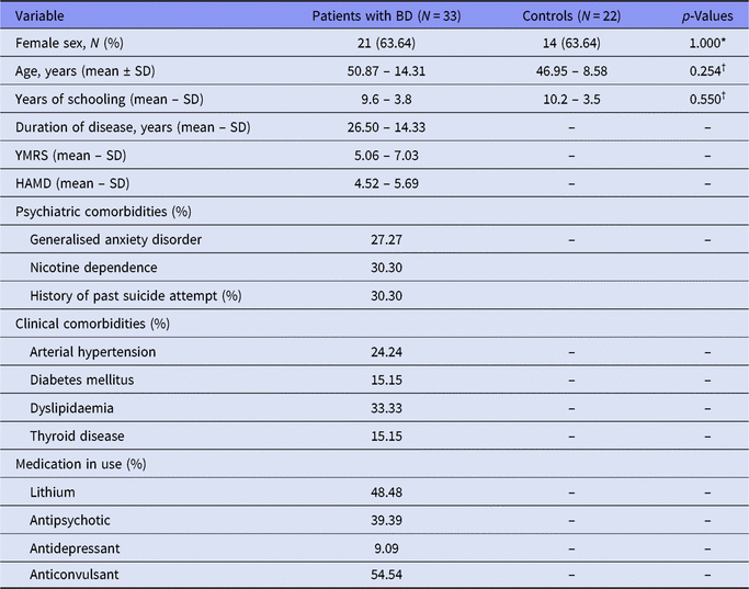
Both groups were similar in terms of age and sex distribution. Table 1 summarises sociodemographic and clinical data of the participants enrolled in this study. Patients with BD had a mean educational level of 9.6 (±3.8) years, and 11 out of the 33 patients (30%) were formally employed at study enrolment. Twenty-one out of the 33 patients were euthymic (64%), while the remaining 12 patients (36%) were experiencing mood episodes (N = 6 in mania/hypomania; N = 6 in depression). The most prevalent comorbid disorders in patients were generalised anxiety disorder (N = 9; 27%) and nicotine dependence (N = 10; 30%). In addition, psychotic symptoms were identified in four patients with BD (12%) at the clinical interview. Regarding patients’ medical history, 28 (85%) had a history of psychosis, 10 (30%) had past suicide attempts, and 6 (18%) had a history of alcohol dependence.
Clinical and demographic features of patients with BD and controls

HAMD, Hamilton Depression Rating Scale; SD, standard deviation; YMRS, Young Mania Rating Scale; BD, bipolar disorder.
* Fisher’s exact test.
† Student’s t-test.
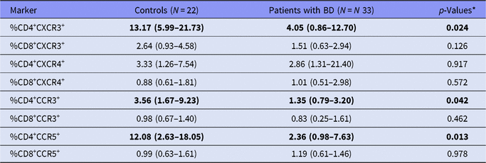
Table 2 presents the results of the immunophenotyping analyses showing the expression of chemokine receptors (CXCR3, CXCR4, CCR3, and CCR5) on different T lymphocytes (CD4+ and CD8+) from patients with BD and controls. The percentage of both CD4+ and CD8+ lymphocytes expressing the chemokine receptor CXCR4 was similar in patients with BD and controls. Likewise, the percentages of CD8+CXCR3+, CD8+CCR3+, and CD8+CCR5+ lymphocytes were similar between patients with BD and controls. Conversely, patients with BD had decreased percentage of CD4+CXCR3+, CD4+CCR3+, and CD4+CCR5+ lymphocytes in comparison with controls. We did not find any correlation between chemokine receptor expression and clinical/demographic parameters.
Percentage of T helper (CD4+) and T cytotoxic (CD8+) lymphocytes expressing chemokine receptors in peripheral blood samples from patients with BD and controls

Percentages are related to total lymphocytes. Values are given as median (25th–75th percentiles). Significant differences are highlighted in bold.
* Mann–Whitney U-test.
Discussion
The main findings of our study were: (i) patients with BD and controls presented similar percentage of CD8+ lymphocytes expressing chemokine receptors CXCR3, CXCR4, CCR3, and CCR5; and (ii) patients with BD presented decreased percentage of CD4+ lymphocytes expressing CXCR3, CCR3, and CCR5.
Growing evidence suggests that a proinflammatory background may contribute to BD neurobiology (Barbosa et al., Reference Barbosa, Machado-Vieira, Soares and Teixeira2014). Increased levels of inflammatory cytokines have consistently been reported in patients with BD (Kunz et al., Reference Kunz, Ceresér, Goi, Fries, Teixeira, Fernandes, Belmonte-de-Abreu, Kauer-Sant'Anna, Kapczinski and Gama2011; Modabbernia et al., Reference Modabbernia, Taslimi, Brietzke and Ashrafi2013). A meta-analysis showed that peripheral blood levels of IL-4, IL-6, IL-10, soluble IL-2 receptor (sIL-2R), sIL-6R, TNF, soluble TNF receptor-1 (sTNFR1), and IL-1 receptor antagonist (IL-1RA) were significantly elevated in patients with BD in comparison with healthy controls. This proinflammatory state seemed to be exacerbated during mood episodes, particularly during mania. Phasic difference was observed for TNF-α, sTNFR1, sIL-2R, IL-6, and IL-1RA (Modabbernia et al., Reference Modabbernia, Taslimi, Brietzke and Ashrafi2013). A more recent meta-analysis assessing peripheral biomarkers for different mood states in BD showed that a combination of high-sensitivity C-reactive protein/IL-6, brain-derived neurotrophic factor/TNF, and sTNFR1 can differentiate specific mood phases (Rowland et al., Reference Rowland, Perry, Upthegrove, Barnes, Chatterjee and Gallacher2018).
Patients with BD have increased frequency of autoimmune diseases in comparison with the general population, and the activation of cell mediated immunity is thought to be part of BD physiopathology (Barbosa et al., Reference Barbosa, Machado-Vieira, Soares and Teixeira2014). We found that patients with BD exhibited changes in the percentage of circulating T helper (CD4+) lymphocytes – but not T cytotoxic (CD8+) lymphocytes – expressing CXCR3, CCR3, and CCR5. These results can be helpful to a better understanding of the immune basis of BD.
The lower number of circulating CD4+ cells expressing CCR3 might reflect a compensatory mechanism for the increased levels of its ligands. Panizzutti et al. (Reference Panizzutti, Gubert, Schuh, Ferrari, Bristot, Fries, Massuda, Walz, Rocha, Berk, Teixeira and Gama2015) reported elevated circulating levels of eotaxin/CCL11, a ligand of the CCR3 receptor, in patients with BD (Panizzutti et al., Reference Panizzutti, Gubert, Schuh, Ferrari, Bristot, Fries, Massuda, Walz, Rocha, Berk, Teixeira and Gama2015). It is worth mentioning that increased eotaxin/CCL11 levels were also found in patients with schizophrenia (Teixeira et al., Reference Teixeira, Reis, Nicolato, Brito-Melo, Correa, Teixeira and Romano-Silva2008). Increased eotaxin/CCL11 levels may be associated with enhanced reactive oxygen species (ROS) production (Tenscher et al., Reference Tenscher, Metzner, Schopf, Norgauer and Czech1996). In addition to activating chemotaxis, both eotaxin/CCL11 and eotaxin-2/CCL24 were capable of inducing the release of ROS by eosinophils (Elsner et al., Reference Elsner, Petering, Kluthe, Kimmig, Smolarski, Ponath and Kapp1998). Another study showed that eotaxin/CCL11 induced microglial production of ROS by upregulating nicotinamide adenine dinucleotide phosphate-oxidase 1 (NOX1), thereby promoting neuronal death (Parajuli et al., Reference Parajuli, Horiuchi, Mizuno, Takeuchi and Suzumura2015). ROS can be used to define oxidative load (Apel & Hirt, Reference Apel and Hirt2004) and oxidative stress, which seem to play a critical role in the biology of BD. Two meta-analyses concluded that thiobarbituric acid reactive substances, nitric oxide, lipid peroxidation, and DNA damage are increased in BD (Andreazza et al., Reference Andreazza, Kauer-Sant’Anna, Frey, Bond, Kapczinski, Young and Yatham2008; Brown et al., Reference Brown, Andreazza and Young2014). Accordingly, the finding of decreased CCR3 activity corroborates previous studies in BD.
We also found that the percentage of CD4+CXCR3+ lymphocytes was decreased in BD in comparison with controls. The interaction between CXCR3 and its ligands plays a role in T helper 1 cell differentiation. The expression of CXCR3 by newly activated CD4+ T cells is associated with their ability to produce IFN-γ and is required for optimal effector cytokine response (Groom et al., Reference Groom, Richmond, Murooka, Sorensen, Sung, Bankert, von Andrian, Moon, Mempel and Luster2012). Here again, we suggest that the decreased percentage of circulating CD4+CXCR3+ lymphocytes in BD might be compensatory to increased circulating levels of its ligands in BD. For instance, circulating levels of CXCL10, one of the most important ligands for CXCR3, were found to be increased in patients with BD in comparison with controls (Brietzke et al., Reference Brietzke, Kauer-Sant’Anna, Teixeira and Kapczinski2009; Barbosa et al., Reference Barbosa, Rocha, Bauer, de Miranda, Huguet, Reis, Zunszain, Horowitz, Pariante and Teixeira2013).
Finally, we found decreased percentage of CD4+CCR5+ lymphocytes in BD patients in comparison with controls. The CCR5 32-bp deletion allele has been described as a susceptibility factor for late-onset schizophrenia (Rasmussen et al., Reference Rasmussen, Timm, Wang, Søeby, Lublin, Fenger, Hemmingsen and Werge2006). Moreover, there are important ligands for CCR5, such as CCL3, CCL4, and CCL5. While the CCL3 gene expression was found to be downregulated in dorsolateral prefrontal cortices of BD patients (Nakatani et al., Reference Nakatani, Hattori, Ohnishi, Dean, Iwayama, Matsumoto, Kato, Osumi, Higuchi, Niwa and Yoshikawa2006), plasma levels of CCL3 were reported to be unchanged in BD (Barbosa et al., Reference Barbosa, Rocha, Bauer, de Miranda, Huguet, Reis, Zunszain, Horowitz, Pariante and Teixeira2013). As we evaluate our findings in the context of available evidence, CCR5 and its ligands should be investigated in more depth in BD.
Our study has several limitations, such as the small number of subjects, cross-sectional design, and lack of information about body mass index. Our relatively small sample size prevented additional analyses, such as assessing whether chemokine receptor expression varies according to mood state. It is worth mentioning that our group has previously shown that chemokine imbalance is related to BD trait and not to mood state (Barbosa et al., Reference Barbosa, Rocha, Bauer, de Miranda, Huguet, Reis, Zunszain, Horowitz, Pariante and Teixeira2013). The fact that we did not correct for multiple comparisons should also be taken into account as a limitation. Also, if more stringent statistical criteria were applied (e.g. Bonferroni correction), the observed differences would have lost significance. The observed differences were relatively small (except for %CD4+CCR5+), and further studies with larger samples are needed to confirm these. As all patients were medicated, the findings might be influenced by patients’ ongoing treatment. By contrast, the strict exclusion criteria and the comprehensive clinical evaluation can be regarded as strengths of the study.
In conclusion, patients with BD have decreased percentage of CD4+CCR3+, CD4+CXCR3+, and CD+4CCR5+ lymphocytes in comparison with controls. Together with previous evidence, our findings reinforce the hypothesis that immune pathways, especially involving CD4+ lymphocytes, are involved in the physiopathology of BD. Future studies should include the evaluation of peripheral expression of chemokine receptors and their ligands to have a deeper understanding of this immune pathway in BD. Longitudinal studies can also provide valuable information about the association between chemokine receptors and illness course and/or mood stage.
Author ORCIDs
Izabela G. Barbosa, 0000-0001-5688-6347.
Acknowledgements
Authors would like to acknowledge the participation of volunteers for their magnificent support. The Neuropsychiatry Program is funded by the Department of Psychiatry and Behavioral Sciences, The University of Texas Health Science Center at Houston. NPR is a Huntington’s Disease Society of America fellowship recipient.
Funding
This work was supported by the Brazilian funding agencies Conselho Nacional de Desenvolvimento Científico e Tecnológico; Coordenação de Aperfeiçoamento de Pessoal de Nível Superior; and Fundação de Amparo à Pesquisa do Estado de Minas Gerais.
Author’s contributions
IGB, MEB, and ALT designed the study, wrote the protocol, and supervised all experiments and analyses. IGB, NPR, and RBH enrolled and evaluated the participants and applied the clinical scales. ELV, FTLG, GEBM, and VAM designed and performed flow cytometry experiments and analysed the data. MAC and NPR performed statistical analyses and wrote the first draft of the manuscript. All authors contributed to and have approved the final manuscript.
Declarations of interest
None.