A patent ductus arteriosus is present in nearly two-thirds of extremely low-birth-weight infants.Reference Chiruvolu, Punjwani and Ramaciotti1 Classically, practitioners have debated whether or not we should be treating these at all,Reference Noori, Friedlich and Seri2 but when treatment is deemed necessary, the debate has shifted to the pros and cons of surgical ligation versus percutaneous device closure. With newer devices and novel approaches, percutaneous closure in infants less than 1000 g is being completed more frequently. The results have been promising, with minimal morbidity and mortality and potentially less short-term post-operative haemodynamic instability.Reference Ogando, Asensio and Sanchez de la blanca3 We report the case of an uncomplicated percutaneous closure of a patent ductus in a 1200-g infant who developed a coarctation of the aorta 4-month post-procedure.
Case report
Our patient was born at 25-week and 3-day gestation with a birth weight of 650 g. At 1 month, an echocardiogram showed a large ductus arteriosus and severe pulmonary overcirculation. In light of these findings and the fact that was unable to wean from the high-frequency oscillator, the decision was made to proceed with percutaneous closure.
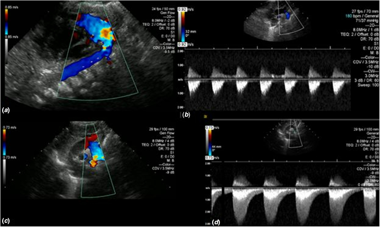
At the time of the procedure, the patient weighed 1200 g. Venous access was obtained with a four French sheath using ultrasound guidance. The duct measured 9.4 mm long and 3.1 mm at the narrowest diameter. A size 5 Medtronic microvascular plug was positioned so that the distal “uncovered” portion of the device was within the lumen of the aorta. Transthoracic echocardiography performed immediately before and after device release showed no residual shunting and no obstruction to flow in the left pulmonary artery or the descending aorta. Follow-up echocardiograms were performed at 1 day, 1 week, and 1 month after closure. They all demonstrated the device in appropriate position without obstruction to flow in the pulmonary artery or aorta (Fig 1a and b). The aortic isthmus measured 3.8 mm (z score 1.4).
Aortic arch with colour immediately post-patent ductus arteriosus device closure showing a wide open arch (a) and a normal Doppler pattern 1-month post-procedure (b). Echo 4-month post-procedure with colour shows aliasing at the site of coarctation (c) and 3-m/s velocity and diastolic antegrade flow continuation (d).
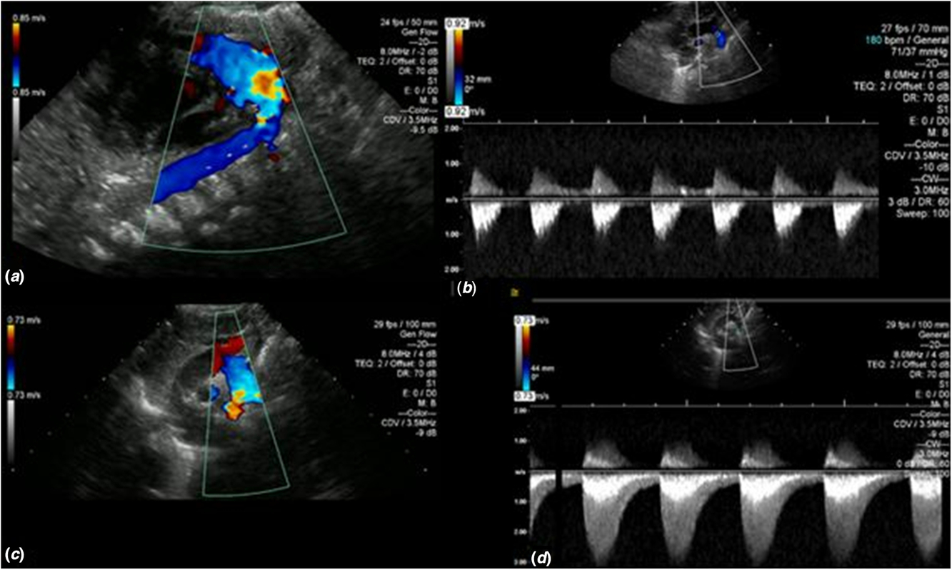
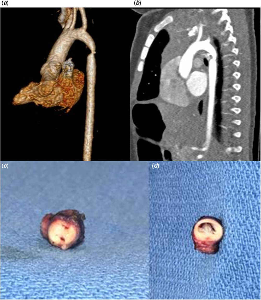
Four-month post-device closure, an echocardiogram was repeated to assess a new murmur. While the device appeared in the appropriate position, there was a moderate narrowing of the descending aorta adjacent to the device with a peak gradient of 35 mmHg (Fig 1c and d). Pulses were described as 1+ in all extremities, and brachiofemoral delay was not documented. He was normotensive; however, the only blood pressures recorded were taken in the left lower extremity. This was thought to represent true native coarctation rather than obstruction from the device given the serial appearance by echocardiography and the mild distal displacement of the left subclavian artery. A CT scan confirmed a moderate-to-severe coarctation in the juxtaductal region with mild displacement of the left subclavian artery; however, contrast did not fill the uncovered portion of the device, which extended into the aorta (Fig 2a and b). The aortic isthmus measured 2 mm (z score −2.99).
3-D reconstruction and CT scan showing moderate-to-severe coarctation (a and b). Path specimen showing the aortic end of the Medtronic microvascular plug has endotheliased and fibrosed to the posterior aspect of the aortic wall and has formed a double-barrel appearance within the aortic lumen (c and d).
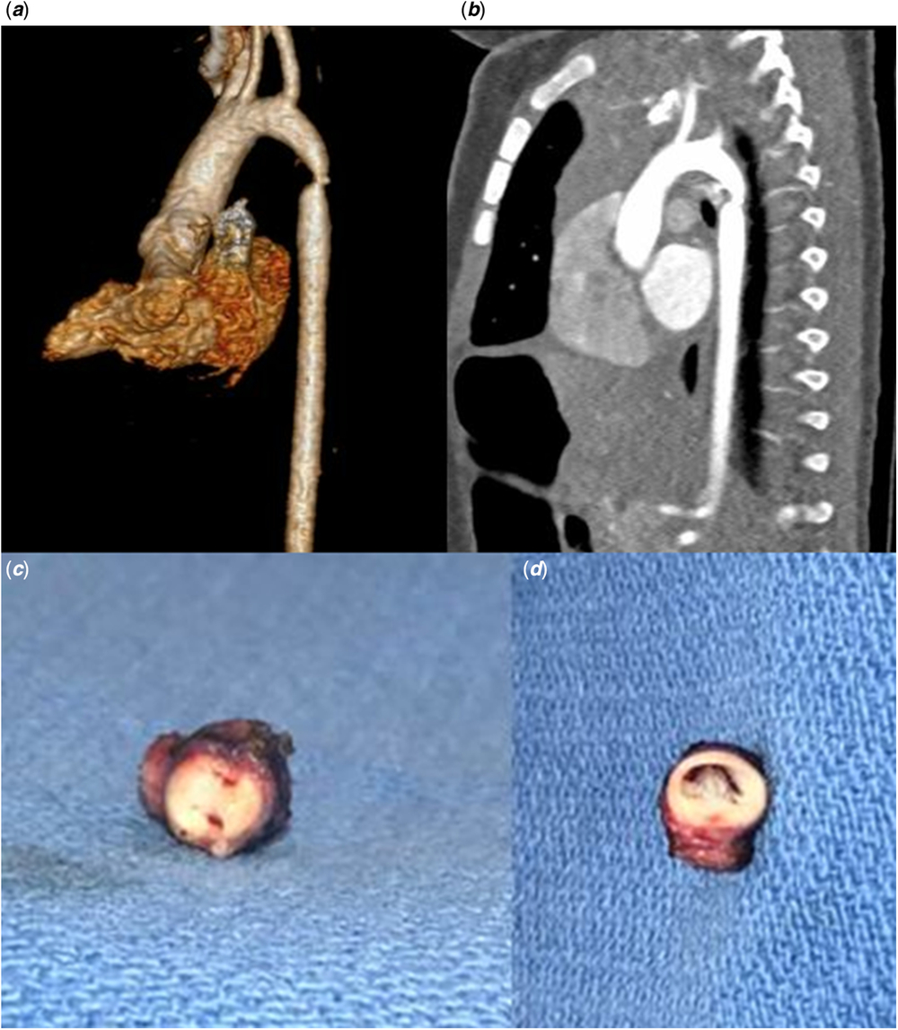
The patient underwent an extended end-to-end repair of the coarctation at 6 months of age. Prior to repair, a proximal right radial arterial pressure was 92/46 compared to a distal lower extremity femoral arterial line pressure of 68/45. The coarctation segment, including half of the device, was resected. Post-operatively, there was no residual gradient from the upper to lower extremities. The aortic wall specimen appeared thick and fibrotic, consistent with a native coarctation. The “uncovered” portion of the device was completely endothelialised. The distal pin of the device appeared to be fibrosed and adherent to the posterior wall of the aorta, creating a double-barrel lumen in the region of the coarctation (Fig 2c and d). Histopathology showed fibrosis consistent with coarctation; however, the pathologist could not determine the likelihood of a reactive fibrosis. Post-operative echo showed a widely patent aortic arch.
Discussion
Over the last several years, centres have reported success using novel devices and techniques for percutaneous closure of a patent ductus arteriosus in infants under 1 kg. The major risks reported have been limited to events that occur during or shortly post-procedure (<24 hours) and include device embolisation, vessel damage, atrial perforation, left pulmonary artery stenosis, and iatrogenic aortic coarctation.Reference Wang-Giuffre and Breinholt4–Reference Zahn, Peck and Phillips7 Despite an angiogram of the aorta during the catheterisation procedure and all subsequent echocardiograms demonstrating a patent aorta up to 1 month following the procedure, our patient developed a significant late coarctation.
This may have been the result of a true native coarctation secondary to late constriction of ductal tissue extending into the aorta, which is supported by the mildly displaced left subclavian artery, the posterior shelf seen on the CT, and gross examination of the aortic specimen. Anecdotally, our surgeons report having seen development of late native coarctation in premature infants after surgical patent ductus arteriosus ligation, so it is reasonable to assume that it can develop after percutaneous device closure in the same population. An alternative explanation is that this was the result of a reactive fibrosis of the aorta triggered by the device. This explanation seems unlikely to completely explain our findings given the frequency with which patent ductus arteriosus devices extend into the aorta and the infrequency with which we see coarctations develop after device closure.
The most surprising finding was the complete endothelialisation of the uncovered distal portion of the device. Previously, it was assumed that blood would flow unobstructed through the uncovered wire at the distal portion of the device and down the descending aorta. In this case, however, the “wire barrel” was completely covered with endothelium, preventing flow through the device. It is unclear if endothelialisation is the natural progression of all Medtronic microvascular plugs or if this was a reactive process triggered by an abnormal aortic wall.
Until now, our experience with over 25 premature infants having undergone percutaneous ductal closure has also been limited to in-procedure morbidity, with only one patient experiencing device embolisation that was successfully retrieved without further complication. Our standard post-procedure protocol has included an echocardiogram at 1-day, 1-week, 1-month, and 1-year post-closure, but this case highlights the importance of closer follow-up for these patients. Our institutional post-procedural protocol now includes an additional echocardiogram 4–6 months after device closure.
Conclusion
Practitioners should be aware of the potential for late presentation of coarctation of the aorta in premature infants who have undergone ductal device closure with the Medtronic microvasular plug and should consider evaluating these patients 4- to 6-month post-procedure.
Acknowledgements
The authors would like to acknowledge Dr John J. Parent for his assistance in editing the manuscript.
Financial Support
This research received no specific grant from any funding agency, commercial, or not-for-profit sectors.
Conflicts of Interest
None.
Ethical Standards
The authors have no relationships or interests that could have direct or potential influence or impart bias on the work to disclose. No research was conducted involving patients and/or animals. The authors assert that all procedures contributing to this work comply with the ethical standards of the relevant national guidelines on human experimentation and with the Helsinki Declaration of 1975, as revised in 2008, and has been approved by the institutional committee.
Contributor’s Statement
Dr R.M.S. drafted the initial manuscript, revised the manuscript, and approved the final manuscript as submitted. Dr M.D.R. reviewed and revised the manuscript and approved the final manuscript as submitted. Dr R.A. reviewed and revised the manuscript and approved the final manuscript as submitted.