Introduction
Beremendiini Reumer, Reference Reumer1984 (Soricidae, Soricinae) is an extinct tribe of soricine shrews. Representatives lived between the early Pliocene and Middle Pleistocene of the Palearctic. Beremendiin shrews have long been regarded as common European fossils (Rzebik-Kowalska, Reference Rzebik-Kowalska, Wójcik and Wolsan1998). New discoveries from Asia (primarily northeastern China) marked the first period of investigations on the diversity of this tribe, although data on the geographic and temporal distribution of Beremendiini were limited and findings for areas outside of Europe were considered to refer to a separate species or even genus (Pidoplitschka, Reference Pidoplitschka1956; Kowalski and Li, Reference Kowalski and Li1963; Gureev, Reference Gureev1979). During the second half of the last century (to 1996), two new Asian genera of beremendiin shrews were identified based on fragmentary remains: Peisorex Kowalski and Li, Reference Kowalski and Li1963 (early Pleistocene in China) and Nectogalinia Gureev, Reference Gureev1979 (late early Pleistocene in Altay); for details, see Kowalski and Li (Reference Kowalski and Li1963), Gureev (Reference Gureev1979), and Flynn and Wu (Reference Flynn and Wu1994). The second period was described by Jin and Kawamura (Reference Jin and Kawamura1996a, Reference Jin and Kawamurab). The authors’ first 1996 publication focused on new materials of Peisorex from the early Pleistocene Haimao locality (northeastern China) based on Peisorex synonymized with Beremendia Kormos, Reference Kormos1934. The second 1996 publication described the new genus Lunanosorex Jin and Kawamura, Reference Jin and Kawamura1996b (late Pliocene in China). Following these two publications, the validity of taxa based on mandibular morphology became questionable (Storch et al., Reference Storch, Qui, Zazhigin, Wójcik and Wolsan1998). Nectogalinia was only identified as a valid genus by McKenna and Bell (Reference McKenna and Bell1997). The most recently identified genus, Dolinasorex Rofes and Cuenca-Bescós, Reference Rofes and Cuenca-Bescós2009 from the early Pleistocene of the Gran Dolina locality in Spain, was examined by Rofes and Cuenca-Bescós (Reference Rofes and Cuenca-Bescós2009). Thus today, three beremendiin genera are generally recognized: Beremendia, Lunanosorex, and Dolinasorex (Rofes and Cuenca-Bescós, Reference Rofes and Cuenca-Bescós2009; Flynn and Wu, Reference Flynn, Wu, Flynn and Wu2017). The validity of Peisorex and Nectogalinia is contested (Storch et al., Reference Storch, Qui, Zazhigin, Wójcik and Wolsan1998; Qiu and Storch, Reference Qiu, Storch, Hoek Ostende, Doukas and Reumer2005).
Over the second half of the last century, beremendiin shrew material from a vast territory of the northern Palearctic has accumulated. Understanding of tribal distribution has improved with paleogeographic data from Jin and Kawamura (Reference Jin and Kawamura1996a), Storch et al. (Reference Storch, Qui, Zazhigin, Wójcik and Wolsan1998), and Jin et al. (Reference Jin, Zhang, Sun and Zheng2009). Storch et al. (Reference Storch, Qui, Zazhigin, Wójcik and Wolsan1998) presented data on the presence of beremendiin shrews in 15 localities in Asia, including Siberia, Kazakhstan, and Mongolia, but they did not provide a morphological description of the identified materials. Thus, the broad territory between European and eastern Asian areas of the tribe can be considered terra incognita for Beremendiini. Kotlia (Reference Kotlia1991) described five isolated teeth of Beremendiini gen. and sp. indet. of the Pliocene Khaigam locality (Kashmir, India). Furió et al. (Reference Furió, Agusti, Mouskhelishvili, Sanisidro and Santos-Cubedo2010) examined the remains of Beremendia fissidens (Petényi, Reference Petényi1864) from the early Pleistocene Dmanisi locality (Georgia). The latter study offers new interpretations of the paleobiology of Beremendia and has raised new questions regarding the functional morphology for hunting and food processing.
The present work describes materials taken from 16 early Pliocene to early Pleistocene localities in Russia (southern Siberia), Kazakhstan, and Mongolia and redescribes the holotype of Nectogalinia altaica Gureev, Reference Gureev1979. Our primary objective is to determine the taxonomic status of these remains relative to European and Chinese materials. We also discuss the paleobiology of Beremendia in terms of its mandible, entoglenoid fossa, and teeth.
Geological setting
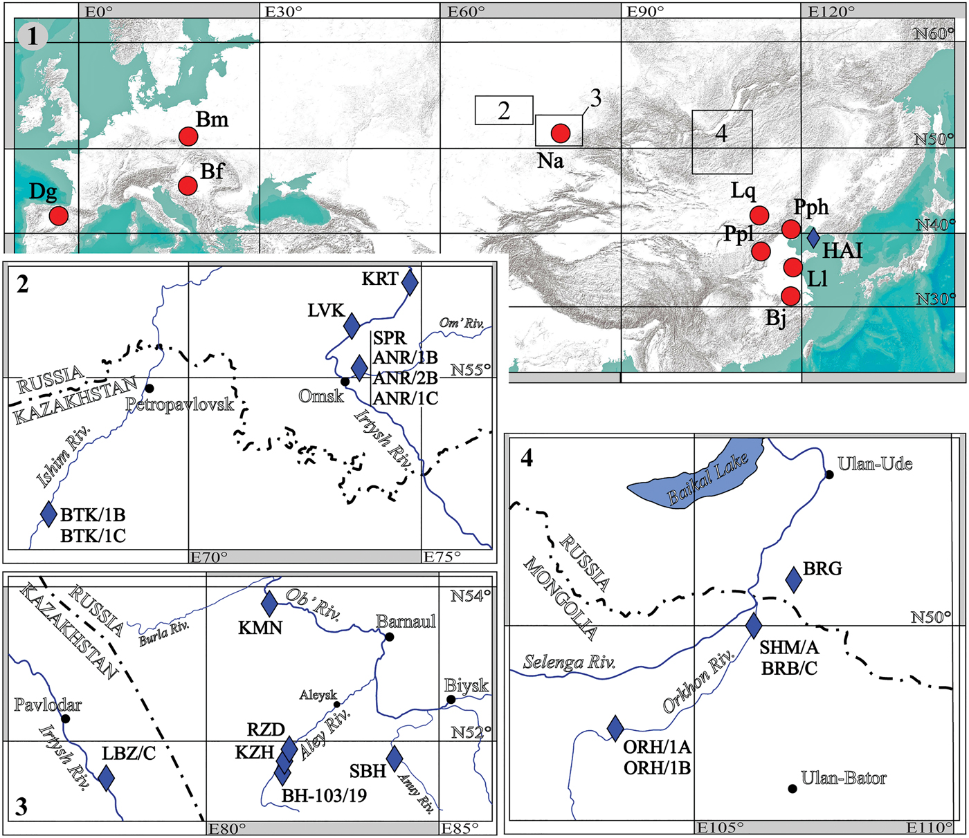
The new fossils of Beremendiini come from 16 early Pliocene to early Pleistocene sites in southwestern Siberia, western Transbaikalia, Altay, northeastern Kazakhstan, and northern Mongolia (Figs. 1, 2; S1): Andreevka 1 (ANR/1B, ANR/1C), Andreevka 2 (ANR/2B), Kamen’-na-Obi (KMN), Kartashovo (KRT), Kizikha (KZH), Livenka (LVK), Razdol'e (RZD), Sibiryachikha (SBH), Speranskoye (SPR), Borehole #103/19 (BH-103/19), Beregovaya (BRG), Bural-Obo (BRB/C), Orkhon 1 (ORH/1A, ORH/1B), Shamar (SHM/A), Biteke 1 (BTK/1B, BTK/1C), and Lebyazhye (LBZ/C). All of these localities were mentioned by Zazhigin and Lopatin (Reference Zazhigin and Lopatin2000, Reference Zazhigin and Lopatin2001), Zazhigin et al. (Reference Zazhigin, Lopatin and Pokatilov2002), and Vasilyan et al. (Reference Vasilyan, Zazhigin and Böhme2017). The lithological and biostratigraphic descriptions of the localities have been published in several articles since the 1960s, including Vangeigeim et al. (Reference Vangeigeim, Beljaeva, Garutt, Dmitrijeva and Zazhigin1966), Devjatkin and Zazhigin (Reference Devjatkin, Zazhigin and Kramarenko1974), Zazhigin (Reference Zazhigin1980, Reference Zazhigin and Logachev1989), Devjatkin et al. (Reference Devjatkin, Zazhigin, Maleeva and Nikiforova1982), Zazhigin and Zykin (Reference Zazhigin, Zykin and Arkhipov1984), Derevyanko and Maloletko (Reference Derevyanko and Maloletko1992), Zykin et al. (Reference Zykin, Zazhigin and Presyazhnjuk1987, Reference Zykin, Zykina and Zazhigin2007), and Zykin (Reference Zykin2012).
Early Pliocene–early Pleistocene localities of Beremendiini: (1) Europe and Asia, with rectangles showing locations of Figures 1.2–1.4; (2) Ishim and Irtysh River basins; (3) Irtysh and Ob’ River basins; (4) Selenga River basin. Red circle = type locality; rhomb = cited locality; ANR/1B = Andreevka 1; ANR/1C = Andreevka 1; ANR/2B = Andreevka 2; Bf = Beremendia fissidens (Petényi, Reference Petényi1864) (type locality Beremend 2); BH-103/19 = Borehole 103/19; Bj = Beremendia jiangnanensis Jin et al., Reference Jin, Zhang, Sun and Zheng2009 (type locality Renzidong); Bm = Beremendia minor Rzebik-Kowalska, Reference Rzebik-Kowalska1976 (type locality Rębielice Królewskie I); BRB/C = Bural-Obo; BRG = Beregovaya; BTK/1B = Biteke 1; BTK/1C = Biteke 1; Dg = Dolinasorex glyphodon Rofes and Cuenca-Bescós, Reference Rofes and Cuenca-Bescós2009 (type locality Gran Dolina TD4-TD6); HAI = Haimao (China; early Pleistocene); KMN = Kamen’-na-Obi; KRT = Kartashovo; KZH = Kizikha; LBZ/C = Lebyazhye; Ll = Lunanosorex lii Jin and Kawamura, Reference Jin and Kawamura1996b (type locality Qipanshan Hill); Lq = Lunanosorex qiui Jin, Sun, and Zhang, Reference Jin, Sun and Zhang2007 (type locality Bilike); LVK = Livenka; Na = Nectogalinia altaica Gureev, Reference Gureev1979 (type locality Razdol'e); ORH/1A = Orkhon 1; ORH/1B = Orkhon 1; Pph = Peisorex pohaiensis Kowalski and Li, Reference Kowalski and Li1963 (type locality Chiachiashan); Ppl = Beremendia [Peisorex] pliocaenica Flynn and Wu, Reference Flynn and Wu1994 (type locality Mazegou); RZD = Razdol'e; SBH = Sibiryachikha Cave; SHM/A = Shamar; SPR = Speranskoye. Also see S1 and S17.

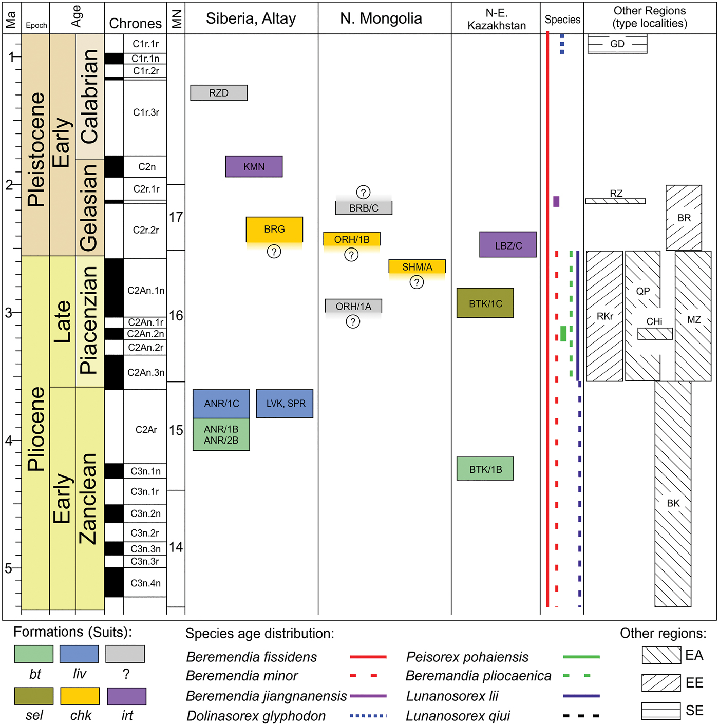
Compiled stratigraphy of early Pliocene–early Pleistocene localities in Siberia, Altay, northern Mongolia, and northeastern Kazakhstan grouped in the formations (bt, chk, irt, liv, sel), age distribution of beremendiin shrews and their type localities (BR, BK, CHi, GD, MZ, QP, RKr, RZ) with related Palearctic regions (EA, EE, SE). BK = Bilike locality (China; early Pliocene; type locality of Lunanosorex qiui Jin, Sun, and Zhang, Reference Jin, Sun and Zhang2007); BR = Beremend 2 locality (Hungary; early Pleistocene; type locality of Beremendia fissidens [Petényi, Reference Petényi1864]); bt = Biteke Formation; CHi = Chiachiashan locality (China; Early Pleistocene; type locality of Peisorex pohaiensis Kowalski and Li, Reference Kowalski and Li1963); chk = Chikoy Formation; EA = eastern Asia; EE = eastern Europe; GD = Grand Dolina locality (Spain; early Pleistocene; type locality of Dolinasorex glyphodon Rofes and Cuenca-Bescós, Reference Rofes and Cuenca-Bescós2009); irt = Irtysh Formation; liv = Livenka Formation; MZ = Mazegou locality (China; early Pliocene; type locality of Peisorex pliocaenicus Flynn and Wu, Reference Flynn and Wu1994); QP = Qipanshan Hill locality (China; late Pliocene; type locality of Lunanosorex lii Jin and Kawamura, Reference Jin and Kawamura1996b); RKr = Rębielice Królewskie I (Poland; late Pliocene; type locality of Beremendia minor Rzebik-Kowalska, Reference Rzebik-Kowalska1976); RZ = Renzidong locality (China; early Pleistocene; type locality of Beremendia jiangnanensis Jin et al., Reference Jin, Zhang, Sun and Zheng2009); SE = southern Europe; sel = Selety Formation. Also see S1, S17, and Figure 1 for locality names.

The ten localities and their sediments with mammal remains are attributed to five regional stratotypic units (Fig. 2): the early Pliocene Kazakh Biteke Formation (Zazhigin and Zykin, Reference Zazhigin, Zykin and Arkhipov1984; Zykin et al., Reference Zykin, Zazhigin and Presyazhnjuk1987, Reference Zykin, Zykina and Zazhigin2007; Zykin, Reference Zykin2012); the early Pliocene southwestern Siberian Livenka Formation (Zazhigin and Zykin, Reference Zazhigin, Zykin and Arkhipov1984; Zykin et al., Reference Zykin, Zykina and Zazhigin2007; Zykin, Reference Zykin2012); the late Pliocene Kazakh Selety Formation (Zazhigin and Zykin, Reference Zazhigin, Zykin and Arkhipov1984; Zykin, Reference Zykin2012); the early Pleistocene Transbaikalian–northern Mongolian Chikoy Formation (Pokatilov, Reference Pokatilov1985); and the early Pleistocene Altay–Kazakh Irtysh Formation (Zykin, Reference Zykin2012). In some localities (KRT, KZH, SBH, and BH-103/19), the fossils were redeposited in the Middle–late Pleistocene sediments. For several localities (RZD, BRB/C, and deposits ORH/1A), the regional stratigraphic context is uncertain.
Materials and methods
Sampling
The material of Beremendiini investigated in this study comprises remains from 16 early Pliocene to early Pleistocene localities presented in the ‘List of localities’ in the Supplemental Material (S1). It includes 56 specimens, primarily consisting of dentary fragments, isolated teeth, and rare skull fragments, including the holotype of Nectogalinia altaica Gureev, Reference Gureev1979 (see Supplemental Materials, S2 and S3).
Terminology
Dental nomenclature follows those of Reumer (Reference Reumer1984), Dannelid (Reference Dannelid, Wójcik and Wolsan1998), and Ziegler et al. (Reference Ziegler, Dahlmann and Storch2007). Cranial and mandibular terms follow Wible (Reference Wible2008). Terminology of the upper and lower unicuspid teeth of shrews remains disputed; for this paper, we use the term ‘antemolar.’ Lower case letters refer to the lower dentition and upper case letters to the upper dentition. In use of the term ‘Lipotyphla,’ we follow Archibald (Reference Archibald2003), Lopatin (Reference Lopatin2006), and Asher and Helgen (Reference Asher and Helgen2010).
Image acquisition and measurements
Pictures of all craniomandibular remains and isolated teeth were taken with a digital camera (Canon EOS 60D) combined with a binocular (LOMO Micromed MSP-1) via an optical adapter (AOT-1C). Features on the images were twice measured with tpsDig ver. 1.40 (Rohlf and Slice, Reference Rohlf and Slice1990) to minimize ‘metering error.’ The statistical analysis was performed on the mean values of these replicates. The measurements were mainly taken according to Reumer (Reference Reumer1984), with some additions (e.g., MRH = mandibular ramus height; MSL = mandibular short length, i.e., the distance between the paraconid of m1 and the lower sigmoid notch of the mandibular ramus; also see Zazhigin and Voyta, Reference Zazhigin and Voyta2018); the measurements are illustrated in the Supplemental Material (S4). All measurements are given in mm. High-resolution images were acquired using an electronic scanning microscope (Zeiss ESEM Quanta 250), with the surfaces covered by platinum sputter coating.
Tooth pigment analysis
To detect the ferriferous pigment of teeth, energy-dispersive X-ray spectroscopy (EDX) was used. EDX represents a method for the elemental analysis or determination of the chemical composition of a sample (for a more detailed description of the technique used for shrew teeth, see Strait and Smith, Reference Strait and Smith2006). The EDX-spectrometer was attached to a Zeiss ESEM Quanta 250 electron microscope. Its technical specifications were: quantification method = normalized stoichiometry; accelerating voltage = 15 kV; acquisition time = 30 s. Quantitative microanalyses were carried out on the surface of the teeth on a ‘pigmented’ and ‘unpigmented’ part of the crown. Results of the elemental analyses of several beremendiin teeth with PPt and VPg are given in the Supplemental Material (S5). The values of the iron content of the Beremendia teeth were compared with those for Sorex isodon Turov, Reference Turov1924 (field no. 201/A86), Crocidura shantungensis Miller, Reference Miller1901 (ZIN 89478), and Anourosorex squamipes Milne-Edwards, Reference Milne-Edwards and Masson1872 (ZIN 38946), as well as published data on Blarina brevicauda (Say, Reference Say and James1823) by Dumont et al. (Reference Dumont, Tütken, Kostka, Duarte and Borodin2014). The results of the EDX analyses represent the averaged (mean) elemental composition (weight %) normalized to 100%, with values of standard deviation (σ, weight %).
Mandibular shape
For analyses of beremendiin mandibular shape and interspecies/intergeneric comparisons, geometric morphometric techniques were used (Rohlf, Reference Rohlf, Marcus, Bello and García-Valdecasas1993, Reference Rohlf1999; Zelditch et al., Reference Zelditch, Swiderski, Sheets and Fink2004). Mandibular shape was described using a configuration of five true landmarks (I and II types sensu Bookstein, Reference Bookstein1991), together with 45 semilandmarks (see S6). Semilandmarks were prepared in tpsDig2 ver. 2.31 (Rohlf, Reference Rohlf2007) with the assistance of the ‘draw background curves’ tool, and the subsequent procedure of an ‘aligning of the curve’ by length (‘resample curve’ tool). Given that most of the hemimandible fossils were damaged, we used bones with preserved coronoid and condylar processes and the portion of the dentary from the anterior margin of the m2 anterior alveolus to the back. The appropriate baselines enabled us to combine the landmark and semilandmark digitizing process (see details in S6) for different species of Beremendiini, Blarinini, and Soricini. The material investigated in the shape analysis study comprised 54 specimens of 16 species (see S7). The special link- and slider-files were prepared in tpsUtil ver. 1.28 (Rohlf, Reference Rohlf2004). The Procrustes superimposition procedure and principal component analyses (PCA; ‘relative warps analyses’) were calculated in tpsRelw ver. 1.35 (Rohlf, Reference Rohlf2003). Following the determination of similarities and differences in mandibular shape among the beremendiin shrews, we attempted to reconstruct the biting/chewing process based on the general outline of the mandible, the shape of the condylar articular surfaces, the shape of the coronoid process, and geometric and ‘volumetric’ characteristics of the entoglenoid fossa. To determine analogies in the trophic specialization of the ‘maximum power of mandibular adduction’ (MPMA), we also used the MPMA-index (calculated as [HC/MSL]*1). This index was calculated for Anourosorex squamipes (ZIN 38946, CMNH 74969), Cryptotis parvus (Say, Reference Say and James1823) (ZIN 71059), Blarina brevicauda (ZIN 65805), Beremendia fissidens (ZIN 104910/1243, 104893/01–02, 104895/hu-01, 104896), and Beremendia minor Rzebik-Kowalska, Reference Rzebik-Kowalska1976 (ZIN 104909).
Statistical analysis
Calculations were performed in PAST ver. 2.04 (Hammer et al., Reference Hammer, Harper and Ryan2001).
Repositories and institutional abbreviations
Specimens and images examined in this study are deposited in the following institutions: Carnegie Museum of Natural History (CMNH), Pittsburgh, Pennsylvania, USA; Geological Institute, Russian Academy of Sciences (GIN), Moscow, Russia; Hungarian Natural History Museum (HNHM), Budapest, Hungary; Institute of Systematics and Evolution of Animals, Polish Academy of Science (ZZSiD), Krakow, Poland; Institute of Vertebrate Paleontology and Paleoanthropology, Chinese Academy of Sciences (IVPP), Beijing, China; and Zoological Institute, Russian Academy of Sciences (ZIN), Saint Petersburg, Russia.
Systematic paleontology
Class Mammalia Linnaeus, Reference Linnaeus1758
Order Lipotyphla Haeckel, Reference Haeckel1866
Family Soricidae Fischer, Reference Fischer1817
Subfamily Soricinae Fischer, Reference Fischer1817
Tribe Beremendiini Reumer, Reference Reumer1984
Genus Beremendia Kormos, Reference Kormos1934
- Reference Kormos1934
Beremendia Kormos, p. 299.
- Reference Gureev1979
Nectogalinia Gureev, p. 458.
Type species
Crossopus fissidens Petényi, Reference Petényi1864.
Other species
Beremendia minor Rzebik-Kowalska, Reference Rzebik-Kowalska1976; Beremendia pliocaenica Flynn and Wu, Reference Flynn and Wu1994; Beremendia jiangnanensis Jin et al., Reference Jin, Zhang, Sun and Zheng2009.
Diagnosis
Blarinini-like condyle with a broad interarticular area and anteriorly placed lower facet; heavily pigmented teeth; upper incisor strongly fissident; upper antemolar row with four teeth (A1–A4); M1 and M2 with a developed protoconule and metaloph; lower incisor acuspulate; entocristid of the lower molars are presented.
Occurrence
Early Pliocene to Middle Pleistocene in Europe and early Pliocene to early Pleistocene in Asia.
Comparisons
Beremendia differs from Lunanosorex in: (1) the straight cutting edge of the first lower incisor (Lunanosorex displays one or two denticulations on this edge; Qiu and Storch, Reference Qiu and Storch2000, pl. 3; Jin et al., Reference Jin, Sun and Zhang2007, figs. 2, 3); (2) the shape of the articular surface of the condylar process (Lunanosorex has a longer upper facet; Jin et al., Reference Jin, Sun and Zhang2007, fig. 2); and (3) the longer and lower positioning of the lateral groove of the first upper incisor (Lunanosorex has a groove length of ~1/3 of the root length, and the distance between its lower edge and the root lower edge corresponds to the groove height; Qiu and Storch, Reference Qiu and Storch2000, pl. 3).
Beremendia differs from Peisorex in the number of upper antemolars (Jin and Kawamura, Reference Jin and Kawamura1996a) and in the presence of a metaloph of M1 and M2. However, conditions of the upper teeth of Peisorex pohaiensis Kowalski and Li, Reference Kowalski and Li1963 are only known from one partial skull obtained from the Haimao locality (Jin and Kawamura, Reference Jin and Kawamura1996a), and for the most part specimens are unavailable. Beremendia and Peisorex do not differ in mandibular characteristics due to the presence of overlapping features.
Beremendia differs from Dolinasorex in: (1) the more posterior positioning of the zygomatic process of maxilla (Dolinasorex exhibits an anterior shift in this process to the level of the M2 metastyle; Rofes and Cuenca-Bescós, Reference Rofes and Cuenca-Bescós2009, fig. 3), (2) its uncompressed upper antemolar row (the P4 parastyle in Dolinasorex comes into contact with the posterobuccal corner of A2); and (3) its moderately developed talonid of m3 (the talonid in Dolinasorex is short).
Teeth and dentary fragments: Beremendia fissidens (Petényi, Reference Petényi1864): (1–3) first upper left incisor from SHM/A (ZIN 104908/1236): (1) buccal view; (2) occlusal view (cut image); (3) lingual view; (4) first upper right incisor from Korotoyak 2 (ZIN 104903/1292); (5, 6) first lower left incisor from KMN (ZIN 104923/1264): (5) medial view; (6) lateral view (cut image); (7) first lower right incisor from ANR/1C (ZIN 104919/1257, i1), medial view; (9) right dentary fragment (cut image) with i1, p4 from SHM/A (ZIN 104908/1222), medial view. Beremendia minor Rzebik-Kowalska, Reference Rzebik-Kowalska1976: (8) right dentary fragment (cut image) with i1–p4 from BRB/C (ZIN 104894), medial view. a1 = first lower antemolar; AAF = area of additional features; p4 = fourth lower premolar; star indicates reversed image. Scale bars = 1 mm.

Remarks
Some authors follow Jin and Kawamura (Reference Jin and Kawamura1996a) in accepting a broad concept for the genus including Chinese forms (e.g., Jin et al., Reference Jin, Zhang, Sun and Zheng2009; Rofes and Cuenca-Bescós, Reference Rofes and Cuenca-Bescós2009; Flynn and Wu, Reference Flynn, Wu, Flynn and Wu2017), which were previously referred to the separate genus Peisorex. Most known fossil material of beremendiins is represented by the presence of the mandible and isolated teeth, which does not permit the determination of the genus. However, we conservatively follow Qiu and Storch (Reference Qiu, Storch, Hoek Ostende, Doukas and Reumer2005) in considering Beremendia and Peisorex as separate valid genera. The validity of the latter is determined at least by the original dental formula (Jin and Kawamura, Reference Jin and Kawamura1996a).
Accordingly, remains obtained from the Chiachiashan (= Jiajiashan) type locality and Haimao Chinese early Pleistocene (Jin and Kawamura, Reference Jin and Kawamura1996a) locality are identified as those of Peisorex pohaiensis. The generic position of the Chinese form ‘pliocaenicus’ (Flynn and Wu, Reference Flynn and Wu1994) remains problematic because its skull structure remains unknown. Before the emergence of new fossil data, this taxon was considered a part of Beremendia (Flynn and Wu, Reference Flynn, Wu, Flynn and Wu2017). The description of the enigmatic ‘Nectogalinia altaica’ is based on a poor description of damaged hemimandible fragments (Gureev, Reference Gureev1979), and Nectogalinia should be placed with Beremendia until new data become available. Thus, Beremendia represents a ‘wastebasket’ taxon for any Beremendia-like remains of the mandible (without the first incisor) or of isolated teeth.
According to Reumer (Reference Reumer1984), both Beremendia fissidens and Beremendia minor are identical in morphology and differ only in size with the latter being considerably smaller. Other species of Beremendia sensu lato taken from Chinese sites (e.g., Peisorex pohaiensis) are of a similar size to Beremendia fissidens or are markedly larger than European species (e.g., Beremendia pliocaenica and Beremendia jiangnanensis; see S8–S12). Thus, fossil remains from any Asian locality generally represented by hemimandible fragments and isolated teeth without clear diagnostic features can be classified morphometrically into one of three size groups: small shrews likely corresponding to Beremendia minor; medium-sized shrews likely corresponding to Beremendia fissidens (not including P. pohaiensis, see ‘Comparisons’ above); and large shrews likely corresponding to Beremendia pliocaenica and Beremendia jiangnanensis. Botka and Mészáros (Reference Botka and Mészáros2014) have described features of the m3 talonid to distinguish Beremendia fissidens and Beremendia minor from the early Pleistocene Somssich Hill 2 locality in Hungary. These differences were not observed in materials from Siberia, Kazakhstan, and Mongolia (detailed comparisons given below). Therefore, original materials used in the present work were metrically separated into small and medium-sized shrews (S13–S14).
Beremendia fissidens (Petényi, Reference Petényi1864)
Figures 3.1–3.7, 3.9, 4, 5.1–5.6, 6.1–6.6, 6.8, 6.11, 7.1–7.4, 12.1, 12.3
- 1896
Crossopus fissidens Petényi, p. 60.
- Reference Gureev1979
Nectogalinia altaica; Gureev, p. 458.
- Reference Storch, Qui, Zazhigin, Wójcik and Wolsan1998
Beremendia minor; Storch et al., p. 97.
Type material
Syntypes, V 61.1585 (HNHM), one skull fragment, two left maxilla fragments, two right maxilla fragments, 13 left hemimandibles, 15 right hemimandibles, one condylar process, eight isolated first lower incisors; Beremend 2, Villány Hills, Hungary; late Pliocene, MN 16 (Botka and Mészáros, Reference Botka and Mészáros2014).
Diagnosis
Mandibular condyle with a broad interarticular area and anteriorly placed lower facet; heavily pigmented teeth; heavily fissident upper incisor; upper antemolar row with four teeth (A1–A4); M1 and M2 with developed protoconule and metaloph; lower incisor acuspulate; entocristid of lower molars presented.
Description
Most remains from Siberian, Kazakh, and Mongolian localities belong to medium-sized shrews (S13–S14). Rare rostral parts of skulls from SHM/A (ZIN 104908/1227, /1228) and BRB/C (ZIN 104909) exhibit the dental formula 1.4.1.3/1.1.1.3, corresponding to Beremendia sensu stricto (to Beremendia fissidens in this case).
Dentition
The teeth are heavily pigmented. The first upper incisor includes a large crown and a notably smaller and moderately curved root. The crown apex is distinctly fissident, and both tips are pigmented. The buccal cingulum is well developed along the margins of the talon with triangular thickening occurring at the talon base; crown margins are positioned above the talon base level without cingulum. The talon is complex: the buccal part forms a hatchet-like vertical cutting ridge, and a distinct lingual part includes a moderately developed medial cusp that extends along the lingual side of the talon as a developed, vertical, cingulum-like, short torus (‘lingual torus’). The pigmented talon cutting ridge is positioned on a relatively unworn tooth from SHM/A (ZIN 104908/1228). The root is of equal thickness along its full length. The lateral side of the root includes a deep triangular groove (‘lateral groove’) that extends along half of the root (unlike in Lunanosorex); the groove is positioned close to the lower edge of the root (Fig. 3.1–3.4).
Only four fragments are known of the premaxilla-maxilla with unicuspid antemolar teeth A1–A4 or their alveoli (see S3). The relative sizes of the antemolars are A1 > A2 > A3>>A4. The first and second upper antemolars (A1, A2) are elongated in occlusal view with a marked posterobuccal shifting of the crown relative to their longitudinal axes. The third upper antemolar is half the size of A2 and more rounded with the same posterobuccal shifting of the crown. A1–A3 with well-developed medial longitudinal ridges from anterior to posterior margins passing through the main cusp and dividing the crown into smaller lingual and larger buccal halves (S15, fig. 1). Together with the incisor talon vertical ridge, medial ridges of A1–A3 form a common longitudinal cutting crest within the anterior part of the upper tooth row (‘complex cutting ridge’). The main cusps of the antemolars are pigmented. The fourth upper antemolar is vestigial, rounded in shape without distinct crown structures, and visibly pigmented. The first three antemolars display well-developed ectocingula and entocingula.
The fourth upper premolar (P4) is subtrapezoidal with a large, well-developed, pigmented, and anteriorly shifted parastyle connected with the paracone base at a well-distinguished parastylar crest. A visible preprotoconal crest extends along the shallow, concave anterior edge of the tooth crown positioned between the parastyle and protocone; together with the postprotoconal crest, it clearly draws contours of the protoconal part of the trigone basin. The tip of the protocone is pigmented. The hypocone condition reveals two morphotypes. Morphotype I includes no hypocone (Fig. 4.1–4.2); the ridge of the hypoconal flange begins at the elevated part with a gradual decline in height. Morphotype II includes a bulged hypocone on the hypoconal flange edge (Fig. 4.4–4.6). The posterior emargination of P4 is well developed. The parastyle is half the height of the paracone in lateral view; the upper outline in lateral view is slightly undulating with visible signs of mesial inflection; the ectocingulum is well developed and broad along the parastyle-paracone bases and becomes narrower with length before reaching the end of the upper edge of the tooth crown (Fig. 4.3).
Skull fragments, Beremendia fissidens (Petényi, Reference Petényi1864): (1–3) right maxilla fragment with P4 from SHM/A (ZIN 104908/1230); (1) occlusal view (cut image); (2) explanatory drawing; (3) P4 (cut image), lateral view; (4, 5) left maxilla fragment with P4–M1 from SHM/A (ZIN 104908/1229): (4) occlusal view; (5) explanatory drawing; (6) fragment of lingual half of P4 (cut image) of specimen ZIN 104908/1229, occlusal view. M1 = first upper molar. Scale bars = 1 mm.

The first and second upper molars (M1, M2) differ in size from front to back; both teeth are subquadrate in outline, but M1 includes a trigon that is narrower than its talon (means: AW(M1) = 2.47 mm vs. PW(M1) = 2.47 mm; see S13), whereas M2 includes a wider trigon (AW(M2) = 2.72 mm vs. PW(M2) = 2.30 mm; see S13). The parastyle of M1 is conical; the paracone is 2/3 of the metacone height; the mesostyle is not bifid and displays a rounded, typically unpigmented tip; the metastyle represents the end of the postmetaconal crest without any bulges or cusplets. Sharp, long, well-pigmented crests connect all cones and styles of the buccal side. The buccal outline of the tooth in occlusal view is slightly concave at the mesostyle level. The protocone is long and broad at its base; the preprotoconal crest forms a well-distinguished protoconule, forming a bulge within the crest. The protoconule bulge and crestal bend form a visible extension of the anterior tooth outline before reaching the protocone level. Simultaneously, the end of the postprotoconal crest is connected to a developed metaloph forming a straight ridge (Fig. 5.3, 5.4). All crests pass through the protoconule, protocone, and metaloph, clearly separating the deep protoconal basin. The hypocone is a weakly distinguished cusp and is conical in the basal extension of the hypoconal flange. The lingual outline of the tooth is slightly stepped due to a shallow notch positioned in front of the hypoconal flange (Morphotype I; see Fig. 5.1, 5.2); it also includes a developed notch in front of the hypoconal flange (Morphotype II; see Fig. 5.5, 5.6). The posterior emargination of M1 is well developed. Cingula are absent, aside from the short and weak entocingulum under the protocone (Fig. 5.1, 5.2).
Upper molars: Beremendia fissidens (Petényi, Reference Petényi1864): (1, 2) first upper right molar from BH-103/19 (ZIN 104924/1271): (1) occlusal view, white arrow marks shallow notch of M1 hypoconal flange outline; (2) explanatory drawing; (3, 4) fragment of lingual part of M1 of specimen ZIN 104924/1271: (3) occlusal view (cut image); (4) explanatory drawing; (5, 6) fragment of lingual half of left M1 from SHM/A (ZIN 104908/1229): (5) occlusal view (cut image), white arrow marks developed notch of M1 hypoconal flange outline; (6) explanatory drawing. Beremendia cf. B. fissidens (Petényi, Reference Petényi1864): (7) first upper right molar from ORH/1B (ZIN 104911), occlusal view; (8, 9) fragment of lingual part of M1 of specimen ZIN 104911: (8) occlusal view (cut image); (9) explanatory drawing. Star indicates reversed image. Scale bars = 1 mm.

In general, the second upper molar is similar to the first but differs in: (1) trigon and talon width relations (AW vs. PW; see above and S13); (2) its relatively bulged metastyles of M2; and (3) its more developed, concave buccal outline M2 in occlusal view. The structure of the third upper molar (M3) remains unknown.
The first lower incisor is straight without denticles on the cutting edge and with a tapered tip bent upward (Fig. 3.6); the tip is heavily pigmented. Longitudinal axes of the incisor crown and root are continuous; the root length (from the lower outline) is ~1/3 of the crown length. The medial side of the incisor root includes a long (~1/2 of the root length), deep groove. The crown medial groove is long and well developed. The posteromedial area of the crown (‘Area of Additional Features’ or AAF) presents two different conditions: Morphotype I with smooth AAF without any folds and grooves (Fig. 3.5, 3.7) and Morphotype II with a groove (Fig. 3.9). The ectocingulid is very short and distinguishable only from the upper part of the crown base from the anterior part of a1 to the level of p4.
The first lower antemolar (a1) is relatively small and moderately procumbent; its crown overlaps with the incisor; the main (protoconid) cusp is heavily anteriorly shifted; the buccal arm of the posterocristid extends posterolaterally from the protoconid along with a surface ending with a cuspule adjacent to the fourth premolar. The posterolingual basin is shallow and delineated. The ectocingulid is well developed, narrow at the base of the protoconid, and gradually broadens toward the posterolateral end of the crown; entocingulids on the two investigated teeth of SHM/A (ZIN 104908/1221, /1223) are rounded, and their shapes and degrees of development remain unknown. The size and shape of the root are also unknown. The main cusp and part of the posterocristid are pigmented.
The fourth lower premolar (p4) is soricine-like in shape with a strong anterior shifting of the high protoconid and a well-developed, short, high posterocristid buccal arm ending at the developed additional cusp (‘cusp’); the posterocristid is slightly buccally shifted from the longitudinal axis of the tooth; an additional cusp is situated farther away before reaching the posterobuccal corner of the tooth (Fig. 6.1, 6.2). The posterolingual basin is shallow and slightly inclined toward the lingual side. In general, the crown is asymmetrical in shape with a shorter lingual half and with a longer, strongly exaenodont buccal half of the tooth. The size and shape of the roots are also unknown. The ectocingulid and entocingulid are well developed. The main cusp and buccal side of the posterocristid are heavily pigmented.
Dentary fragments: Beremendia fissidens (Petényi, Reference Petényi1864): (1, 2) fragment of the right dentary from SHM/A (ZIN 104908/1222): (1) medial view (cut image); (2) explanatory drawing; (3) fragment (cut image) of the right dentary from SBH (ZIN 104893/02), occlusal view; (5) fragment (cut image) of the right dentary from SBH (ZIN 104893/01), lateral view; (6) third lower right molar (cut image) from SBH (ZIN 104893/02), occlusal view; (8, 11) third lower right molar from SBH (ZIN 104893/02): (8) lateral view; (11) explanatory drawing of lingual side of the talonid, white arrow marks gap of entocristide. (4) Nectogalinia altaica Gureev, 1979, holotype from RZD (GIN 664/201), diagrammatic image of m1 and m2 of occlusal view. (7, 9, 10) Beremendia minor Rzebik-Kowalska, Reference Rzebik-Kowalska1976, third lower right molar from BRB/C (ZIN 104894): (7) occlusal view (cut image); (9) explanatory drawing of lingual side of the talonid; (10) lingual view (cut image). a = line indicating longitudinal axis of tooth crown; a1 = first lower antemolar; b = left-turned arrow indicating anterior tilt of protocristid of m1 relative to a; c = right-turned arrow indicating weak tilt of protocristid of m2 relative to a; d = height of m3 entocristid; e = height of entocingulid of m3 relative to d; i1 = first lower incisor; p4 = fourth lower premolar; star indicates reversed image. Scale bars = 1 mm.

The lower molars are graded in size: the first molar is the largest, the second is slightly smaller, and the third is even smaller (Fig. 6.3). Teeth m1 and m2 differ in their absolute dimensions (means: L(m1) = 2.58 mm vs. L(m2) = 2.26 mm) and include trigonids of comparable lengths: the trigonid of m1 is ~70–71% the total length of the tooth, whereas that of m2 is ~68–69% the total length. The trigonid of m1 is narrower than that of m2 (S13). The paraconid of m1 is anteriorly shifted, and the protoconid is large and anteriorly shifted; hence, the protocristid is slightly inclined relative to the longitudinal axis of the tooth crown. The metaconid forms a small cusp with its tip extending higher than the paraconid tip. The hypoconid forms a large cusp. The well-defined hypolophid ends without a bulge or cusplet and with a distinct but narrow gap between its base and that of the entoconid (Fig. 6.3). The entoconid is a well-developed conical cusp that reaches the base of the metaconid; its height from the upper margins of the entocingulid is twice that (or slightly more) of the entocingulid. The precingulid, ectocingulid, posterocingulid, and entocingulid are well developed as narrow, clearly delineated belts; the lower tooth outline undulates with visible inflections at the protoconid level on both the buccal and lingual sides. The main conids, cristids, and trigonid basin are pigmented.
In general, the morphology of the second lower molar is similar to that of the first with two main differences: (1) having relatively comparable m2 trigonid (TRW) and talonid (TAW) widths (means: TRW = 1.30 mm vs. TAW = 1.31 mm; see S13) and (2) the nearly perpendicular positioning of the protocristid of m2 relative to the longitudinal axis of the crown (Fig. 6.4).
The lower third molar (m3) includes a developed trigonid and a smaller talonid; the talonid is narrow with a shallow, rounded basin; the borders of the basin are formed by a continuously ‘oblique cristid–hypolophid–entocristid’ without a distinct hypoconid or entoconid; the entocristid does not reach the metaconid base (Fig. 6.6), and the talonid basin is opened by a narrow gap (Fig. 6.11). Only the precingulid and ectocingulid are well defined (Fig. 6.8); the others are weak and smooth. The buccal sides of the trigonid cusps and cristids and the buccal side of the talonid are pigmented.
Skull fragments
Fragments of a rostral part of the skull were collected from two Mongolian localities: SHM/A (N = 4) and BRB/C (N = 1) (see S3). The maxillary fragments with preserved infraorbital canal areas include a canal with wide lateral walls; the anterior margins of the wall are rounded and situated above the P4-M1 junction (S15, Fig. 1); the posterior margin is wider than the anterior margin and is also rounded and situated above the M2 metacone cusp (or is shifted slightly posteriorly) (see S15, Fig. 1). The lower part of the wall within the upper molar roots is strongly sculptured, including convexities and concavities within the bone above and between the roots; in the inferoposterior corner of the wall there is a well-distinguished undulating row of attached muscle (Blarina-like condition; likely M. levator nasi inferior; Nikolskaya, Reference Nikolskaya1965). Its lacrimal foramen is positioned at the M1 mesostyle and lies at the anterior third of the wall of the infraorbital canal. The relatively small zygomatic process of the maxilla of SHM/A (ZIN 104908/1234) extends laterally; its tip is an acute, obliquely oriented, short crest without any bulges situated behind the M2-M3 junction; the process is half the length of the postmetacrista of M2. The preserved fragment of nasal and frontal bones of BRB/C (ZIN 104909/1241) includes a relatively large, paired frontal foramina (Sorex-like condition); the foramina are located immediately behind the border of the cribriform plate.
Mandible
The mandibular body is robust and wide (high); its ventral margin includes a weak ledge situated within the center of the m1 crown; due to the presence of this ledge the dentary includes a gradually tapered contour from the anterior end to the posterior end. Moreover, the ventral margin includes two digastric muscle attachments with the first taking the form of a small, smooth spicule (slightly bulged facet) positioned under the end of m3 and shifted buccally from the vertical plane; the second includes a weak longitudinal facet positioned between the end of the symphysis (level of m1 talonid) and the spicule (Fig. 7.1). The mental foramen is positioned within a slightly tilted groove under the hypoconid of m1 (from the anterior end to the hypoconid). The mandibular ramus is moderately long and large; its anterior margin is tilted posteriorly and bends in the upper part so that the tip of the coronoid process is heavily anteriorly tilted. The coronoid process includes a well-developed coronoid spicule and two well-delineated external fossae for the temporal muscle (‘external fossa’) positioned anterior and posterior to the spicule (Fig. 7.3, 7.4); the internal fossa of the temporal muscle (‘internal fossa’) is deep and clearly delineated. The base of the condylar process in medial view includes a wide and shallow subtriangular fossa with a moderately distinguished pterygoid spicule. The condylar process is distinct; the posterior subtriangular surface includes a broad interarticular area; the upper articular facet is short and tilted; the lower facet is longer than the upper facet and lacks a free lateral end due to the positioning of the dorsal margin of the angular process (i.e., the facet rests against the base of the angular process; see Fig. 7.2). The angular process is short and robust with an obtusely pointed, medially bent tip. The mandibular foramen is large, connects to the internal fossa, and is positioned at the toothrow baseline.
Dentary fragments: Beremendia fissidens (Petényi, Reference Petényi1864): (1) diagrammatic image of the right mandible dentary fragment from SHM/A (ZIN 104908/1222), lateral view; (2) left condylar process (cut image) from KMN (ZIN 104923/1263), articular view; (3, 4) right mandibular ramus of specimen from SBH (ZIN 104893/01): (3) lateral view (cut image); (4) explanatory drawing. Beremendia minor Rzebik-Kowalska, Reference Rzebik-Kowalska1976: (5, 6) right mandibular ramus of specimen from BRB/C (ZIN 104894): (5) lateral view (cut image); (6) explanatory drawing; (7) diagrammatic image of the right (reversed image) condylar process from BRB/C (ZIN 104894), articular view; (8) diagrammatic image of the right mandible dentary fragment from BRB/C (ZIN 104894), lateral view; (9) anterior part (cut image) of the right dentary from BRB/C (ZIN 104894), lateral view; (10, 11) lower teeth a1–m1 of the right dentary from BRB/C (ZIN 104894): (10) occlusal view (cut image); (11) explanatory drawing. a = area of a digastric muscle attachment (‘weak longitudinal facet’); a1 = first lower antemolar; b = anterior portion of the lateral temporal fossa; c = a posterior portion of the lateral temporal fossa; i1 = first lower incisor; m1, m2, m3 = first, second, and third lower molars; p4 = fourth lower premolar. Scale bars = 1 mm.

Nectogalinia altaica
The holotype from RZD (GIN 664/201) includes a fragment of the right hemimandible with m1 and m2. The teeth include wide and well-distinguished cingulids and include visible PPt (see S5, fig. 4, table 4). The third lower molar is missing, but the anterior root is present in its alveolus. The lateral wall of the first lower incisor is heavily damaged; the alveoli of a1 and p4 and the posterior alveolus of m3 are filled with solid sand. The symphysial area of the dentary is damaged; the spicule within the ventral margins is markedly expressed; the facet of the digastric muscle is well distinguished along the lower side of the dentary and is shifted posteriorly on the lateral side. Complete coronoid and condylar processes represent the mandibular ramus, but the angular process has broken off (see S15, fig. 2).
Variation
The studied remains of Beremendia fissidens show variations in tooth pigmentation, characteristics, and dimensions.
Tooth pigmentation
Teeth collected from Mongolian (SHM/A, BRB/C, ORH/1A, ORH/1B) and Siberian (BRG) localities are markedly pigmented (VPg). The tips of the upper and lower incisors, the anterior part (cutting edge) of the upper incisor talon, the tips of the upper and lower antemolars, the main cusps and structures of upper molariform teeth, and the main conids and cutting edges of cristids of the lower molars bear rich dark and reddish-brown pigments. The left P4 and M1 of a specimen collected from SMA/A (ZIN 104908/1229) display an extremely colorful parastyle with a parastylar crest and protocone. The trigon of M1 running from the parastyle, protocone, and trigon basin to the anterior bulge of the hypoconal flange crest is colored with dark reddish-brown pigment. The other teeth collected from SMA/A (ZIN 104908/1233), BRB/C (ZIN 104909/1241), and ORH/1B (ZIN 104911) are only pigmented at the tips of cusps without (or with limited) pigmentation within the trigon basin.
Other teeth display relatively unicolored cusps with a more or less clearly expressed, weak PPt. This pattern is attributed to the chemical leaching of the actual pigment during fossilization, although traces of the pigmented and unpigmented enamel border are retained, which could have occurred due to the replacement of ferruginous pigments with other substances. This assumption was partially confirmed through the use of energy-dispersive X-ray spectroscopy (EDX). EDX analyses of several fossil teeth of Beremendia relative to some Recent taxa (see S5, S7) reveal the influence of chemical leaching caused by pigmentation (and more precisely by the ferruginous part of the pigment; see S5). The analyses show an absence of iron in different parts of the tooth crowns with PPt for specimens collected from KRK/2 (ZIN 104903/1292), RZD (holotype of Nectogalinia altaica, GIN 664/201), and BH-103/19 (ZIN 104925/1271) (see S5, figs. 2, 4, 5, tables 2, 4, 5). The left upper first incisor collected from SHM/A (see S5, fig. 3, table 3) with VPg contains iron in varying amounts from 45.6–58.63%.
Teeth
The dimensions of the upper teeth (BL(P4) = 2.70–3.04 mm; BL(M1) = 2.27–2.71 mm; see S8) and lower teeth (L(m1) = 2.41–2.76 mm; L(m2) = 2.10–2.37 mm; see S13) vary considerably. Some specimens taken from RZD (ZIN 104897) and ORH/1A (ZIN 104910) include an ectocingulid with a straight lower edge or an entocingulid (GIN 664/202) of m1 and m2. The first lower incisors collected from KMN (ZIN 104923/1264) and BTK/1C (ZIN 104915/1312) include small posteromedial corners within the crowns (AAF, Morphotype I; see Fig. 3.5, 3.7). Other specimens of SHM/A (ZIN 104908/1221–1223, /1226) with preserved lower incisor crowns include a folded AAF (Morphotype II; Fig. 3.9). The fourth upper premolars of specimens of SHM/A (ZIN 104908/1227, /1230) lack a distinct hypocone or show visible signs of elevation along the hypoconal flange ridge (Morphotype I; Fig. 4.1, 4.2); specimens collected from SHM/A (ZIN 104908/1229), BRB/C (ZIN 104909/1241), and BH-103/19 (ZIN 104924/1270) display a distinct hypocone as a bulged hypoconal flange ridge or a slightly shifted and lingually small separated cusp (Morphotype II; Fig. 4.4–4.6). The first upper molar of BH-103/19 (ZIN 104924/1271) includes a very weakly developed notch at the lingual edge of the tooth (Morphotype I; Fig. 5.1, 5.2); other M1 teeth collected from SHM/A (ZIN 104908/1229), BRB/C (ZIN 104909/1241), and LBZ/C (ZIN 104922/1261) display a more or less developed notch (Morphotype II; Fig. 5.5, 5.6); in addition, the ectocingulum of M1 is developed to varying degrees (compare Fig. 5.2, 5.6).
Comparisons
The remains of Asian Beremendia fissidens investigated here are mostly comparable in size (e.g., the length of m1 and m2) to Beremendia fissidens collected from European sites (S8, S9). However, two fossils—the holotype of Nectogalinia altaica (GIN 664/202) and a specimen collected from the Sibiryachikha Cave (ZIN 104893/01)—include a smaller m1 (Fig. 8, ‘Na_h’ and ‘893/01’). The coefficient of variation (CV, %) for m1 length for the investigated material is 5.03% larger than that of materials collected from European localities: Osztramos 7, 4.37%; Csarnóta 2, 4.88%; Villány 3, 4.10%; Tegelen, 3.71%; and Osztramos 3/2, 4.71% (calculated with S8 measurements). The mandibular dimensions of the Asian fossils are comparable to (or slightly smaller than) their European counterparts (MRH(Asia) = 5.88–6.84 mm vs. MRH(Europe) = 5.93–7.48 mm; see S8, S13). Qualitative characteristics of the Asian specimens vary considerably or at least more than those of specimens from European sites (e.g., Botka and Mészáros, Reference Botka and Mészáros2014, figs. 10–12; see S7). The most visible differences are: (1) two morphotypes of hypocone conditions of the fourth upper premolar (European materials include only the second morphotype; Fig. 4); (2) two morphotypes of the lingual margin outline of the first upper molar (European materials include only the second morphotype; Fig. 5); and (3) two morphotypes of AAF of the first lower incisor (European materials include only the first morphotype with a slightly developed, short groove; Fig. 3.7). Furthermore, the European fossils include a weaker or even absent ectocingulid of the first lower incisor than that observed in Asian material.
Measurements of the m1 and m2 of Beremendiini samples: (1) common graph for eight species: Beremendia fissidens (Petényi, Reference Petényi1864) (Bf); Beremendia jiangnanensis Jin et al., Reference Jin, Zhang, Sun and Zheng2009 (Bj); Beremendia minor Rzebik-Kowalska, Reference Rzebik-Kowalska1976 (Bm); Dolinasorex glyphodon Rofes and Cuenca-Bescós, Reference Rofes and Cuenca-Bescós2009 (Dg); Lunanosorex lii Jin and Kawamura, Reference Jin and Kawamura1996b (Ll); L. qiui Jin, Sun, and Zhang, Reference Jin, Sun and Zhang2007 (Lq); Na = Nectogalinia altaica Gureev, Reference Gureev1979; Peisorex pohaiensis Kowalski and Li, Reference Kowalski and Li1963 (Pph); Beremendia [Peisorex] pliocaenica Flynn and Wu, Reference Flynn and Wu1994 (Ppl); (2) detail of graph with variability area of European samples of Beremendia fissidens (dark gray polygon as in 1), original material of Bf (three- and four-digit numbers), Ll, Pph, and Ppl. Bf-B5 = Beremendia fissidens from Beremend 5 locality (Hungary; late Pleistocene); Bm_RKr = Beremendia minor from Rębielice Królewskie I (Poland; late Pliocene); Bm_SH = Beremendia minor from Somssich Hill 2 locality; h = holotype; h1 = holotype dimensions from original description of Pph (IVPP V2671); h2 = holotype of Pph redimensions by Jin and Kawamura (Reference Jin and Kawamura1996a); h4 = holotype dimensions from original description of Ppl (IVPP V8900); h5 = redimensions of Ppl holotype by Jin and Kawamura (Reference Jin and Kawamura1996a) (see S7); L(m1) = m1 length; L(m2) = m2 length; Ll_HX = L. lii from Houxushan Hill locality (China; late Pliocene); Ll_QP = L. lii from Qipanshan Hill locality (China; late Pliocene); Pph_HAI = P. pohaiensis from Haimao locality (China; early Pleistocene); r = redimensions herein of replicated Pph holotype (see S6). Also see S2.

The lengths of lower molars in the investigated specimens of Beremendia fissidens are larger than those of Beremendia minor (S9; Fig. 8). Qualitative differences observed between Beremendia fissidens and Beremendia minor in the shape of the m3 hypolophid (Beremendia fissidens with “straight posterior margin < m3 talonid basin>”) and high entoconid (Beremendia fissidens; the “entoconid is lower than that of the smaller species”) described by Botka and Mészáros (Reference Botka and Mészáros2014, p. 91) were not observed in the Asian material. Seven specimens with a preserved m3 from BRB/C (ZIN 104894), SHM/A (ZIN 104908/1221–1224), ORH/1A (ZIN 104910), and ANR/1C (ZIN 104918/1256) include a rounded posterior margin of the m3 talonid basin (state of Beremendia minor according to Botka and Mészáros, Reference Botka and Mészáros2014) and an entoconid varying in height depending on the degree of wear. However, Beremendia minor from the Asian material includes a longer entocristid of m3, which forms a complete lingual wall around the trigonid basin; in contrast, Beremendia fissidens from Asia includes a slightly shorter entocristid of m3 and a small gap between its end and the base of the metaconid (not forming a full lingual wall around the basin; Fig. 6.11). However, these aspects of the m3 trigonid shape require additional investigation with more material. In addition, the mandibular ramus of Beremendia minor collected from BRB/C (ZIN 104894) includes a relatively longer coronoid spicule and another configuration of facets for the temporal muscle (M. temporalis, pars ventralis; Cornette et al., Reference Cornette, Tresset and Herrel2015), with an anterior rather than posterior facet; the latter is shifted heavily toward the posterior margin of the coronoid process (Fig. 7.5, 7.6, compare ‘b’ and ‘c’). The posterolingual basin of p4 of Beremendia minor is not shifted lingually unlike that observed for Beremendia fissidens (Fig. 7.10, 7.11). However, these results are strictly based on observations of a single Mongolian specimen, and European Beremendia minor can exhibit other features along the lateral surface of the mandibular ramus.
The examined specimens of Beremendia fissidens are smaller than Beremendia jiangnanensis (S6); the former species possess includes more complex anterior parts of the upper antemolars (Beremendia jiangnanensis includes “no parastyle on A1 and A2” according to Jin et al., Reference Jin, Zhang, Sun and Zheng2009, p. 159).
Beremendia fissidens from Asian localities is slightly smaller than the holotype of Beremendia pliocaenica (Fig. 8, ‘Ppl’; S11). Qualitative features of the mandible/lower teeth for the differentiation of Beremendia fissidens and Beremendia pliocaenica are not known.
Remarks
The examined Asian specimens of Beremendia fissidens exhibit variability in morphological features (e.g., morphotypes of the first lower incisor, fourth upper premolar, and first upper molar). Because all of these features can be found in different parts of the geographic range, we assume the intraspecific nature of such variability. Species diagnosis based on new morphological features is possible only after conducting a comprehensive study of additional Asian materials.
Beremendia cf. B. fissidens (Petényi, Reference Petényi1864)
Figure 5.7–5.9
Description
Beremendia cf. B. fissidens includes a single isolated first upper molar from ORH/1B (ZIN104911/1244). This tooth is generally comparable in size and shares features with the M1 of Beremendia fissidens from Asian material. However, this tooth also presents some significantly different features, e.g., (1) an entocingulum developed around the base of the protoconid (Fig. 5.7) and (2) a weakly developed metaloph forming a concave, ‘low’ ridge (Fig. 5.8, 5.9).
Remarks
Given that the weakly developed metaloph of M1 is likely typical of the M1 of Peisorex pohaiensis (see Jin and Kawamura, Reference Jin and Kawamura1996a, fig. 3B) and because such metaloph conditions were not observed in other available materials of Beremendia fissidens, we excluded this specimen from Beremendia fissidens.
Beremendia minor Rzebik-Kowalska, Reference Rzebik-Kowalska1976
Figures 3.8, 6.7, 6.9, 6.10, 7.5–7.11
- Reference Rzebik-Kowalska1976
Beremendia minor Rzebik-Kowalska, p. 369.
- Reference Zazhigin and Logachev1989
Shikamainosorex sp.; Zazhigin, p. 18, 21.
- Reference Storch, Qui, Zazhigin, Wójcik and Wolsan1998
Beremendia minor; Storch et al., p. 101.
Holotype
ZZSiD MF/1513, the fragment of right mandibular ramus with p4–m3 in situ and partly damaged mandibular ramus without an angular process; Rębielice Królewskie I, Poland; late Pliocene, MN 16 (Rzebik-Kowalska, Reference Rzebik-Kowalska1976).
Diagnosis
See Rzebik-Kowalska (Reference Rzebik-Kowalska1976), Reumer (Reference Reumer1984), and ‘Emended diagnosis’ by Botka and Mészáros (Reference Botka and Mészáros2014, p. 92).
Description
The rare material investigated, represented by a complete hemimandible and two dentary remains (see S2, S3), can be referred to as Beremendia minor given its small size (Fig. 8; S13, S14).
Dentition
The first lower incisor is straight without denticles on the cutting edge; in general, its morphology corresponds with that of the lower incisor of Beremendia fissidens. The lingual side includes a moderately developed groove on the AAF (Morphotype II, see Fig. 3.8). Ectocingulids are absent.
The first lower antemolar is small and relatively long: its main cusp (protoconid) is relatively longer than that found in Beremendia fissidens, and so the crown of a1 appears to be more compact. The tooth includes a visible posterolingual basin and broad cingulids. The fourth lower premolar is soricine-like in outline. The posterolingual basin is not tilted or shifted lingually. Cingulids are developed (Fig. 7.9–7.11).
The lower molars differ in size. The dimensions of the lower molars are as follows: m1 lengths range from 1.99–2.37 mm; m2 lengths range from 1.71–2.06 mm. The length of m2 is ~85–86% that of m1. To compare, the relative lengths of m1 and m2 of Beremendia fissidens range from 80–86% (calculated from means, see S8, S9); individual relative values of the investigated Asian specimens range from 83–90% (calculated from values for S14). The trigonid of m1 is ~68% the total length of the tooth, and the trigonid of m2 is 66–68% the total length of the tooth. Thus, m2 is shorter than m1 due to the length of the talonid (at least for the ZIN 104894 specimen). The m1 and m2 differ slightly in width (TRW, TAW; see S13). The third lower molar includes a developed trigonid and a smaller talonid. The longer entocristid of m3 forms the lingual wall of the trigonid basin; the entocristid is slightly longer than the ectocingulid (Fig. 6.9, 6.10). All major tips of the lower teeth are pigmented (S5, fig. 1) except in the case of the talonid cusps and cristids of m3.
Mandible
The mandibular body includes a well-developed spicule along the lower margin and facets for attachment of the digastric muscle (as in Beremendia fissidens; Fig. 7.8). The mental foramen lies anterior to the hypoconid of m1. The mandibular ramus includes a relatively straight anterior margin of the coronoid process. The coronoid process includes a long coronoid spicule; the posterior facet for the attachment of the temporal muscle is narrow and shorter than the anterior facet. The mandibular foramen is large and round. The superior pterygoid fossa within the medial base of the condylar process is broad and delineated; its size is comparable to that of the internal fossa. The superior pterygoid spicule is developed as a sharp, cone-like bulge. The short, massive angular process is fully preserved; its tip is bent upward and medially; both sides bear well-delineated facets: an inferior pterygoid fossa on the medial side is formed as a long, moderately deep depression (M. pterygoideus medialis insertion sensu Dötsch, Reference Dötsch1986) and an ‘inferior masseteric facet’ (M. masseter insertion sensu Dötsch, Reference Dötsch1986).
Variation
Teeth from ANR/1C display PPt. The hemimandible of BRB/C displays reddish VPg. EDX analyses reveal the presence of iron at varying levels of 1.15–25.92% (S5, fig. 1, table 1). Dimensions of the lower molars vary moderately but range within the sizes of m2 of Beremendia minor (see Fig. 8). The ventral margins of the dentaries of BRB/C reveal a well-developed spicule; the ANR/1C specimen (ZIN 104918/1255) with a partly preserved ventral margin lacks a bulging spicule.
Comparisons
Beremendia minor is smaller in size than other species of the genus (see Beremendia fissidens above).
Remarks
In general, the dental and mandibular morphology of the material collected from BRB/C is typical of Beremendia. Features that complicate definitive species identification include: (1) an m3 talonid morphology that does not correspond with features found in European fossils described by Botka and Mészáros (Reference Botka and Mészáros2014), and (2) a strong tendency toward ‘sculpturization’ within the mandibular body (i.e., development of a ventral margin spicule) and mandibular ramus (i.e., development of a coronoid spicule). The paucity of Asian fossils available does not allow us to confirm species diagnoses.
Mandibular shape of Beremendiini and of other Soricinae samples
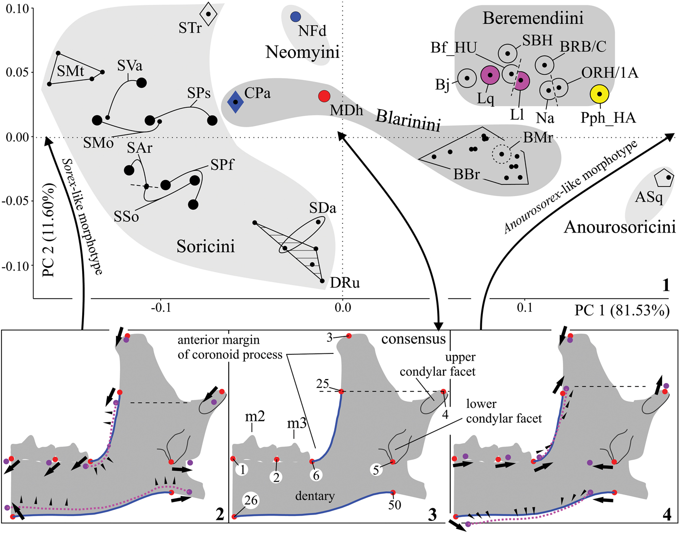
Dentaries are the most common fossil remains of this group. Indeed, among our Asian specimens, hemimandibles represent 45% of the investigated specimens; isolated teeth represent 39% (calculated on the basis of list S3). Comparisons of morphological characteristics of the mandible and lower teeth of specimens of Nectogalinia altaica (GIN 664/201) and Peisorex pohaiensis (cast of holotype, ZIN 104899) and of specimens of Beremendia collected from Asian and European (ZIN 104895) sites reveal similarities, e.g., a lack of variability in most characteristics of Beremendia fissidens, Beremendia minor (except for size differences), Beremendia pliocaenica, Beremendia jiangnanensis (except for size differences), and Peisorex pohaiensis. To identify the morphological relationships of Beremendiini by mandibular shape, we performed a morphometric analysis. First, for the estimation of variation limits observed among Beremendiini, we examined members of Soricini, Neomyini, Anourosoricini, and Blarinini (S7). Our results reveal the distinct positioning of Beremendiini among species of other tribes of Soricinae (Fig. 9.1). In comparison to the morphometrically variable soricine species, which includes members of valid (Sorex Linnaeus, Reference Linnaeus1758, Otisorex De Kay, Reference De Kay1842) and invalid (monotypic subgenus Stroganovia Yudin, Reference Yudin1989 includes Sorex [Stroganovia] daphaenodon Thomas, Reference Thomas1907; Sorex [?] trowbridgii Baird, Reference Baird and Henry1857, subgenus undetermined) subgenera as well as extinct genera (Drepanosorex Kretzoi, Reference Kretzoi1941, includes also Drepanosorex rupestris Zaitsev and Baryshnikov, Reference Zaitsev and Baryshnikov2002), species of Beremendia, Lunanosorex, and Peisorex form a relatively distinctive group.
Results of mandibular shape analysis of the samples of Soricini, Neomyini, Anourosoricini, Blarinini, and Beremendiini tribes: (1) principal component plot for extant and extinct shrews (first versus second axes); (2) shape difference vectors in the negative part of the first axis (‘Sorex-like morphotype’); (3) consensus (average) configuration; (4) shape difference vectors in the positive part of the first axis (‘Anourosorex-like morphotype’). ASq = Anourosorex squamipes Milne-Edwards, Reference Milne-Edwards and Masson1872; BBr = Blarina brevicauda (Say, Reference Say and James1823); Bf_HU = Beremendia fissidens (Petényi, Reference Petényi1864) from Beremend 5; Bj = Beremendia jiangnanensis Jin et al., Reference Jin, Zhang, Sun and Zheng2009; BMr = Blarinoides mariae Sulimski, Reference Sulimski1959 from Węże 1; BRB/C = Beremendia minor Rzebik-Kowalska, Reference Rzebik-Kowalska1976 from BRB/C (ZIN 104894); CPa = Cryptotis parvus (Say, Reference Say and James1823); CS = consensus; DRu = Drepanosorex rupestris Zaitsev and Baryshnikov, Reference Zaitsev and Baryshnikov2002 from Treugolnaya Cave type locality; Ll = Lunanosorex lii Jin and Kawamura, Reference Jin and Kawamura1996b from Qipanshan Hill type locality (by Jin and Kawamura, Reference Jin and Kawamura1996b); Lq = L. qiui Jin, Sun, and Zhang, Reference Jin, Sun and Zhang2007 from Bilike type locality (by Jin et al., Reference Jin, Sun and Zhang2007); MDh = Mafia dehneli (Kowalski, Reference Kowalski1956) from Podlesice locality; Na = Nectogalinia altaica Gureev, Reference Gureev1979; NFd = Neomys fodiens (Pennant, 1771); ORH/1A = Beremendia fissidens from ORH/1A (ZIN 104910); Pph_HA = Peisorex pohaiensis Kowalski and Li, Reference Kowalski and Li1963 from Haimao locality (by Jin and Kawamura, Reference Jin and Kawamura1996a); SAr = Sorex araneus Linnaeus, Reference Linnaeus1758; SBH = Beremendia fissidens from SBH (ZIN 104893/01); SDa = Sorex daphaenodon Thomas, Reference Thomas1907; SMo = Sorex monticolus Merriam, Reference Merriam1890; SMt = Sorex minutus Linnaeus, Reference Linnaeus1758; SPf = Sorex pacificus Coues, Reference Coues1877; SPs = Sorex palustris Richardson, Reference Richardson1828; SSo = Sorex sonomae Jackson, Reference Jackson1921; STr = Sorex trowbridgii Baird, Reference Baird and Henry1857; SVa = Sorex vagrans Baird, Reference Baird and Henry1857; short black arrows = landmark shifting vectors; black triangles = semilandmarks shifting vectors; gray mandible, red points, and blue solid lines = consensus configuration; violet points and pink dashed lines = outline shifting relative consensus. Numbers 1–50 are landmarks and semilandmarks (see S6).

The results of the PCA are as follows: shape transformation along the first and second principal components accumulate ~93% of the variation. For the first component (81.53% of the variation), the mandibular shape transforms from that of a Sorex-like morphotype (or Sorex-like mandibular outline; most closely related to Sorex monticolus Merriam, Reference Merriam1890) via consensus configuration, most closely related to Mafia dehneli (Kowalski, Reference Kowalski1956), to that of an Anourosorex-like morphotype (most closely related to Anourosorex squamipes). The Sorex-like morphotype differs from the other in its more slender dentary, in the markedly shorter articular surface of the condylar process that connects to the anteroinferior position of the upper condylar facet, and in the more posterior position of the lower condylar facet. Moreover, the anterior margin of the coronoid process is tilted anteriorly and the lower molar base is positioned lower down (compare Fig. 9.2, 9.3). On the opposite side of the axis, the Anourosorex-like morphotype includes a strong, robust dentary with a shorter than average configuration and tapering from front to back; the articular surface of the condyle is markedly longer due to the stronger posterosuperior shifting of the upper condylar facet and due to the anterior shifting of the lower facet whereas the anterior margin of the coronoid process is tilted posteriorly (Fig. 9.4). Along the second principal component (11.60% of variation), the Neomys-like morphotype, most closely related to Neomys fodiens (Pennant, 1771), is characterized by a shorter coronoid process (the ventral margins of the dentary are shifted upward whereas the coronoid apex is shifted downward and slightly forward) than that of the ‘average’ configuration. The Drepanosorex-like morphotype (closest to Drepanosorex rupestris and Sorex daphaenodon) on the opposite side of the component differs from the others in its longer, broader coronoid process, in the lower positioning of the upper condylar facet, and in the more aligned ventral margins of the dentary (a more horizontal margin).
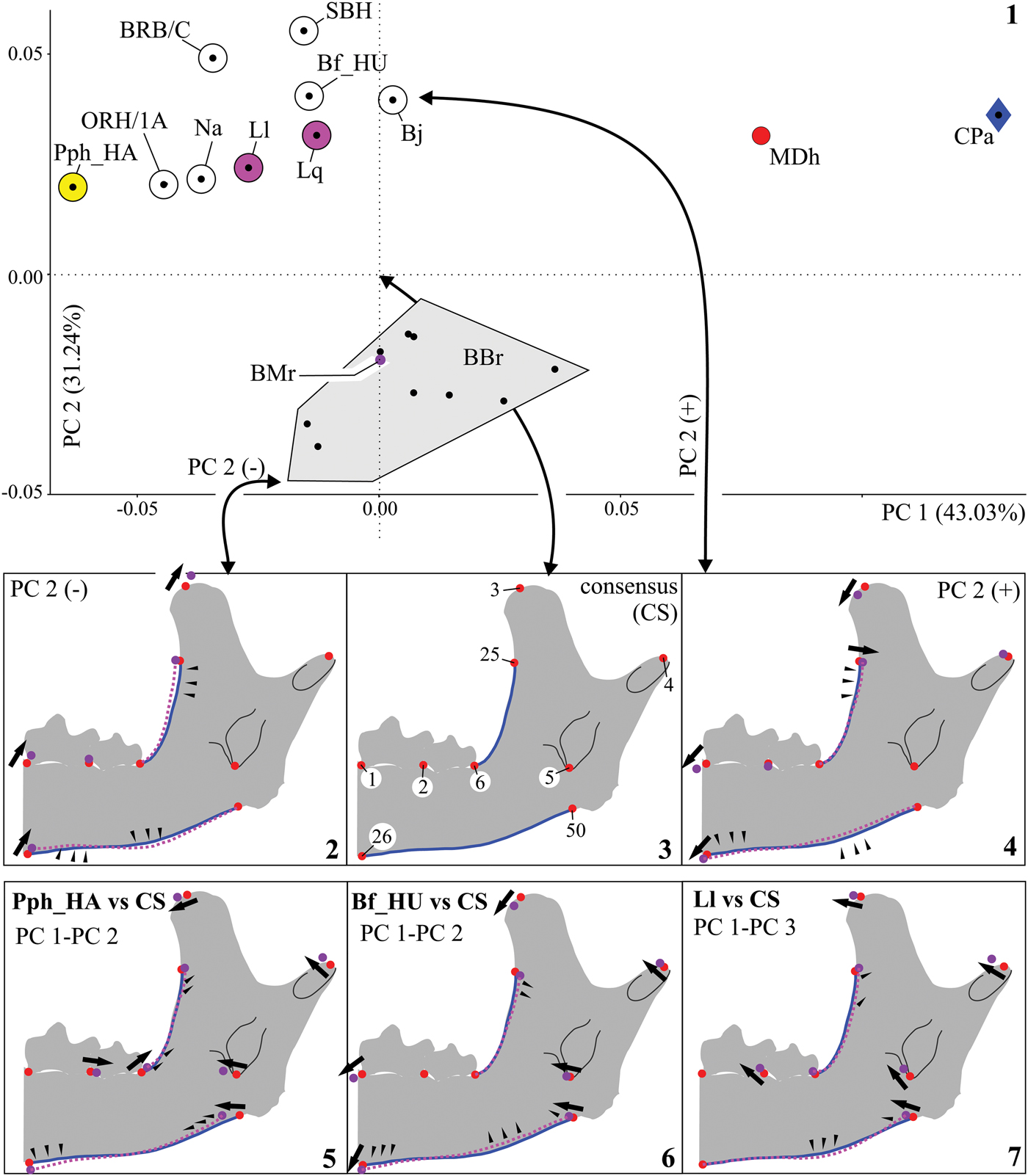
The second PCA of shape variations was performed for Beremendiini and Blarinini species (Fig. 10.1). The results of the second PCA confirm: (1) the separate position of the Beremendiini relative to species of Blarina Gray, Reference Gray1837, Blarinoides Sulimski, Reference Sulimski1959, Mafia Reumer, Reference Reumer1984, and Cryptotis Pomel, Reference Pomel1848; and (2) the narrow limits of Beremendiini variation relative to those of Blarinini. For the first principal component (43.03% of variation), the mandibular shape transformed from Beremendiini/(Blarina/Blarinoides) outlines into Mafia and Cryptotis outlines. The first group roughly corresponds to the Anourosorex-like morphotype (see Fig. 9.1) with a broad condylar process and a short, tapered dentary; Mafia and Cryptotis are positioned close to the Sorex-like morphotype area with a shorter condylar surface and more slender, longer dentary. Specific differences in the mandibles of Beremendiini and Blarinini are found from the second principal component (31.24% of the variation). Blarina brevicauda is positioned within the negative part of the axis (Fig. 10.1). The Blarina-like morphotype (closest to ZIN 65805) differs from the average configuration in the more undulating ventral margins of the dentary (Blarina has a shallow notch positioned along the margin below m2), in its longer coronoid process, and in stronger anterior tilt of its anterior margins (compare Fig. 10.2, 10.3). The Beremendia-like morphotype on the opposite side of the axis differs from the average in the straight and markedly tilted ventral margins of the dentary, with the posterior tilt of the anterior margins of the coronoid process, and with the anterior bending of the coronoid process tip (Fig. 10.4). Both species of Lunanosorex are separated in the negative part of the third principal component (5.70% of the variation), thus diverging from the consensus in terms of its slightly more undulating ventral dentary margins, the anterior shifting of its coronoid tip, and anterosuperior shifting of its upper condylar facet (Fig. 10.7). Our paired comparisons of the consensus configurations of Peisorex pohaiensis (from Haimao) and Beremendia fissidens (from Beremend 5) reveal the following differences: P. pohaiensis demonstrates a posterior shifting of m3 (see landmarks 2 and 6), a slight shortening of the dentary (see landmarks 26 and 50), and an anterior shifting (tip bending) of the coronoid tip (landmark 3) (Fig. 10.5). In contrast, Beremendia fissidens exhibits an anteroinferior shifting of the coronoid tip and other shifts similar to those of Peisorex but weakly expressed (Fig. 10.6).
Results of mandibular shape analysis of the samples of Blarinini and Beremendiini tribes: (1) principal component plot for extant and extinct shrews (first versus second axes); (2) shape difference vectors in the negative part of the second axis (‘Blarina-like morphotype’); (3) consensus (average) configuration; (4) shape difference vectors in the positive part of the second axis (‘Beremendia-like morphotype’); (5) shape difference vectors of Peisorex pohaiensis Kowalski and Li, Reference Kowalski and Li1963 from Haimao vs. consensus configuration (CS), within first/second axes space; (6) shape difference vectors of Beremendia fissidens (Petényi, Reference Petényi1864) from Beremend 5 vs. consensus configuration, within first/second axes space; (7) shape difference vectors of Lunanosorex lii Jin and Kawamura, Reference Jin and Kawamura1996b from Qipanshan vs. consensus configuration, within first/second axes space. Abbreviations and symbols as for Figure 9.

Discussion
General remarks
Our investigation of original material taken from Siberian, Kazakh, and Mongolian localities confirms the extensive geographic dispersal of members of Beremendia from Europe to eastern Siberia (the locality of Beregovaya) and northern Mongolia (localities Orkhon 1, Bural-Obo, and Shamar). Published data on Chinese fossil materials reveal the presence of two large species: Beremendia pliocaenica (Mazegou site; Flynn and Wu, Reference Flynn and Wu1994) and Beremendia jiangnanensis (Renzidong; Jin et al., Reference Jin, Zhang, Sun and Zheng2009). Thus, Beremendia includes at least four species distributed from Europe to eastern Asia during the Pliocene and to the end of the early Pleistocene in Asia and surviving until the Middle Pleistocene in Europe (Rzebik-Kowalska, Reference Rzebik-Kowalska, Wójcik and Wolsan1998). The validity of Peisorex and the taxonomic position of P. pohaiensis (and probably Beremendia pliocaenica) remain unclear despite the taxonomic work of Jin and Kawamura (Reference Jin and Kawamura1996a). Well-preserved skull remains of P. pohaiensis collected from the Haimao site (Jin and Kawamura, Reference Jin and Kawamura1996a), the analysis of Qiu and Storch (Reference Qiu, Storch, Hoek Ostende, Doukas and Reumer2005), and our findings on diagnostic M1 taken from the Mongolian locality ORH/1B (ZIN 104911) again raise questions surrounding the validity of Peisorex with defined diagnostic skull features and upper teeth: (1) three upper antemolars (A1–A3), and (2) M1 and M2 with or without a weakly developed metaloph.
However, questions surrounding taxonomic validity cannot be solved because our opinions regarding the ‘diagnostic features’ of Peisorex from Haimao are based on sketches by Jin and Kawamura (Reference Jin and Kawamura1996a), and we have thus not observed the original material. However, we can identify a definite resemblance between the M1 of Haimao teeth and the M1 identified from ORH/1B (with the likely exception of a metaloph positioned at the base of the metacone of Haimao M1). Second, the main feature of Peisorex (the reduced number of upper antemolars) might be attributed to a lack of preserved teeth. The effect of an absence of small/vestigial upper antemolars in the posterior position is better recognized in Crocidurinae (Natori and Shigehara, Reference Natori and Shigehara1997; Koyasu et al., Reference Koyasu, Kawahito, Hanamura, Oda, Merrit, Churchfield, Hutterer and Sheftel2005). For samples of Suncus murinus (Linnaeus, Reference Linnaeus1766) collected from Okinawa Island (Japan), 27.4% of 95 specimens were found to not include A4, and 18.9% specimens lacked left and right A4 (Natori and Shigehara, Reference Natori and Shigehara1997). The extant soricine species available might follow a more stable dental formula. Feldhamer and Stober (Reference Feldhamer and Stober1993) described five North American soricine shrews and revealed the following: Blarina brevicauda (N = 199) did not lack upper antemolars; for Blarina carolinensis (Bachman, Reference Bachman1837) (N = 118), 6.7% (N = 8) of specimens lacked A5, among which only one specimen lacked A5 on both sides; Cryptotis parvus (N = 125) only exhibited one unilaterally missing A4; Sorex longirostris Bachman, Reference Bachman1837 (N = 41) only exhibited one unilaterally missing A5; and Sorex hoyi Baird, Reference Baird and Henry1857 (N = 93) did not show an absence of A5 although other antemolars were missing (3.2%). Therefore, based on data for Soricinae, we believe the bilateral loss of supposed A4 in Haimao P. pohaiensis to be highly unlikely (Jin and Kawamura, Reference Jin and Kawamura1996a). However, we must not forget that this conclusion is based on only one known skull. Furthermore, among three skull remains and 10 maxilla fragments of Beremendia jiangnanensis, A4 has been found only in two specimens (Jin et al., Reference Jin, Zhang, Sun and Zheng2009, p. 155) with the authors claiming that “A4 is usually absent” without describing the nature of this effect; is it ontogenetic or due to fossilization? It is likely that the variation observed in the number of upper antemolars is a distinctive feature of the Chinese species. New fossil materials taken from Chinese and Mongolian sites must be examined to test this hypothesis. Pons-Monjo et al. (Reference Pons-Monjo, Moyà-Solà and Furió2012) identified a similar feature from extinct Nesiotites Bate, Reference Bate1945 (Soricidae).
Morphological features of European and Asian Beremendia
The main question that arises with the discovery of new fragmentary materials from a broad territory concerns whether one can distinguish intraspecies variability from interspecies variability. A distinctive feature could indicate a new taxon and perhaps a shift in state frequencies known of the studied part of a group distribution. Published data for Beremendia sensu lato for different parts of Europe, including information collected from the Russian Plain by Agadjanian (Reference Agadjanian2009) and from China, allowed us on the one hand to measure the unsuitability of the mandible and lower teeth for taxon diagnoses and on the other hand to identify a wide variety of features among Beremendia species. The use of these data shaped our perspective regarding the variability in the qualitative and quantitative features of the Asian Beremendia analyzed here—most specimens were associated with medium-sized and widely distributed Beremendia fissidens and small Beremendia minor. Deviations in ‘shapes’ and ‘sizes’ (see Variation, above) correspond with the intraspecific variability of Beremendia fissidens aside from the probable conformity between deviated M1 of Beremendia cf. B. fissidens and P. pohaiensis and small Beremendia minor. The Asian specimens display morphotypes of: (1) the first lower incisor, (2) fourth upper premolar, and (3) first upper molar. These morphotypes can be found in different parts of the Beremendia range, e.g., the size and depth of the groove on the posteromedial corner of the i1 crown vary in Beremendia fissidens/Beremendia minor (for European conditions of the Morphotype I ‘short shallow groove,’ see Botka and Mészáros, Reference Botka and Mészáros2014, figs. 14, 16). Given that the original material (Fig. 3.5, 3.7, 3.9) and European specimens display both morphotypes, the state with a smooth AAF from KMN and BTK/1C should be attributed to rare cases of variation in Beremendia fissidens, which is especially the case because a ‘smooth AAF’ is not related to geographic or temporal specificities: the first tooth was collected from Altay (KMN, early Pleistocene) whereas the second was collected from Mongolia (BTK/1C, late Pliocene). The morphotypes of the P4 hypocone states are also representative of different regions and ages, including the European sites examined by Reumer (Reference Reumer1984, p. 106). The rare morphotype of the lingual outline of M1, namely the ‘shallow notch of lingual margins,’ was once found from original material (Fig. 5.1), but Reumer's figures present both states across European sites (Reumer, Reference Reumer1984, pl. 33, figs. 1a, 2, 3) for Beremendia fissidens. The length and development of the coronoid spicule discussed above relative to Beremendia fissidens and Beremendia minor is a variable feature in ancient species collected from the European sites (Reumer, Reference Reumer1984, pl. 34, figs. 3c, 4b). It is likely that the size of the spicule and the composition of external fossa facets can be related to the development of chewing muscles and indirectly to foraging activity. The limited development of the metaloph together with a heavily delineated and relatively long entocingulum of M1 of Beremendia cf. B. fissidens has not been observed in the European material. Indeed, Reumer (Reference Reumer1984, p. 106) noted that the “protocone is situated on a U-shape crest, which connects it to both the paracone and metacone < M1–M2 > .” In other words, the crest connects the base of the paracone to the preprotocrista and connects the base of the metacone to the metaloph. Thus, these M1 states (Fig. 5.7–5.9) can be compared to Chinese taxa with similar upper molars.
Mandibular shapes in Beremendiini
Analyses of shape variability (Figs. 9, 10) reveal the original outline of the mandible of Beremendiini with a short mandibular body, notable signs of posterior tapering, and a long (massive) condylar process. Most shape similarities were found between Beremendiini and Blarinini (more precisely, Blarina brevicauda). Both morphotypes (see Fig. 10.2, 10.4) were found to be more closely related to one other than to other morphotypes (Fig. 9). This result confirms the likely presence of a trophic analogy between Beremendia and Blarina and explains the presence of similarities in facet shapes for attaching the ‘M. levator nasi inferior’ (see above description of the skull fragments) and other similarities in the “structure of the articular condyle, only differing in the superior pterygoid fossa” and in the “wear pattern of the teeth” described by Furió et al. (2010, p. 932).
However, questions remain regarding the limited shape variability of beremendiins. The PCA results (Figs. 9, 10) reveal considerable differences between Soricini and Blarinini and fewer in the case of Beremendiini. The intratribal range of the latter is comparable to the intrageneric/intraspecies range of Blarina. We offer two possible explanations for this. First, with the need to synonymize beremendiin genera, morphometric differences within the tribe are so minor that all specimens should be defined within a common genus. Most authors investigating Beremendiini, including Jin and Kawamura (Reference Jin and Kawamura1996a), Jin et al. (Reference Jin, Zhang, Sun and Zheng2009), and Flynn and Wu (Reference Flynn, Wu, Flynn and Wu2017), consider Peisorex to be a junior synonym of Beremendia; yet, Lunanosorex and Dolinasorex are still valid genera (Jin et al., Reference Jin, Sun and Zhang2007; Rofes and Cuenca-Bescós, Reference Rofes and Cuenca-Bescós2009). The second thesis is related to the likely high trophic specialization of Beremendiini and to the resulting similarities in mandibular shapes found between different members. Additionally, and indirectly from mandibular shapes, we identify probable similarities in trophic niches of beremendiin shrews and Blarina.
Mandibular mobility and ‘biting-chewing’ model
Shrews bear unique chewing apparatuses expressed in details of the articular surface of the condylar process and specifically in the presence of upper and lower articular facets with an interarticular area in between (Fearnhead et al., Reference Fearnhead, Shute and Bellairs1955). Contrary to the ‘simple’ ball- (e.g., Tenrecidae) or roller-like (e.g., Talpidae) condyle, a feeding process with a ‘complex’ shrew-like condyle can be separated into a ‘bite phase’ (grasping) and ‘chewing phase’ and into certain irregularities (Dötsch, Reference Dötsch1986). The simultaneous ipsilateral use of both facets is theoretically impossible due to the entoglenoid fossa composition (‘internal relief’). Any mammalian group displays complex chewing cycles in connection with its feeding specialization and morphology: different vertical and horizontal movements (Ungar, Reference Ungar2010). Expanding with the range of different mandibular movements, diverse mammalian taxa (some primates; most perissodactyls, hyracoids, and wombats; and various artiodactyls) have an unfused mandibular symphysis (Lieberman and Crompton, Reference Lieberman and Crompton2000). Shrews also display an unfused symphysis. At the same time, the mobility of the symphysis renders the modeling of shrew mandibular movements more difficult. Fearnhead et al. (Reference Fearnhead, Shute and Bellairs1955) described the morphology of the entoglenoid area and mandibular ramus, the histology of the craniomandibular joints of Crocidura Wagler, Reference Wagler and Oken.1832 and Sorex, and some evolutionary aspects. Based on their conclusions, we propose a model of Beremendia mandibular mobility that first considers tooth and jaw morphology and then assumes similarities with a potentially modern trophic analog.
The size of the condylar process (condylar surface) denotes the relative size of a shrew's food, which is only an assumption applied for model construction, for species with carnivore-like specialization and corresponding features (see below) but that hunt small prey (insect larvae, earthworms, etc.), e.g., the extant Anourosorex squamipes. However, A. squamipes can feed on vertebrates (frogs, toads, and lizards) because its masticatory apparatus includes all of the necessary adaptations; in van Dam's (Reference Van Dam2004, p. 741) opinion, “frogs, toads, and lizards constituted a main part of the diet of fossil Anourosoricini, implying a more terrestrial way of life.” In this sense, Beremendia might have represented an extreme variant with specialization for feeding on larger prey. An analysis of both shrew features (e.g., ‘short muzzle’ and ‘powerful lower jaw’) shows that Beremendia is the most specialized following A. squamipes (generally Anourisoricini; Van Dam, Reference Van Dam2004) from the value of the MPMA-index (0.36–0.38 vs. 0.46) and based on the sizes of the condylar process and mandible. Beremendia is thus firmly related to the index values for Blarina (Table 1).
The mandible dimensions (MSL, HC) and MPMA-Index values of six soricine and crocidurine shrews. Measurements are presented as the mean and range.

Notes: * — n = 5 (ORH/1A, ZIN 104910; RZD, GIN 664/201; SBH, ZIN 104893/01, /02; B5, ZIN 104895/hu-02); ** — n = 1 (BRB/C, ZIN 104909).
Thus, Blarina can be seen as a modern ‘trophic analog’ of Beremendia with a similar masticatory apparatus of comparable shape (Figs. 9, 10) and MPMA-index values based on published conclusions (e.g., Furió et al., Reference Furió, Agusti, Mouskhelishvili, Sanisidro and Santos-Cubedo2010). The differences observed between Beremendia and Blarina in terms of mandibular shape (landmarks 3, 6–25, 26–50, see Fig. 10.2, 10.4) are mainly connected to different means/positions of muscle attachments and accordingly with differences in power vector directions followed during chewing actions. However, the morphotype shapes display very slight differences in condylar process shapes or, more precisely, in average similarities in the relative positioning of the higher point of the upper condylar facet (fourth landmark) and of the medioinferior point of the lower condylar facet (fifth landmark). Accordingly, Furió et al. (2010, p. 932) wrote: “[b]oth genera share a similar structure of the articular condyle.” A visual comparison of their articular surfaces reveal similarities in the size and relative positioning of condylar facets and in the widths of the interarticular area (S16, fig. 1). The large condyle requires a broad entoglenoid fossa surface. Analyses of the craniomandibular joints of Beremendia and Blarina reveal a free (i.e., empty) space for condylar movement, i.e., the upper facets were shorter than the longitudinal surface (most of the length) of the upper articular fosses. The upper condylar facet of Beremendia fissidens from B5 (ZIN 104895/hu-02) was 2.02 mm long, and the upper articular fossa was 3.06 mm long; the dimensions for Beremendia brevicauda (ZIN 65809) were 1.57 mm and 2.62 mm, respectively. Thus, the upper condylar facet might have moved within the entoglenoid fossa (S16, fig. 2). The craniomandibular articulation of Blarina can be characterized by three movements: (1) the upper condylar facet shifts between the anteromedial and posterolateral corners within an articular fossa; (2) the upper condylar facet shifts along the external surface of an upper articular facet; and (3) the anterior shifting of the lower condylar facet is only limited by the anteromedial corner of a lower articular fossa. This characterization challenges the consensus on the simultaneous ipsilateral use of the upper and lower condylar facets and explains the simultaneous ipsilateral shifting of the upper and lower condylar facets relative to the articular surfaces of the entoglenoid fossa (as determined by manual shifting).
MSS-model (‘Mandible Sliding and Swinging’).—According to Dötsch (Reference Dötsch1986), the masticatory sequence can be divided into several phases; the main ones involve ‘grasping’ and ‘chewing’ with some irregular movement as the main phase depends on the traits of a food item. Based on Dötsch's (Reference Dötsch1986, Reference Dötsch, Merritt, Kirkland and Rose1994) results and our analyses, we separated ‘grasping’ and ‘chewing’ activity into subphases with an illustration of the relative positioning of the entoglenoid fossa and condyle surfaces:
(a) Phase ‘quiescence’ (PQ): upper and lower teeth are in occlusion. Condylar facets come into contact with articular facets of the entoglenoid fossa of both hemimandibles (Fig. 11.2).
(b’) Phase ‘grasping’/subphase ‘mandible depression’ (GP/sPD): the mouth opens to grasp a food item. The upper condylar facet is positioned within the upper articular fossa and prevents opening. As the mouth opens, the upper condylar facets slide toward the external surface of the upper articular facets with lateral movements (see Fig. 11.3) due to symphysis mobility, an absence of the zygomatic arches, and M. pterygoideus medialis activity. Finally, the angular process pulls medially and rotates the hemimandible around its longitudinal axis; at this point the coronoid tip swings laterally. The lower condylar facet shifts anterolaterally; tooth rows diverge laterally in opposite directions (see the positioning of the incisor tip in Fig. 11.7).
(b”) Phase ‘grasping’/subphase ‘holding’ (GP/sPH): the mouth is closed through the contraction of M. temporalis as the primary adductor. The upper condylar facet reaches the external surface of the upper articular facet; the lower condylar facet slides posteriorly (Fig. 11.4).
(с) Phase ‘chewing’ (CP): slide movements of the mandible are repeated in the anterior and posterior mandibular positions (Fig. 11.5). These movements from cycle to cycle result in the shearing and crushing of a food item between the upper and lower teeth. Anterior movement (protraction) creates a ‘gap’ between the rows while sliding forward whereas posterior movement (retraction) clamps the rows while sliding backward. Depending on the prey size, the upper condylar facet can slide along the external surface of the upper articular fossa into the fossa. Sliding is carried out with M. temporalis, M. digastricus, and M. pterygoideus medialis and lateralis. These muscles exert different forces and can involve additional mandibular sliding or swinging (e.g., ‘lateral movements’ sensu Dötsch, Reference Dötsch, Merritt, Kirkland and Rose1994). In general, all main phases of mandibular movements are connected to slide tilting surfaces move relative to one another and to the swinging of hemimandibles around the longitudinal axis to open the mouth. Electromyographic and cinematographic studies by Dötsch (Reference Dötsch1986, 1994) confirm our model because the electromyographic detection of muscle action corresponds with the mentioned phases/subphases. Most questionable is the ‘mandible depression’ subphase (GP/sPD) because we cannot be sure of the relationship between the lateral mandibular movements and activity of the M. pterygoideus medialis (Fig. 11.6) or of the movement of the upper condylar facet beyond the entoglenoid fossa. Dötsch (Reference Dötsch, Merritt, Kirkland and Rose1994) confirmed the relation between mouth opening and M. digastricus activity and did not mention the pterygoid muscle. However, Dötsch described lateral mandibular swinging: “[d]uring this grasping behavior, the jaws may engage in lateral movements” (Dötsch, Reference Dötsch1986, p. 30) and the “[l]arger rotation of the lower jaw at the inferior jaw articulation produced stronger outward deflection of the coronoid process on the working side during jaw opening” (Dötsch, Reference Dötsch, Merritt, Kirkland and Rose1994, p. 236).
MSS-model plots: (1) diagrammatic image of rostrum of Blarina brevicauda (Say, Reference Say and James1823), lateral view with the colored left dentary (gray) and entoglenoid fossa (black); (2) diagrammatic image of ‘upper tooth row’ (dark gray), border of entoglenoid fossa (orange line) with surrounding bone (hatched), and dentary (specimen ZIN 104894) of Beremendia during ‘quiescence’ phase, lateral view; condylar facets displayed from medial side (red and blue); (3) same, Beremendia during ‘grasping’ phase, subphase ‘mandible depression’; (4) same, Beremendia during ‘grasping’ phase, subphase ‘holding’; (5) same, Beremendia during ‘chewing’ phase; (6) schematic drawing of Beremendia/Blarina right dentary parts (coronoid process tip, condylar facets, angular process), position between states of the PQ and GP/sPD phases, posterior view; (7) schematic drawing of Beremendia upper (gray) and lower first incisors during PQ (red outline) and GP/sPD (blue outline) phases, anterior view. A1–A4 = upper antemolar teeth; I1 = first upper incisor; i1 = first lower incisor; P4, M1, M2 = upper molariform teeth (schematically as dark gray rectangles); p4, m1, m2 = lower molariform teeth; black multiple arrows = direction of muscle action for ‘abduction’ and ‘retraction’ of dentary; blue arrow = direction of the upper condylar facet shifting anteriorly and laterally during GP/sPD phase (in 3), and posteriorly and medially during GP/sPH phase (in 4); wide green arrows = the directions of the upper and lower condylar facets ‘roundtrip’ movements during CP phase (in 5); orange curved arrows = direction (lateral/medial movements) of the swinging of a dentary along the longitudinal axis during particular phases (in 3, 4, and 6); red doubled arrows = directions of the ‘roundtrip’ movements during CP phase (in 5); wide red arrow = main dentary movements (depression, pressing on food); violet multiple arrows = direction of muscle action for ‘adduction‘ and ‘protraction’ of dentary (in 5).

Remarks on beremendiin paleobiology
The MSS-model does not cover the full complexity of various movements and subphases, e.g., the independent movement of left and right hemimandibles, or all of the variants of lateral movements (forces of different muscles). However, the model describes the central probabilities of extinct Beremendia in terms of the consumption of different sizes of prey. We posit that Beremendia was an ‘active gatherer’ that ate prey of a range of sizes from small and soft insect larvae to hard coleopterans and mollusks and small vertebrates. Given the morphological similarities observed between Beremendia and Blarina, e.g., the developed muscles of ‘nose movers’ (attachment to M. levator nasi inferior, see S15, fig. 1), dark-colored tooth enamel, condyle sizes, mandibular shapes, and relatively large body, we can assume the presence of a ‘trophic’ and likely ‘ecological’ analogy between them. Thus, Blarina food items can be used for the careful reconstruction of potential dietary items of Beremendia. According to Laerm et al. (2007, p. 66), the diet of Blarina included “[a]nnelids, gastropods, lepidopteran larvae, chilopods, arachnids, other invertebrates, and small quantities of plant material” and it also preyed “on small rodents, amphibians, and reptiles.” Maier (Reference Maier, Merrit, Churchfield, Hutterer and Sheftel2005) also published data on two natural ‘predator-antipredator’ interactions occurring between Blarina brevicauda and eastern spadefoot toad Scaphiopus holbrookii Harlan, Reference Harlan1835 derived from a series of laboratory trials. The results revealed the ‘active predator’ behavior of Blarina, which did not attempt to attack toads despite its size and means of active protection (Maier, Reference Maier, Merrit, Churchfield, Hutterer and Sheftel2005, p. 362, 363). Although the success of hunting a toad is not defining, it is important to note that a shrew can kill a large vertebrate. Thus, Beremendia might have served as an active gatherer of relatively small prey and a hunter of relatively large prey. Furió et al. (Reference Furió, Agusti, Mouskhelishvili, Sanisidro and Santos-Cubedo2010) have discussed the benefits of fissident upper incisors, which helped the shrew forage for ‘intractable’ food items with hard shells. The authors noted that fissident teeth provided the opportunity to control bite force and to pierce hard-shelled prey with the lower incisor (Blarina bears nonfissident first upper incisors). The MSS-model allows us to assume additional traits required for the grasping of intractable prey; during GP/sPD and/or GP/sPH, the hemimandibles and tooth rows can diverge, and tips/cutting edges of the lower incisors can assume a broad V-shaped configuration (Fig. 11.7), forming a broad trench opposite the fissident upper incisors for holding hard-surfaced prey. The movement of prey along the tooth rows and initial attempts at shearing could be performed with anteroposterior mandibular movements characterizing the CP phase (Fig. 11.5) and the specific shape of the lower incisor cutting edge and upper incisor hatchet-like talon. Blarina bore similar characteristics of lower incisor cutting edges but differed in the shape of the upper incisor talon from that of Beremendia (compare Fig. 12.1, 12.2). The conical medial cusp of Blarina’s talon applied rough crushing. The food item shearing of shrews was carried out using the lower incisors, antemolars, premolars, and upper antemolar rows. The upper antemolars of Beremendia and Blarina bear crown longitudinal ridges arranged to form a ‘complex cutting ridge of I1–A3’ (Fig. 12). Beremendia displays a useful saw-like outline for the rapid cutting/shearing of prey before transporting food to the main mastication molar complex. Beremendia can cover all available types of natural Pliocene–Pleistocene biotopes similar to habitat-tolerant Blarina brevicauda, which has been collected from grasslands, in various successional stages of disturbed habitats, and in all seral stages from coniferous and deciduous hardwood forests and saturated wetlands (Laerm et al., Reference Laerm, Ford, Chapman, Trani, Ford and Chapman2007). Beremendia could have occupied biotopes reconstructed from the late Pliocene palynological assemblage in Zhangcun, Shanxi (China; Qin et al., Reference Qin, Ferguson, Zetter, Wang, Syabryaj, Li, Yang and Li2011), close to water bodies, open grasslands, shrubs, broad-leaved forests, coniferous forests, and intermediate habitats.
Diagrammatic images of upper teeth in context of the ‘cutting ridges’ possibilities: Beremendia fissidens (Petényi, Reference Petényi1864), specimen from SMR/A, ZIN 104908/1228: (1) fragment of the left upper tooth row, with marked ‘cutting ridges’ area (light blue), and schematic ‘transverse’ profile bends (blue line below), occlusal view; (3) fragment of premaxilla/maxilla, with ‘longitudinal’ profile bands, lateral view. Blarina brevicauda (Say, Reference Say and James1823), Recent specimen from Wisconsin, USA, (ZIN 65805): (2) fragment of the left upper tooth row, with marked ‘cutting ridges’ area (light blue), and schematic ‘transverse’ profile bends (blue line below), occlusal view; (4) fragment of premaxilla/maxilla, with ‘longitudinal’ profile bands, lateral view. (5) Technical tools (saw and raker set of saw teeth) with similar functions as ‘cutting ridges.’ Light pattern (1, 2) and dark pattern (3, 4) = complex cutting ridge as a combination of antemolar crests. A1–A5 = upper antemolar teeth; I1 = first upper incisor. Scale bars = 1 mm.

Conclusions
Beremendiini is an extinct group of soricine shrews that were widely distributed during the Pliocene and Pleistocene. Their distribution in China has been investigated, yet, northern Asian data remain poorly studied (Gureev, Reference Gureev1979; Storch et al., Reference Storch, Qui, Zazhigin, Wójcik and Wolsan1998). We analyzed all available Beremendiini material collected from 16 early Pliocene to early Pleistocene localities in Russia (Siberia), Kazakhstan, and Mongolia and found two beremendiin species: Beremendia fissidens and Beremendia minor. Northern Asian Beremendia displayed considerable variations in terms of their size and characteristics, but most of the different states were observed in European or Chinese specimens. Some of the most critical traits found include the uniformity of the mandibular shape and broad variations in the qualitative characteristics of the mandible and lower teeth. Mandibular outlines were found to be similar among members of Peisorex, Beremendia, and Lunanosorex. The limits of the shape variations observed among genera are comparable to intraspecific variations observed for Blarina. A discussion of this question led us to assume the presence of broad trophic possibilities directly connected to the morphology of the masticatory apparatus: a large condyle; a short, large, tapered jaw; and anterior teeth better adapted for shearing and cutting than for crushing. Given that fossil remains generally include hemimandibles and isolated teeth, we were unable to assess several traits of skulls. However, we suspect that distinguishing the skull characteristics could support the recognition of Beremendia jiangnanensis from the Renzidong site and Peisorex pohaiensis from the Haimao site. Until discoveries of the beremendiin skulls are made, questions concerning the number of northern Asian species that developed and the validity of the genera will remain unaddressed.
Acknowledgments
This study was completed within the framework of the Federal themes of Zoological Institute no. АААА-А19-119032590102-7, ‘Phylogeny, morphology, and systematics of placental mammals,’ and Geological Institute ‘Paleontological grounds for the stratigraphic scale of the upper Cenozoic of northern Eurasia.’ This study was partly funded by Project no. 19-04-00049 of the Russian Foundation for Basic Research. The study partly used the collection materials of the Zoological Institute of the Russian Academy of Sciences (http://www.ckp-rf.ru/usu/73561/). It was supported by the Federal Agency for Scientific Organizations program for the support of Bioresource Collections. Authors are grateful to L.J. Flynn and M. Furió for reviewing our manuscript and contributing to its improvement, and A.O. Averianov for text and ideas discussed.
Accessibility of supplemental data
Data available from the Dryad Digital Repository: https://doi.org/10.5061/dryad.01kh74c. Supplementary material sets include: (S1) List of the 16 early Pliocene–early Pleistocene localities; (S2) List of analyzed specimens of Beremendia from 16 early Pliocene–early Pleistocene localities; (S3) List of the analyzed bones and isolated teeth of Beremendia from 16 early Pliocene–early Pleistocene localities; (S4) Measurements performed for a morphometric description of the material; (S5) Results of elemental analyses of shrew teeth using the EDX method; (S6) Configuration of five true landmarks and 45 semilandmarks used for mandibular shape analyses; (S7) List of the analyzed specimens of other fossil and Recent taxa; (S8) Dental and bone measurements of Beremendia fissidens from five European sites; (S9) Dental and bone measurements of Beremendia fissidens and Beremendia minor from three European sites; (S10) Dental and bone measurements of Peisorex pohaiensis and Beremendia jiangnanensis from Chinese sites; (S11) Dental measurements of Beremandia pliocaenica; (S12) Dental and bone measurements of Lunanosorex lii, L. qiui, and Dolinasorex glyphodon; (S13) Dental and bone measurements of investigated material of Beremendia fissidens, Beremendia cf. B. fissidens, and Beremendia minor from 16 early Pliocene–early Pleistocene localities of Russia (Siberia), Kazakhstan, and Mongolia; (S14) Fifty-seven individual measurements of investigated specimens of Beremendia fissidens, Beremendia cf. B. fissidens, and Beremendia minor from 16 early Pliocene–early Pleistocene localities in Russia (Siberia), Kazakhstan, and Mongolia; (S15) Characters of Beremendia skull rostral part and upper teeth; (S16) Characters of the condylar articular surfaces and entoglenoid fossa of Blarina and Beremendia; and (S17) List of abbreviations.