INTRODUCTION
The genus Hepatozoon (Eucoccidiorida, Hepatozoidae) includes about 340 species, which develop in reptiles, amphibians, rodents, birds or mammals. In spite of the wide variety of natural hosts involved in the life cycles of these protozoa (Smith, Reference Smith1996), all Hepatozoon species share similar biology and a rather unique transmission pathway: the ingestion of infectious sporozoites (Smith, Reference Smith1996). Indeed, once the definitive host (i.e. an haematophagous invertebrate) is ingested by a vertebrate intermediate host, sporozoites reach the blood or lymphatic circulation (Baneth et al. Reference Baneth, Samish and Shkap2007). Merogony takes place in different target tissues, including lymphatic organs, muscles or the bone marrow, and micromerozoites penetrate erythrocytes of all vertebrates, but mammals and birds (Ferguson et al. Reference Ferguson, Kirk Hillier and Smith2012), in which gametogony occurs in leucocytes (Baneth et al. Reference Baneth, Samish and Shkap2007). The life cycle completes when the invertebrate definitive host ingests blood cells parasitized by gamonts, which undergo sexual reproduction (syzygy) and sporogony in the vector haemocoel or in its gut wall, finally maturing into sporozoites enclosed within sporocysts in an oocyst (Smith, Reference Smith1996).
Amongst Hepatozoon protozoa affecting carnivores, Hepatozoon canis James, 1905 is the most extensively studied and widespread species (Baneth, Reference Baneth2011), parasitizing dogs (Canis familiaris), cats (Felis catus) (Baneth, Reference Baneth2011), foxes (Vulpes vulpes, Cerocyon thous) (Alencar et al. Reference Alencar, Kohayagawa and Santarém1997; Gabrielli et al. Reference Gabrielli, Kumlien, Calderini, Brozzi, Iori and Cancrini2010; Hodžić et al. Reference Hodžić, Alić, Fuehrer, Harl, Wille-Piazzai and Duscher2015; Tolnai et al. Reference Tolnai, Sréter-Lancz and Sréter2015), jackals (Canis aureus, Canis mesomelas) (McCully et al. Reference McCully, Basson, Bigalke, De Vos and Young1975; Duscher et al. Reference Duscher, Kübber-Heiss, Richter and Suchentrunk2013; Farkas et al. Reference Farkas, Solymosi, Takács, Hornyák, Hornok, Nachum-Biala and Baneth2014), wild dogs (Lycaon pictus) (Matjila et al. Reference Matjila, Leisewitz, Jongejan, Bertschinger and Penzhorn2008), hyenas (Crocuta crocuta) and lions (Panthera leo) (Kelly et al. Reference Kelly, Marabini, Dutlow, Zhang, Loftis and Wang2014; Williams et al. Reference Williams, Berentsen, Shock, Teixiera, Dunbar, Becker and Yabsley2014). The distribution of H. canis encompasses large areas of tropical, subtropical and temperate regions (Baneth, Reference Baneth2011), generally overlapping the dispersion range of the cosmopolitan brown dog tick Rhipicephalus sanguineus sensu lato (Acari, Ixodidae), its main arthropod vector (Baneth, Reference Baneth2011; Giannelli et al. Reference Giannelli, Ramos, Dantas-Torres, Mencke, Baneth and Otranto2013a , Reference Giannelli, Ramos, Di Paola, Mencke, Dantas-Torres, Baneth and Otranto b ; Ramos et al. Reference Ramos, Giannelli, Carbone, Baneth, Dantas-Torres and Otranto2014). However, the detection of H. canis in carnivores well outside the areas inhabited by R. sanguineus (e.g. Slovakia, Czech Republic, Austria, Hungary) (Majláthová et al. Reference Majláthová, Hurníková, Majláth and Petko2007; Duscher et al. Reference Duscher, Kübber-Heiss, Richter and Suchentrunk2013; Tolnai et al. Reference Tolnai, Sréter-Lancz and Sréter2015; Mitková et al. Reference Mitková, Hrazdilová, Steinbauer, D'Amico, Mihalca and Modrý2016) has reinforced the hypothesis that additional ixodid ticks are involved in the life cycle and transmission of this protozoon.
In addition to the main vector, Amblyomma ovale, Rhipicephalus microplus, Haemaphysalis longicornis and Haemaphysalis flava ticks have been confirmed as definitive hosts for H. canis (Murata et al. Reference Murata, Inoue, Taura, Nakama, Abe and Fujisaki1995; Rubini et al. Reference Rubini, Paduan, Martins, Labruna and O'Dwyer2009; de Miranda et al. Reference de Miranda, de Castro, Olegário, Beletti, Mundim, O'Dwyer, Eyal, Talmi-Frank, Cury and Baneth2011; Demoner et al. Reference Demoner, Rubini, Paduan Kdos, Metzger, de Paula Antunes, Martins, Mathias and O'Dwyer2013). Conversely, the amplification of H. canis DNA in Ixodes ricinus, Ixodes canisuga, Ixodes hexagonus, Dermacentor reticulatus, Dermacentor marginatus and Rhipicephalus turanicus (Hornok et al. Reference Hornok, Tánczos, Fernández de Mera, de la Fuente, Hofmann-Lehmann and Farkas2013; Latrofa et al. Reference Latrofa, Dantas-Torres, Giannelli and Otranto2014; Najm et al. Reference Najm, Meyer-Kayser, Hoffmann, Pfister and Silaghi2014) accounted for their potential vector competence (Hamšíková et al. Reference Hamšíková, Silaghi, Rudolf, Venclíková, Mahríková, Slovák, Mendel, Blažejová, Berthová, Kocianová, Hubálek, Schnittger and Kazimírová2016). However, the possibility that these tick species could have acquired H. canis DNA from an infected animal during their blood feeding (Giannelli et al. Reference Giannelli, Ramos, Dantas-Torres, Mencke, Baneth and Otranto2013a , Reference Giannelli, Ramos, Di Paola, Mencke, Dantas-Torres, Baneth and Otranto b ), makes this question still unanswered. For instance, the development of H. canis in R. turanicus, an ixodid tick morphologically similar and genetically close to R. sanguineus (Dantas-Torres et al. Reference Dantas-Torres, Latrofa, Annoscia, Giannelli, Parisi and Otranto2013), has been hypothesized (Kamani et al. Reference Kamani, Baneth, Mumcuoglu, Waziri, Eyal, Guthmann and Harrus2013; Latrofa et al. Reference Latrofa, Dantas-Torres, Giannelli and Otranto2014; Aktas, Reference Aktas2014), but never demonstrated.
The present study reports on the sporogonic development of H. canis in R. turanicus specimens, which were collected from a naturally infected fox from southern Italy. In addition, data on the in vitro infection of canine leucocytes with sporozoites obtained from mature H. canis sporocysts developed in this ixodid species are provided.
MATERIALS AND METHODS
Sample collection
Ticks were collected from a young male fox, aging about 1 year, hospitalized at the Department of Veterinary Medicine of the University of Bari, following a road accident. The animal was rescued in the countryside of Valenzano municipality (41·043781N, 16·884203E, Bari province, southern Italy) and submitted to clinical investigations for a suspected fracture of the right tibia. The fox was infested by ixodid ticks and diagnosed as infected by Hepatozoon sp., following the detection of gamonts in blood smears, which were stained with the May-Grünwald Giemsa Quick Stain (Bio Optica, Milano, Italy). The level of parasitaemia was estimated based on the percentage of peripheral blood neutrophils containing intracellular gamonts. The blood sample was frozen, until molecularly analysed for the identification of the parasite at the species level.
Tick identification and maintenance
A total of 19 engorged ticks were detached from the animal hair coat. Specimens were placed in plastic vials, secured with a cotton plug, and immediately identified at the stage and species level according to their morphology as R. turanicus (n = 6, i.e. two males and four females), I. hexagonus (n = 12, including six nymphs and six females) and Haemaphysalis erinacei (one female) (Manilla, Reference Manilla1998; Walker et al. Reference Walker, Keirans and Horak2000). In the case of R. turanicus specimens, the morphology of the adanal plates, accessory shields, spiracular plates and the genital opening were carefully examined (Dantas-Torres et al. Reference Dantas-Torres, Latrofa, Annoscia, Giannelli, Parisi and Otranto2013). The ticks were placed in an incubator under controlled conditions (i.e. 20 ± 3 °C, RH > 80% for I. hexagonus and H. erinacei; and 26 ± 1 °C, RH > 70% for R. turanicus), allowing the oviposition of females and moult of nymphs (Giannelli et al. Reference Giannelli, Ramos, Dantas-Torres, Mencke, Baneth and Otranto2013a , Reference Giannelli, Ramos, Di Paola, Mencke, Dantas-Torres, Baneth and Otranto b ). Specimens were daily monitored and, when egg batches were laid, about 50 eggs were separated for subsequent DNA extraction (see below).
Detection of Hepatozoon in dissected ticks
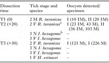
Ticks were dissected at different days post-collection (dpc) from the fox hair coat and examined for Hepatozoon oocysts, prioritizing specimens that completed the oviposition. Specimens of R. turanicus (two for each time-point) were analysed at the day of collection (T1), at 20 dpc (T2) and at 30 dpc (T3), whereas I. hexagonus specimens were examined at T2 and T3 (six ticks per each dissection time) and the female of H. erinacei at T3 (Table 1). Ticks were individually placed on slides containing a drop of saline solution and dissected by means of a sterile scalpel. They were incised through the spiracular plate and all the gut content, including the haemolymph, was observed under a light microscope at different magnifications. Oocysts were morphologically identified (Baneth et al. Reference Baneth, Samish and Shkap2007; Giannelli et al. Reference Giannelli, Ramos, Di Paola, Mencke, Dantas-Torres, Baneth and Otranto2013b ) and measurements (i.e. oocyst diameter and surface) were recorded for 15 specimens for each stage with an image analysis software (Leica®, LAS 4·1). In the case of immature oocysts, the ratio between the surface of the central nucleus and that of the oocyst was compared with Student's t-test. Differences were regarded significant when P < 0·05. Finally, all the dissected ticks were placed individually in sterile tubes with phosphate-buffered saline and stored at −20 °C, until molecular analysis.
Number and developmental stage of H. canis oocysts (immature, IM or mature, M) detected in engorged ixoidid ticks (males, M; females, F; nymphs, N) examined at different dissection times, expressed as days post-collection from the fox

a All females collected were allowed to oviposit, being dissected only when egg laying was concluded/interrupted.
b None of I. hexagonus nymphs moulted to the adult stage.
Experimental infection of canine leucocytes with sporozoites
The infectivity of H. canis sporozoites to canine leucocytes was assessed by experimentally infecting dog monocytes. Briefly, cells were isolated from the blood of a donor dog in good clinical conditions and molecularly negative for other canine pathogens, including H. canis, after obtaining the owner consent. Blood samples were collected from the brachial vein in ethylenediaminetetraacetic acid (EDTA) tubes. The buffy coat was separated using Ficoll-Hypaque (Lymphedex, innotrain Diagnostik GmbH, Germany) and the leucocytes were cultured in RPMI 1640 medium, supplemented with 10% fetal calf serum (FCS) and antibiotics (penicillin 5000 IU mL−1, streptomycin 2500 µg mL−1, amphotericin B10 µg mL−1). Cells were kept in a short-term culture at 37 °C. After 24 h, the medium was removed and the cells washed twice with FCS-free medium and inoculated with 100 µL tick homogenates, containing approximately 1000 H. canis previously activated sporozoites. Indeed, the oocysts (mechanically ruptured during dissection) and the sporocysts were suspended into 100 µL saline solution centrifuged at 250g for 10 min; the pellet was suspended in RPMI 1640 medium with 5% (w/v) fresh chicken bile and incubated at 37 °C for 30 min. Finally, the excysted sporozoites were concentrated by centrifugation (250g for 10 min), the supernatant containing the bile discarded, the pellet washed three times with saline solution, and the final aliquot was suspended in 100 µL RPMI 1640 culture medium. After an adsorption of 60 min at 37 °C, the inoculum was replaced with the FCS-free medium. The slides were removed 36 h after the inoculation and stained with the Diff Quick® (Bio Optica Spa, Italy) and examined under a light microscope. The infection procedure was performed in three short-term cultures.
Molecular analyses
DNA from the fox blood, ticks and eggs were extracted using a commercial kit (Qiagen, DNeasy Blood & Tissue Kit, Milan, Italy), following the manufacturer's instructions. Samples were tested by a conventional PCR for the detection of Hepatozoon (Inokuma et al. Reference Inokuma, Okuda, Ohno, Shimoda and Onishi2002). In addition, the identification of Rhipicephalus ticks was confirmed by generating and analysing partial mitochondrial cox1 (600 bp) gene sequences, as already described elsewhere (Dantas-Torres et al. Reference Dantas-Torres, Latrofa, Annoscia, Giannelli, Parisi and Otranto2013). The PCR amplification was carried out in a total volume of 50 µL, including 100 ng of genomic DNA, 10 mm Tris–HCl (pH 8·3) and 50 mm KCl, 2·5 mm MgCl2, 250 µ m of each dNTP, 50 pm of each primer and 1·25 U of AmpliTaq Gold (Applied Biosystems, Foster City, CA, USA). The reactions were run in a thermal cycler (2720, Applied Biosystems, Foster City, CA, USA). Negative (no DNA template, negative reference blood samples) and positive controls (Hepatozoon DNA from a positive tick) were included in all PCR reactions. Amplicons were resolved in ethidium bromide-stained agarose (Gellyphor, EuroClone, Milan, Italy) gels (1·5%) and sized by comparison with Gene Ruler™ 100-bp DNA Ladder (MBI Fermentas, Vilnius, Lithuania) as molecular marker, and finally gels were photographed using Gel Doc 2000 (BioRad, Hercules, CA, USA). All amplicons were resolved in GelRed-stained (2%) agarose (Biotium, California, USA) gels and sized by comparison with markers in the 1 kb DNA Ladder (MBI Fermentas, Vilnius, Lithuania). Gels were photographed using the GelLogic 100 gel documentation system (Kodak, New York, USA). Amplicons were purified and sequenced, in both directions using the same primers as for PCR, employing the Big Dye Terminator Cycle Sequencing Kit (v.3.1, Applied Biosystems, Foster City, California, USA) in an automated sequencer (ABI-PRISM 377). Sequences were compared with those available in the GenBank™ database, using Basic Local Alignment Search Tool (BLAST-http://blast.ncbi.nlm.nih.gov/blast.cgi).
RESULTS
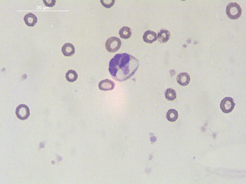
The Hepatozoon species found in the fox blood and in all positive tick specimens was molecularly identified as H. canis, showing 18S rRNA sequences 100% overall nucleotide BLAST identity with those of H. canis deposited in GenBank™ (accession number KJ605145). The level of parasitaemia in the fox was 60% (Fig. 1). All ticks survived during the observation period were dissected according to the study plan. Only three specimens of R. turanicus and two of I. hexagonus laid egg batches. None of the I. hexagonus nymphs moulted to adults.
Gamont of H. canis within fox monocyte (Diff quick stain, scale bar = 20 µm).

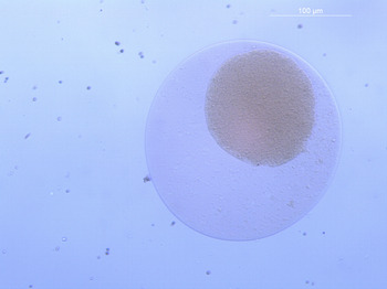
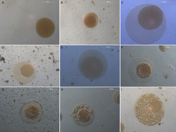
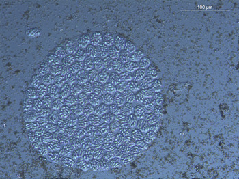
The results of tick dissection for each follow-up point and tick specimen are reported in Table 1. More than 500 H. canis oocysts were detected in R. turanicus ticks, whereas the remaining ixodid species were negative. Immature oocysts were observed in ticks dissected soon after the collection (T1). They measured 201 ± 72·8 × 138·8 ± 48·6 µm and displayed an amorphous central structure, condensed in a plasmatic matrix, similar to poached eggs (Fig. 2). Conversely, oocysts undergoing a different degree of maturation (Fig. 3) were detected in R. turanicus at T2, and included mature and undeveloped oocysts, lacking any sporocysts and sporozoites. In the latter, the ratio between the surface of the central nucleus and that of the oocyst (range: 32·5–82·4%) was significantly correlated with the oocyst diameter (t-test, P < 0·05). Mature oval-shaped oocysts measured 259·9 ± 36·1 × 246·1 ± 33·9 µm (Fig. 4) and contained a variable number of sporocysts, whose dimensions were 32·1 ± 4·7 × 20·2 ± 2 µm. In ticks dissected at T3, only mature oocysts were detected, along with free sporocysts and sporozoites, with the latter being elongated in shape and measuring 15·5 ± 4·1 × 3 ± 0·6 µm in diameter.
Immature H. canis oocysts detected in the haemocoel of a male R. turanicus (scale bar = 100 µm) at T1.

Footage showing the sporogony of H. canis in a female R. turanicus at T2. Following sygyzy, a zygote develops into a small immature oocyst (A); the oocyst membrane separates from the sporont (B) and its diameter increases along with complete development (C); the sporont nucleus becomes granular (D) and ameboid processes depart from the centre (E), budding into a protrusion or sporoblast (F), that gives rise to a developing sporocyst (G). This stage progressively reaches its final club shape (G), and detaches itself from the sporont mass (H). Developing sporozoites mature within the sporocyst wall (I) (scale bar = 100 µm).

Mature H. canis oocyst (scale bar = 100 µm) from a fed R. turanicus female at T3.

The positivity of all R. turanicus ticks examined at each time-point was molecularly confirmed, with all specimens being PCR-positive for H. canis and molecularly identified as R. turanicus (cox1 sequences showed 100% homology to R. turanicus sequences deposited in GenBank™, accession number KF145153.1) None of the I. hexagonus and H. erinacei specimens examined was positive for H. canis, as well as the egg batches collected from all female ticks that oviposited.
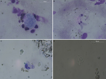
Following exposure to chicken bile, sporozoites were activated and displayed gliding and flexion–extension movements, often clustering together. These cells were featured by a diaphanous body and an eccentric nucleus (Fig. 5). The percentage of monocytes infected with H. canis in the three short-term cultures after 36 h from the inoculation was 34, 18 and 58%, respectively, with a mean 36·7% cells parasitized by H. canis sporozoites, which were observed within the leucocyte cytoplasm (Fig. 5).
Hepatozoon canis sporozoites following exposure to chicken bile and cell infection (scale bars = 20 µm). When not activated, sporozoites were observed within the sporocyst walls (A), or free in the cell medium (B). Early stage penetration of canine leucocytes by sporozoites (C and D).

DISCUSSION
Results of this study demonstrate that R. turanicus is a suitable vector for H. canis, as corroborated by its sporogonic development to reach the sporozoite stage, and by following the successful experimental infection of canine leucocytes. So far, only a few ixodid species (i.e. R. sanguineus s.l., A. ovale, R. microplus, H. longicornis, H. flava) have been considered as definitive hosts for H. canis (Murata et al. Reference Murata, Inoue, Taura, Nakama, Abe and Fujisaki1995; de Miranda et al. Reference de Miranda, de Castro, Olegário, Beletti, Mundim, O'Dwyer, Eyal, Talmi-Frank, Cury and Baneth2011; Demoner et al. Reference Demoner, Rubini, Paduan Kdos, Metzger, de Paula Antunes, Martins, Mathias and O'Dwyer2013), whereas others (e.g. I. ricinus or D. reticulatus) have been accounted as potential vectors (Hornok et al. Reference Hornok, Tánczos, Fernández de Mera, de la Fuente, Hofmann-Lehmann and Farkas2013; Latrofa et al. Reference Latrofa, Dantas-Torres, Giannelli and Otranto2014; Najm et al. Reference Najm, Meyer-Kayser, Hoffmann, Pfister and Silaghi2014) but their role has never been demonstrated. Besides the results of previous surveys, which highlighted the presence of the pathogen DNA in R. turanicus (Kamani et al. Reference Kamani, Baneth, Mumcuoglu, Waziri, Eyal, Guthmann and Harrus2013; Aktas, Reference Aktas2014; Latrofa et al. Reference Latrofa, Dantas-Torres, Giannelli and Otranto2014) or in other ‘cryptic’ species included in the R. sanguineus complex (Latrofa et al. Reference Latrofa, Dantas-Torres, Giannelli and Otranto2014), the potential development of H. canis in R. turanicus has never been evaluated, until now. The detection of immature and mature H. canis oocysts in R. turanicus accounts for the transtadial transmission of this pathogen, as it most likely occurred from nymphs to adults. In addition, the potential for the interstadial transmission from larvae to nymphs cannot be ruled out, as recently found for R. sanguineus (Giannelli et al. Reference Giannelli, Ramos, Di Paola, Mencke, Dantas-Torres, Baneth and Otranto2013b ). Due to the opportunistic collection of the infected fox, it was not possible to define the exact time when H. canis sygyzy and sporogony occurred in R. turanicus ticks. Nonetheless, the finding of mature oocysts in ticks detached after 20 days indicates that the pathogen requires at least 1 month for reaching its infective stage in R. turanicus. This corroborates observations drawn in R. sanguineus nymphs, in which H. canis fully matures in about 30 days (Giannelli et al. Reference Giannelli, Ramos, Di Paola, Mencke, Dantas-Torres, Baneth and Otranto2013b ). Conversely, the developmental time reported in A. ovale and R. sanguineus adults ranges from 11 to 14 days and from 27 to 53 days, respectively (Baneth et al. Reference Baneth, Samish and Shkap2007; Rubini et al. Reference Rubini, Paduan, Martins, Labruna and O'Dwyer2009). The possibility that the pathogen development is affected by the moulting time of the tick species should be taken into account.
The morphology and size of H. canis stages detected in R. turanicus is consistent with that found in R. sanguineus s.l. (i.e. 240–300 µm) (Baneth et al. Reference Baneth, Samish and Shkap2007; Giannelli et al. Reference Giannelli, Ramos, Dantas-Torres, Mencke, Baneth and Otranto2013a , Reference Giannelli, Ramos, Di Paola, Mencke, Dantas-Torres, Baneth and Otranto b ), A. ovale (210–306 µm) (Rubini et al. Reference Rubini, Paduan, Martins, Labruna and O'Dwyer2009) and H. flava/H. longicornis (300 µm) (Murata et al. Reference Murata, Inoue, Taura, Nakama, Abe and Fujisaki1995). Interestingly, the detection of a wide dimension range for mature oocysts (i.e. 259·9 ± 36·1 × 246·1 ± 33·9 µm) could be related to their abundance in the infected ticks. While a correlation probably exists between the number, the diameter of oocysts and the tick body dimension, results may also indicate that parasite growth can be slowed due to crowding, as already suggested for Hepatozoon griseisciuri in its vector Haemogamasus reidi (Redington and Jachowski, Reference Redington and Jachowski1971).
The detection of H. canis in R. turanicus might have relevant implications for understanding the distribution of this tick-borne pathogen in areas where R. sanguineus s.l. is not present. Unlike the brown dog tick, R. turanicus display a wider host preference, with the immature stages often feeding on rodents or lagomorphs, and adults on domestic and wild mammals, including cattle, sheep, goat and wildlife (e.g. hedgehogs, hares, jackals) (Manilla, Reference Manilla1998; Walker et al. Reference Walker, Keirans and Horak2000). Rhipicephalus turanicus is predominantly an exophilic tick, and its adaptability to different environmental and ecological conditions has favoured its setting and spreading in Mediterranean and dry regions, that, outside the African continent, include large parts of Asia and continental Europe (Nijhof et al. Reference Nijhof, Bodaan, Postigo, Nieuwenhuijs, Opsteegh, Franssen, Jebbink and Jongejan2007; Waner et al. Reference Waner, Keysary, Eremeeva, Din, Mumcuoglu, King and Atiya-Nasagi2014; Toma et al. Reference Toma, Khoury, Bianchi, Severini, Mancini, Ciervo, Ricci, Fausto, Quarchioni and Di Luca2015; Çetinkaya et al. Reference Çetinkaya, Matur, Akyazi, Ekiz, Aydin and Toparlak2016; Millán et al. Reference Millán, Proboste, Fernández de Mera, Chirife, de la Fuente and Altet2016). For example, R. turanicus rapidly colonized the island of Cyprus during the last 40 years, where it now seems to play an important role as spreader of zoonotic tick-borne pathogens, including Coxiella burnetii, Anaplasma and Rickettsia species (Chochlakis et al. Reference Chochlakis, Ioannou, Papadopoulos, Tselentis and Psaroulaki2014). Similarly, this tick species was introduced in Austria, in areas where R. sanguineus s.l. ticks are not endemic (Sixl, Reference Sixl1972), but H. canis infection has been reported in wildlife (Duscher et al. Reference Duscher, Kübber-Heiss, Richter and Suchentrunk2013, Reference Duscher, Leschnik, Fuehrer and Joachim2014). Altogether, results suggest that R. turanicus might contribute to the spreading of this pathogen, taking part in its ‘sylvatic’ life cycle, as supported by the simultaneous detection of H. canis and R. turanicus in the red fox here examined. In fact, in view of their free-roaming behaviour, increasing population density and regular visits to sub-urban areas (Uspensky, Reference Uspensky2014), foxes have been indicated as bridging hosts of several pathogens of domestic dogs, including Echinococcus multilocularis, Angiostrongylus vasorum and H. canis (Otranto et al. Reference Otranto, Cantacessi, Dantas-Torres, Brianti, Pfeffer, Genchi, Guberti, Capelli and Deplazes2015a , Reference Otranto, Cantacessi, Pfeffer, Dantas-Torres, Brianti, Deplazes, Genchi, Guberti and Capelli b ). In addition, a new species of Hepatozoon, (i.e. Hepatozoon silvestris sp. nov.) has been recently described in wild felids, enforcing the concept of pathogen circulating between wild and domestic populations, when the same ecological niches are shared (Hodžić et al. Reference Hodžić, Alić, Prašović, Otranto, Baneth and Duscher2016).
The infection of canine leucocytes with H. canis sporozoites indicates that this procedure may represent a valid alternative to experimental infection of laboratory-raised animals for investigating the biology of this canine tick-borne pathogen. The development of Hepatozoon protozoa in primary and continuous cell lines has been poorly investigated, with the exception of early attempts on H. griseisciuri (Hendrick and Fayer, Reference Hendrick and Fayer1973) and Hepatozoon rarefaciens (Ball and Chao, Reference Ball and Chao1973), which were cultured in mite and mosquitoes cells, respectively. The development of H. canis life cycle in vitro might provide interesting clues for defining its pathogenic role, for exploring the immunology and treatment of canine hepatozoonosis (De Tommasi et al. Reference De Tommasi, Giannelli, de Caprariis, Ramos, Di Paola, Crescenzo, Dantas-Torres, Baneth and Otranto2014) and also for providing a solid infrastructure for in-depth studies on its biology. In particular, the use of tick cell lines (Passos, Reference Passos2012) may support the discovery of association between Hepatozoon species and additional ixodid ticks implicated in their transmission, a hypothesis that deserves further investigations.
ACKNOWLEDGMENT
The authors would like to thank Rossella Panarese (University of Bari) for her support during the study.
FINANCIAL SUPPORT
This research received no specific grant from any funding agency, commercial or not-for-profit sectors.