Introduction
The control of fish reproduction is a key issue for both basic and applied developmental sciences. One of the first steps to manipulate fish reproduction is to understand the mechanisms involving sperm–oocyte interactions that result in fertilization and subsequent embryo development. This information is important to achieve good fertilization rates (Bobe & Labbé, Reference Bobe and Labbé2010). Successful fertilization in teleost fish depends on a complex chain of adaptive mechanisms and species-specific sperm–oocyte interactions. The main steps during fertilization process include: (1) activation of sperm motility and reaching the oocyte; (2) entrance of spermatozoon through the micropyle; (3) binding of sperm to the oocyte surface; (4) penetration of sperm into the ooplasm; (5) fusion of the pronuclei of the oocyte and sperm; and (6) activation of the oocyte and embryonic development (Coward et al., Reference Coward, Bromage, Hibbitt and Parrington2002; Hirohashi et al., Reference Hirohashi, Kamei, Kubo, Sawasa, Matsumoto and Hoshi2008). The stage at which the sperm enters the ooplasm is one of the critical steps in development and is conditioned by sperm and oocytes characteristics.
Teleost oocytes present a single micropyle, which is a small channel that provides external access to the ooplasm. The small inner diameter of the micropyle permits a single spermatozoon to penetrate and achieve fertilization, and then closes preventing polyspermy. Additional mechanisms that prevent polyspermy also occur, such as the cortical reaction, increase in the perivitelline space, and formation of the fertilization cone (Ginsburg, Reference Ginsburg1961, Reference Ginsburg1963; Coward et al., Reference Coward, Bromage, Hibbitt and Parrington2002; McMillan, Reference McMillan2007).
Fertilization success is also affected by sperm quality. In fish, the fertilization ability of the spermatozoan is measured a priori by motility. Parameters regarding motility are well known to correlate with fertilization success, including the percentage of motile spermatozoa and other motility traits (Stolz & Neff, Reference Stolz and Neff2006; Rudolfsen et al., Reference Rudolfsen, Figenschou, Folstad and Kleven2008). The duration of sperm motility is important, because the length of this parameter is normally very short for the majority of freshwater teleosts as observed in salmonids (30 s; Scott & Baynes, Reference Scott and Baynes1980; Morisawa & Morisawa, Reference Morisawa and Morisawa1986), catfish (30–40 s; Mansour et al., Reference Mansour, Lahnsteiner and Patzner2002), and characids (40–90 s for most of species; Viveiros & Godinho, Reference Viveiros and Godinho2009).
Fertilization success is also influenced by the interaction between oocytes and sperm. The amount of sperm necessary to maximize the fertilization rates (inseminating dose) depends on the characteristics of the gametes, including the diameter of the oocyte and sperm motility. In teleosts, the inseminating dose was evaluated in some species including Brycon insignis (314,481 spermatozoa per oocyte; Shimoda et al., Reference Shimoda, Andrade, Vidal, Godinho and Yasui2007), Misgurnus anguillicaudatus (687,65 spermatozoa per oocyte; Yasui et al., Reference Yasui, Arias-Rodrigues, Fujimoto and Arai2009), Clarias gariepinus (15,000 spermatozoa per oocyte; Rurangwa et al., Reference Rurangwa, Roelants, Huyskens, Ebrahimi, Kime and Ollevier1998) and Oncorhynchus mykiss (200,000 spermatozoa per oocyte; Silveira et al., Reference Silveira, Kavamoto, Rigolino and Tabata1988). However, such variation among species is not fully understood, although some authors have hypothesized that egg diameter and motility parameters are the main factors affecting inseminating dose.
This study aimed to evaluate inseminating dose and its relationship with oocyte ultrastructural and sperm motility analysis parameters in a teleost fish. We used the yellowtail tetra Astyanax altiparanae (Garutti & Britski, 2000) as a model.
Materials and methods
All procedures were performed in agreement with the Guide for the Care and Use of Laboratory Animals of University of São Paulo, Brazil.
Gamete sampling and fertilization
All fertilization procedures and gamete handling were performed according to our previous work (Yasui et al., Reference Yasui, Senhorini, Shimoda, Pereira-Santos, Nakaghi, Fujimoto, Arias-Rodrigues and Silva2015). Briefly, fish were induced to spawn using a single dose of Ovopel [(d-Ala6, Pro9-NEt) – mGnRH + metoclopramide] (3 mg of pellet kg−1 for males and females) and then maintained in a 200 L tank at 25–30°C. Gametes were sampled 10–12 h afterwards. Fish were anesthetized with menthol at 0.1 mg L−1 and the genital area was carefully dried. Sperm sampling was achieved by stripping and the sperm was immediately collected using a 1000 µl Eppendorf micropipette (Eppendorf, Germany). The sampled sperm was transferred to a 1.5 ml tube containing 400 µl of modified Ringer solution (128.3 mM NaCl, 23.6 mM KCl, 3.6 mM CaCl2, 2.1 mM MgCl2) and homogenized by gentle pipetting. This solution maintains the viability of the sperm up to 3 days at 2.5°C (Yasui et al., Reference Yasui, Senhorini, Shimoda, Pereira-Santos, Nakaghi, Fujimoto, Arias-Rodrigues and Silva2015). After each sampling, we immediately measured sperm motility and sperm concentration using a haemocytometer.
Oocytes were stripped on a 100 mm Petri dish covered by a plastic film (Saran wrap) and then used for fertilization trials. All procedures were performed at room temperature (~27°C).
Evaluation of inseminating dose
Oocytes were pipetted (50 µl, 295.2 ± 7.4 oocytes) into six Petri dishes (100 mm diameter) covered by a plastic film (Saran wrap) using a 200 µl cut tip (inner diameter 3 mm). After pipetting, each egg mass was immediately inseminated with 50 µl of diluted sperm. The sperm was diluted using Ringer's solution to give concentrations of 28,125, 56,250, 112,500, 225,000, 450,000 and 1,125,000 spermatozoa per ml.
After insemination, the gametes were activated by addition of 950 µl distilled water, and the mixture on the Petri dish was immediately homogenized vigorously by hand for 30 s. Dilution with water gave rise to inseminating doses of 100, 200, 400, 800, 1600 and 10000 spermatozoa per oocyte ml−1 (four replications for each dose were done). Considering the variation in the number of oocytes pipetted in each replication, the insemination dose was individually analyzed for each sample.
Evaluation of sperm motility using CASA analysis
In this experiment, we used sperm samples from five adult males (SL = 6.8 ± 0.72 cm; TW = 19.697 ± 3.04 g). For evaluation of sperm motility, we used an automated system Sperm Class Analyzer – SCA® (Microptic, Barcelona, Spain) connected to a microscope (Nikon NI, Tokyo, Japan). In order to measure the sperm motility in our studying species, we set the software using the following parameters:
-
1. particle area: 2–90 microns2;
-
2. progressivity: 80% of STR;
-
3. circular: 50% LIN;
-
4. connectivity: 14;
-
5. VAP points: 5;
-
6. warning to six fields;
-
7. containing 5000 sperms;
-
8. 20% concentration;
-
9. images in the report: 15;
-
10. number of images: 25;
-
11. images per second: 24;
-
12. slow < 2 microns s−1;
-
13. medium < 5 microns s−1;
-
14. rapid < 15 microns s−1.
One microlitre of sperm was placed on a Makler chamber (Selfi-Medical Instruments, Haifa, Israel) previously coated with bovine serum albumin (BSA) at 0.1%. The sperm was then activated by 30-fold dilution with distilled water and motility parameters were measured at 15, 30, 60, 90, 120 and 135 s post-activation.
We measured the following parameters: linearity (LIN); beat cross-frequency (BCF); amplitude of lateral head (ALH); curvilinear velocity (VCL); straight line velocity (VSL); average path velocity (VAP); wobble (WOB); and straightness (STR).
Scanning electron microscopy oocyte analysis
The samples were fixed in Karnovsky solution (2.5% glutaraldehyde + 2.5% paraformaldehyde) and post-fixed in osmium tetroxide at 1% for 2 h, washed in 0.1 M sodium phosphate buffer pH 7.4, and then dehydrated in a graded series of ethanol. Then the samples were dried at the critical point in a dryer with liquid CO2, the samples were mounted onto stubs and coated in gold–palladium ions (Denton Vacuum Desk II), and subsequently scanned in a scanning electron microscope (SEM: Jeol – JSM 5410, Akishima, Tokyo, Japan).
Statistics
Data are shown as mean ± standard error. Regression analysis was performed to estimate the inseminating dose using a ‘linear response plateau’ model. Sperm parameters were checked for normal distribution using the Lilliefors’ test and then compared USING analysis of variance (ANOVA) followed by Tukey multiple range test. The statistical analyses were performed using SAEG 9.1 and Statistica 10 for Windows® at a probability of 0.05.
Results
Inseminating dose
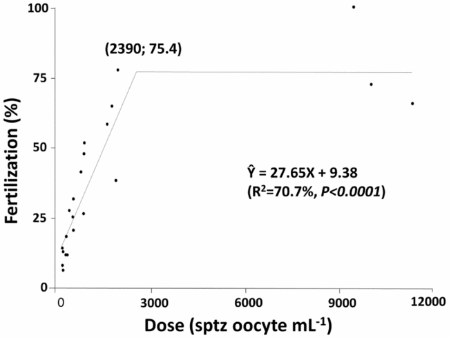
Regression analysis of the inseminating dose is shown on Fig. 1 and revealed that an increase in insemination dose promotes a linear increase in the fertilization rate up to a concentration of 2390 spermatozoa per oocyte per ml; at this point the fertilization rate was maximum, reaching 75.4%. After this point, the fertilization rate reached a ‘plateau’ in which an increase in insemination dose did not affect the fertilization rates (Fig. 1).
Insemination dose of Astyanax altiparanae. The results indicated that an insemination dose above 2390 sperm per oocyte does not increase fertilization rates above 75.4%.

Sperm motility
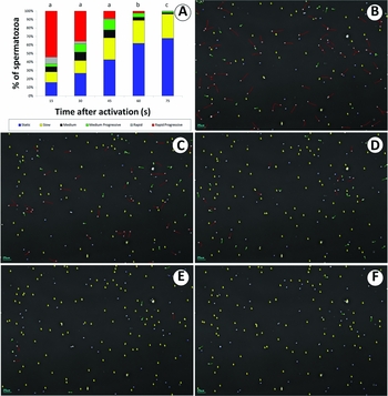
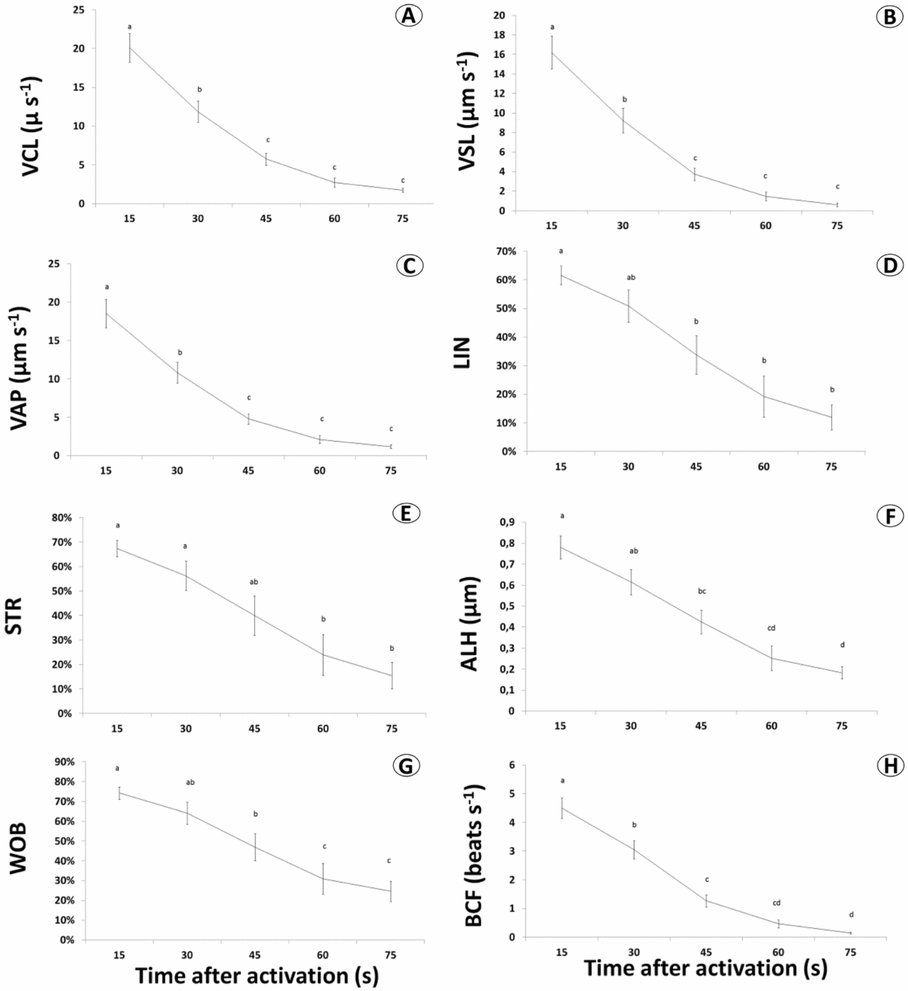
Sperm motility of Astyanax altiparanae is shown on Fig. 2. A quantitative analysis expressed by total motility, including all the motile cells, revealed that the sperm motility at 15 s after activation was 83.9 ± 3.1% and decreased significantly to 54.5 ± 5.5% only at 45 s (P = 0.0020). However, a more detailed analysis of sperm velocities indicated a rapid decrease in sperm quality. A general analysis of sperm motility indicates that the percentage of rapid progressive cells decreased abruptly (P = 0.0005) from 15 s (54.1 ± 3.3%) to 30 s (33.3 ± 2.0%). At 75 s, the percentage of sperm motility decreased to 29.4 ± 2.9% with predominantly ‘slow’ cells (26.1 ± 2.6%) and also non-motile cells (61.9 ± 8.7%). In agreement with these data, a decrease in velocity parameters was found from 15 to 30 s on curvilinear velocity (VCL) (20.1 ± 1.9 µm s−1 to 11.9 ± 1.4 µm s−1, P = 0.0005), progressive linear velocity (VSL) (16.2 ± 1.7 µm s−1 to 9.2 ± 1.3 µm s−1, P = 0.0008) and average velocity of trajectory (VAP) (18.5 ± 1.8 µm s−1 to 10.8 ± 1.3 µm s−1, P = 0007). Beat cross-frequency (BCF) also decreased from 4.5 ± 0.3 beats s−1 at 15 s to 3.1 ± 0.3 beats s−1 (P = 0.0034). This variation is also expressed on the motility analysis showing sperm traces (Fig. 3 A–H). At 15 s (Fig. 2 B), predominantly rapid progressive cells (red traces) were found but at 75 s (Fig. 2 F) most of the cells were static (blue dots) and slow cells (yellow traces).
Sperm motility of Astyanax altiparanae. Types of motility (A) measured at 15 s (B), 30 s (C), 45 s (D), 60 s (E) and 75 s (F) post-activation. On the right, spermatozoa traces during motility evaluation emphasizing all the cells recognized and analyzed by the software. Spermatozoa were classified as fast progressive (red traces, > 15 microns s−1), medium progressive (green traces, < 5 microns s−1), slow movement (yellow traces, < 2 microns s−1) and non-motile cells (blue dots).

Parameters related to sperm motility of Astyanax altiparanae. Curvilinear velocity (VCL) (A), progressive linear velocity (VSL) (B), average velocity of trajectory (VAP) (C), linearity (LIN) (D), straightness (STR) (E), amplitude of lateral head (ALH) (F), wobble (WOB) (G) and beat cross-frequency (BCF) (H) were measured at 15 s, 30 s, 45 s 60 s and 75 s post-activation.

Parameters related to linearity were maintained for longer periods. Linearity (LIN) (Fig. 3 D) decreased from 61.6 ± 3.2% at 15 s to 33.8 ± 6.7% at 45 s (P = 0.0175). STR (Fig. 3 E) was 67.4 ± 3.4% at 15 s and decreased only at 60 s to 23.9 ± 8.4% (P = 0.0012). Wobble (WOB) (Fig. 3 G) decreased significantly from 74.2 ± 3.1% at 15 s to 46.9 ± 6.8% at 45 s (P = 0.0280).
Ultrastructural analysis of oocytes
Astyanax altiparanae oocytes presented several grooves surrounding the micropyle (Fig. 4 A–F). Such grooves were observed under the stereomicroscope after egg hydration (Fig. 4 A) and also by scanning electron microscopy (SEM) (Fig. 4 B–F). The micropyle presented an approximate diameter of 7.57 µm. The average length of grooves from the micropyle in inactivated oocytes was 257,111 µm. The diameter of the oocyte before activation was 695,119 ± 49.5 µm, and after then was 1,170,909 µm. Interestingly, the grooves were centred in the micropyle region in which there is a projection giving rise to a central ‘ridge’ (Fig. 4 A).
Micropylar region of Astyanax altiparanae in fertilized (A) and unfertilized oocytes (B–F). In (A), developing embryo at blastula stage with chorion after hydration, showing the location of the grooves. General viewing of oocyte showing the micropyle surrounded by grooves by standard error of the mean (SEM) (B). Grooves conformation and a ‘ridge’ containing the micropyle in the central area (C). Detail of the ridge area containing the micropyle (D). Lateral view of the ridge (E). Micropylar channel (F).

Discussion
In fish, several sperm parameters correlated positively with successful fertilization, such as velocity, motility and beating frequency (which is also indicative of velocity) (Kime et al., Reference Kime, Van Look, McAllister, Huyskens, Rurangwa and Ollevier2001; Casselman et al., Reference Casselman, Schulte-Hostedde and Montgomerie2006; Gasparini et al., Reference Gasparini, Simmons, Beveridge and Evans2010; Galego et al., Reference Galego, Pérez, Asturiano and Yoshida2013), spermatozoon size (Gomendio & Roldan, Reference Gomendio and Roldan2008), sperm viability (Hunter & Birkhead, Reference Hunter and Birkhead2002). However, many authors indicated sperm velocity as the main factor affecting fertilization (Gage et al., Reference Gage, Macfarlane, Yeates, Ward, Searle and Parker2004; Hosken et al., Reference Hosken, Taylor, Hoyle, Higgins and Wedell2008; Rudolfsen et al., Reference Rudolfsen, Figenschou, Folstad and Kleven2008; Gasparini, et al., Reference Gasparini, Simmons, Beveridge and Evans2010). In freshwater teleosts, sperm motility is limited to a few seconds after activation (30–50 s for most species, see Cosson, Reference Cosson2004). According to Tuset et al. (Reference Tuset, Dietrich, Wojtczak, Slowinska, Monserrat and Ciereszko2008), fertilization success was related to the following variables: duration of sperm movement, linearity of movement, and sperm concentration.
Astyanax altiparanae spermatozoa remain motile for longer periods presenting good fertilization ability as expressed by sperm parameters. This characteristic improves the fertilization ability as the spermatozoa may have a chance to search for the micropyle for a longer period.
Regarding oocytes, the diameter is also related to fertilization as larger oocytes present an increased area in which sperm will need to seek the micropyle. Such fertilization efficacy is mainly expressed via the insemination dose (i.e. spermatozoa:oocyte ratio). A positive correlation was found within teleost species regarding oocyte diameter such as the Esox lucius (diameter of the oocyte = 2000 µm, inseminating dose = 700,000 sptz oocyte−1, Marciel, Reference Marciel1981), Brycon insignis (diameter of the oocyte = 1300 µm, insemination dose = 314,481 spermatozoa per oocyte, Shimoda et al., Reference Shimoda, Andrade, Vidal, Godinho and Yasui2007), Oncorhynchus mykiss (diameter of the oocyte = 4000 µm, inseminating dose = 200,000 spermatozoa per oocyte; Silveira et al., Reference Silveira, Kavamoto, Rigolino and Tabata1988), Hippoglossus (diameter of the oocyte = 3000 µm, inseminating dose = 1,600,000 spermatozoa per oocyte; Suquet et al., Reference Suquet, Billard and Cosson1995), Scophthalmus maximus (diameter of the oocyte = 1000 µm, insemination dose = 6000 spermatozoa per oocyte; Suquet et al., Reference Suquet, Billard and Cosson1995) and in our study species the diameter is also reduced (diameter of the oocyte = 695,119 µm, insemination dose = 2.390 spermatozoa per oocyte).
Other factors may affect the fertilization ability. Yanagimachi et al. (Reference Yanagimachi, Cherr, Matsubara, Andoh, Harumi, Vines, Pillai, Griffin, Matsubara, Weatherby and Kenneth2013) reported chemical attractants in the micropylar region that facilitates the sperm entrance and attempt fertilization in teleost fishes and insects. The authors suggested that although the sperm moved randomly in the water, a previous contact with the oocyte made the sperm to move on the oocyte surface and seek the micropyle. This type of sperm orientation is not unique to fish and has long been reported for other species and named as a contact reaction of spermatozoa (Lillie, Reference Lillie1919). In the medaka Oryzias latipes, similar attractants were reported by Iwamatsu et al. (Reference Iwamatsu, Ishijima and Nakashima2005), although Ishijima et al. (Reference Ishijima, Hamaguchi and Iwamatsu1993) did not observe any alteration in sperm velocity close to the micropylar area.
The number of micropyles and its diameter may also affect fertilization success. Teleosts present a single micropyle that differs from the multimicropylar oocytes within condrosteans (Linhart & Kudo, Reference Linhart and Kudo1997). Theoretically, the diameter of the micropyle may affect sperm entrance because a larger micropyle increases the probability of sperm entrance. However, teleosts present low variation in micropyle diameter as in Oryzias latipes (5.30 µm; Ishijima et al., Reference Ishijima, Hamaguchi and Iwamatsu1993), Fundulus heteroclitus (4–5 µm; Kuchnow & Scott, Reference Kuchnow and Scott2006), Saurida elongate (3.7 µm; Hirai, Reference Hirai1988), Oplegnathus fasciatus (6.2 µm; Hirai, Reference Hirai1988), Oncorhynchus mykiss (4 µm; Yanagimachi et al., Reference Yanagimachi, Cherr, Pillai and Baldwin1992) and the Astyanax altiparanae, in the present study (7.57 µm). As far as we know, there is no relationship between the diameter of the micropyle and fertilization success.
In other species, including Barbus conchonius (Amanze & Iyengar, Reference Amanze and Iyengar1990) structures called ‘ridges’ and ‘stretch marks’ were found in the micropylar area that were similar to the grooves found in Astyanax altiparanae and also in Astyanax bimaculatus (Rizzo et al., Reference Rizzo, Sato, Barreto and Godinho2002). Such structures may physically guide the sperm to the micropyle and this mechanism may improve fertilization success. In the grass goby Zosterisessor ophiocephalus, follicular cells produce a forest of attachment filaments up to 600 µm in length that surround the micropyle (Giulianini & Ferrero, Reference Giulianini and Ferrero2000) and is also a mechanism for sperm guidance.
In our study, it was also possible to describe a conical elevation on the central region of those grooves that surrounded the micropyle. Zebrafish Brachydanio rerio eggs also presented such a ridge in the micropylar area, as described by Hart (Reference Hart1990). In this study we found for the first time a combination of both structures. The ridges and grooves indicated that the sperm is guided by grooves to a ridge where the micropyle is found in the central region. In this mechanism, hydration physically blocks oocyte fertilization by two mechanisms. The first mechanism is similar to that in other species in which the micropyle is separated from the surface of the oocyte by increasing the perivitelline space. In the second mechanism, increase in the perivitelline space will reduce the area that contains the grooves, therefore reducing sperm guidance. The latter mechanism was also observed by Riehl & Patzner (Reference Riehl and Patzner1991) in catfish Sturisoma aureum, in which the grooves occupied almost the entire region of the oocyte.
In conclusion, we found that Astyanax altiparanae has one of the lowest insemination dose values among teleosts described in the literature. This fertilization efficacy is explained by: (1) reduced oocyte diameter; (2) increased micropyle diameter; (3) increased duration of sperm motility; and (4) the presence of grooves on the surface of the oocyte that guide sperm to the micropyle. In addition, we found a new morphology in oocytes that combined grooves and the micropyle ridge. Our findings are innovative in our species of study and bring new perspectives on the fertilization ability and subsequent insemination dose for fish with external fertilization.
Acknowledgements
We acknowledge CEPTA-ICMBio for kindly providing facilities and experimental fish and the Faculty of Medicine of Ribeirão Preto, University of São Paulo (USP), Brazil for the scanning electron microscopy analysis.
Funding
This work was supported by the São Paulo Research Foundation (FAPESP, Young Investigators Award Grant #2010/17429-1, Young Researcher Scholarship #2011/11664-1, and MSc Scholarship #2012/18961-4).