Introduction
Otology can be an unforgiving choice of specialty. The margin for surgical error is small, and the consequences of misjudgement may be devastating to the patient, with deafness, facial paralysis, disequilibrium, cerebrospinal fluid leak and meningitis as potential sequelae. Such ‘high stakes’ are reflected in the disproportionate anxiety reported by junior surgical trainees when joining the otology service for the first time, and in the concern expressed by many of our patients during the pre-operative consenting process.
Otological surgical skills are founded traditionally on many hours of supervised study in the temporal bone laboratory, and evolve as the trainee develops a three-dimensional appreciation of the healthy human temporal bone.
In the early stages of training, many juniors struggle to grasp the precise orientation of closely related anatomical structures. Persistence is encouraged, to ensure that good habits learned in the temporal bone laboratory foster the development of safe surgical technique in the operating theatre.
Once the trainee has an appreciation of what is normal, it is important to understand how the temporal bone may be altered by disease. The commonest destructive disease process encountered in the temporal bone is cholesteatoma. Effective tympano-mastoid surgery requires an understanding of how this disease can be removed safely, without compromising important structures and without leaving residual disease. For many, the first opportunity to appreciate the pathway of spread and the destructive capacity of a cholesteatoma is in the operating theatre itself.
For the patient newly diagnosed with cholesteatoma, there are also many questions to be answered before consent for surgery is given. Most patients will have few symptoms yet face a necessary consenting process disclosing a daunting list of potential complications, including unaidable hearing loss, permanent imbalance and facial weakness.
As surgeons, it is our responsibility to convey the risk of untreated cholesteatoma in terms that are meaningful to the patient, allowing them to make an informed choice from the options available to them.
Some surgeons use pen and paper to illustrate their explanations, while others use anatomical models or discuss computed tomography (CT) images in the hope that a realistic impression of risk and benefit is presented. The oto-endoscope and operating microscope alone may not always be helpful. Although these offer the patient a view of their ear canal, tympanic membrane and any retraction pocket that might exist, a relatively normal superficial appearance often belies the extent of deeper invasion and can confuse more than clarify the need for surgical intervention.
In this paper, we describe the application of contemporary segmentation software to CT scans of patients with cholesteatoma. Interactive, three-dimensional computer models were created, allowing the observer (either trainee surgeon or patient) to gain an immediate appreciation of the site and extent of cholesteatoma spread and its relationship to important adjacent structures.
Materials and methods
Axial and coronal CT scans were obtained for patients with cholesteatoma. The images, in a Digital Imaging and Communications in Medicine standard single-file format, were imported into Amira® version 4.1.2 software (Visage Imaging, Carlsbad, California, USA). This is a software platform for advanced three-dimensional visualisation and volume modelling, which allows images to be segmented semi-automatically by selecting the different regions of interest and assigning a particular colour to each structure. This was performed using a Cintiq 21UX tablet with interactive pen display (Wacom, Saitama, Japan). This liquid crystal display monitor has a completely flat surface, allowing smooth pen-on-screen interaction which increases efficiency. With practice, a basic study can be completed in approximately one hour.
Results and analysis
Case one: small, epitympanic cholesteatoma passing medial to the ossicles
This patient presented with near-normal hearing, recurrent otorrhoea with only a temporary response to topical antibiotic and steroid drops, and an attic pit visible on otoscopic examination.
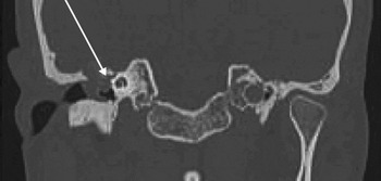
Computed tomography showed a small epitympanic mass suggestive of a small attic cholesteatoma (Figure 1). The rest of the middle-ear space was normal, as was the other ear.
Coronal computed tomography scan showing a small epitympanic cholesteatoma (arrow) (case one).

As the patient's ear had been dry since referral by his general practitioner, he was concerned that surgical exploration of his ear was being proposed, especially for such an apparently innocuous opacity on oto-endoscopy and imaging.
The most difficult part of the consenting process was to explain the probability that surgical exploration, although likely to succeed in making the patient's ear ‘safe’, would worsen his hearing, as the cholesteatoma was surrounding the ossicular chain and this would probably require the chain to be dismantled to remove the disease adequately.
After some discussion, the patient agreed to surgical exploration by atticotomy and attic reconstruction with autologous tragal cartilage. A cholesteatoma was removed together with the head of the malleus and the incus. An interposition ossiculoplasty (malleus handle to stapes head) was inserted, with an excellent hearing outcome.
At the time of writing, the patient had been followed up for over three years, and had shown no change in the hearing result and no evidence of recurrent or residual disease.
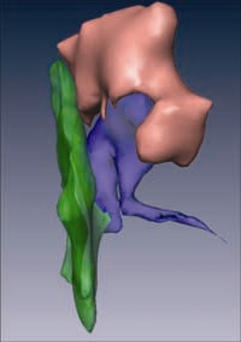
Prompted by difficulties experienced with this case, we used Amira software to segment this patient's CT images. This was quickly achieved. We concentrated our attention on the tympanic membrane, ossicles and the cholesteatoma mass, to produce a three-dimensional representation of the epitympanic invasion. The resultant three-dimensional model (Figure 2) was shown to the patient post-operatively. He commented on how much better his pre-operative understanding of the condition would have been had he seen this model at the time of consent.
Three-dimensional model rotated to show epitympanic disease wrapping around the head of the malleus and the body of the incus (case one).

Case two: large cholesteatoma complicated by lower motor neuron facial palsy at presentation
This patient was referred urgently having developed an acute lower motor neuron facial palsy, with a longer history of persistent but neglected otorrhoea and gradually failing hearing.
Examination confirmed a partial facial palsy and a featureless, deep meatus with infected, keratinous debris and granulations.
Computed tomography showed opacification of the middle-ear space and mastoid, with thinning of the tegmen and apparent dehiscence of the facial nerve in the tympanic segment (Figure 3).
Coronal computed tomography scan showing a large cholesteatoma with apparent dehiscence of the tympanic segment of the facial nerve and thinning of the tegmen (arrow) (case two).

Surgical exploration was planned following a frank discussion with the patient, who was surprised to be informed that his newly acquired facial weakness was related to his ear disease.
In this case, we segmented a number of additional structures in the three-dimensional model. Using the Digital Imaging and Communications in Medicine format images, the labyrinth, internal auditory canal, ossicular chain, facial nerve, tensor tympani tendon, carotid artery and sigmoid sinus were mapped, in addition to outlining the boundaries of the cholesteatoma. The lateral view clearly showed gross detail of the anterior epitympanic extension, illustrating the need to access the root of the zygoma to address the disease (Figure 4).
Three-dimensional model showing a large cholesteatoma with anterior epitympanic extension (arrow) (case two).

The segmentation software also allowed the pinna and the surface anatomy of the skull to be selected and made more or less transparent. This feature was useful in explaining basic anatomy to the patient, before conveying the depth of the disease process and the need for surgical treatment.
Tegmental erosion could be seen graphically when the model was rotated to the middle fossa view (Figure 5), and the relationship of the cholesteatoma to the facial canal became clear. We felt that this model was pivotal in enabling this patient to appreciate the seriousness of his condition, and in facilitating informed consent before surgery.
Three-dimensional model rotated to show middle fossa view of tegmental erosion (arrow) (case two).

Case three: large cholesteatoma with labyrinthine invasion
This patient presented with a history of otorrhoea and bouts of disequilibrium. He had been particularly dizzy after having his ear microsuctioned by the referring physician. His hearing had been reasonable until quite recently, but was now deteriorating.
Otological examination revealed an attic pocket filled with keratin. The fistula sign was positive.
Computed tomography confirmed complete opacification of a poorly aerated mastoid and invasion of the lateral semicircular canal (Figure 6). A segmented computer model was prepared, presenting an excellent, three-dimensional representation of this process (Figure 7).
Axial computed tomography scan showing a large cholesteatoma with tegmental erosion and labyrinthine invasion (case three).

Case three. (a) Three-dimensional model showing surface anatomy of the left pinna. Attic presentation of cholesteatoma can be seen through the ear canal. (b) Three-dimensional model with increased transparency of surface features. (c) Three-dimensional model with total transparency of surface soft tissues, revealing extensive mastoid involvement. (d) Three-dimensional model rotated to middle fossa view, showing invasion of the lateral semicircular canal.

Our trainees have found this type of model especially useful in comprehending the proximity of important structures in the temporal bone, and in understanding why multiple complications often occur at the same time. In case three, the cholesteatoma had breached the tegmen, invaded the lateral canal and extended into the anterior epitympanum, a site often addressed poorly by surgeons and consequently at risk of residual disease.
In this patient, the three-dimensional models enabled the consenting process to focus on the high risk of post-operative sensorineural loss, imbalance and cerebrospinal fluid leak, and the need for tegmental repair.
Discussion
The past few years have seen a surge of interest in temporal bone modelling. Consequently, there has been a substantial improvement in the quality of three-dimensional models available.Reference Ni, Sha, Dai and Li1–Reference Mason, Applebaum, Rasmussen, Millman, Evenhouse and Panko5
The three-dimensional models presented in this paper are not intended to compete with the fine detail evident in other reconstructions. Instead, by concentrating our attention on the relationship of real cholesteatomas to a few relevant structures, a valid appreciation of the clinical condition can be conveyed to students and patients alike.
A temporal bone can be segmented semi-automatically in approximately one hour. With planning, it is possible either to prepare personalised models for patients prior to their out-patient clinic visit, or to use generic examples of models showing common patterns of spread and complications (available from our website, http://ear-lab.medicine.dal.ca).
The three-dimensional models of the three cases described above offer a crude but visually engaging demonstration of cholesteatoma in the temporal bone, a condition the nature, extent and potential danger of which is difficult to convey to either students or patients without pictures.
We aimed to introduce the observer gradually to the complex anatomy of this region. The observer was first encouraged to recognise the familiar appearance and location of the external ear and the surface anatomy of the skull (Figure 7a), before external features were made more transparent (Figure 7b) to reveal deeper structures. Rotation, tilt and zoom features could also be used to improve the three-dimensional viewing experience.
Alternatively, different structures could be highlighted sequentially against the ‘transparent’ temporal bone. Finally, a cholesteatoma could be superimposed to give an impression of the extent of disease and its correlation with the clinical presentation. Using this stepwise approach, we hoped to encourage a gradual appreciation of how a cholesteatoma relates to the complex, three-dimensional anatomy of the temporal bone.
The use of computer-generated, three-dimensional anatomical models is now widespread. Although there have been previous, conflicting reports of their usefulness in the classroom, a recent and notable study which applied three-dimensional modelling to the middle and inner ear indicated a significant improvement in the performance of medical students on an ear anatomy test, after taking the three-dimensional tutorial.Reference Garg, Norman, Eva, Spero and Sharan6, Reference Nicholson, Chalk, Funnell and Daniel7 As Nicholson et al. suggest, there may be a significant novelty value to interactive models, which students find engaging, and they are perhaps thus more inclined to spend time on the subject matter, compared with controls.Reference Nicholson, Chalk, Funnell and Daniel7 It has already been acknowledged that students find computer-assisted anatomy lessons enjoyable.Reference Nieder and Nagy8
The patients' perspective
There is some debate as to what should constitute informed consent for ear, nose and throat surgery.Reference Albera, Argentero and Bonziglia9 The issue is particularly sensitive regarding patients requiring surgery for cholesteatoma; such patients may have few pre-operative symptoms, yet require surgery with a daunting list of potentially disfiguring and disabling complications. The medicolegal implications of poorly communicated consent should also not be underestimated.Reference Cherry and Weir10
Despite increased public scrutiny and abundant warnings from medicolegal defence organisations regarding the need for good consenting practice, evidence suggests that the process is often inadequate.Reference Haddow and Crowther11
Much has been written on the challenges of communicating pre-operative information to patients in a relevant, sympathetic and accurate fashion.Reference Dawes and Kitcher12 However, while it is important to warn patients of potential complications, it is equally important that they are not dissuaded from necessary intervention by an aggressively defensive approach.
Adjunctive measures have been introduced to augment the communication process.
It has been shown that care must be exercised in offering written information regarding surgical interventions, and that something more than a simple information sheet is probably required.Reference Kubba13, Reference Brown, Massoud and Bance14
The use of rough sketches in the out-patient clinic and conventional anatomical models of the ear can be disorientating and unfamiliar to the novice. However, a recent study assessing the ability of children to understand, assimilate and remember research information endorsed the use of simple language and pictures to reinforce verbal direction.Reference Tait, Voepel-Lewis and Malviya15
The widespread availability of computers in the clinic room provides the opportunity to employ more interactive formats to engage the patient in their consenting process. Such applications have already met with some success in other areas of otology, in which there has been a need to communicate concepts that at first appear difficult to grasp.Reference Morris and Rothera16
• The use of computer-generated, three-dimensional anatomical models is widespread in undergraduate medical education
• Interactive models may improve recall on simple tests
• This paper describes the creation of personalised, interactive, three-dimensional anatomical models from computed tomography data, to show the location and spread of cholesteatoma within the human temporal bone
• These models are useful not only for otology training but also to enhance the process and outcome of informed consent in patients
Conclusion
We prepared three-dimensional computer images, using patients' CT data, and found that these provided a crude but user-friendly representation of the spread of cholesteatoma within the temporal bone. Such three-dimensional images offer a relatively cheap and accessible resource, both for training junior surgeons and also for informing patients of their pre-operative condition so that they are able to make informed decisions about their care.
The three-dimensional models for the cases described in this paper can be viewed at the Ear and Auditory Research Laboratory website (http://ear-lab.medicine.dal.ca).
Acknowledgements
The authors wish to thank the Capital Health Research Fund, Halifax, Nova Scotia, Canada, whose financial support made this project possible.